Cited by 7

The DOI matches its Crossref record.

Structural Basis for the IgE-Binding Cross-Reacting Epitopic Peptides of Cup s 3, a PR-5 Thaumatin-like Protein Allergen from Common Cypress (Cupressus sempervirens) Pollen

Barre, Annick, Sénéchal, Hélène, Nguyen, Christophe, Granier, Claude, Poncet, Pascal, Rougé, Pierre·2023·Allergies

Abstract

The present work was aimed at identifying the IgE-binding epitopic regions on the surface of the Cup s 3 allergen from the common cypress Cupressus sempervirens, that are possibly involved in the IgE-binding cross-reactivity reported between Cupressaceae species. Three main IgE-binding epitopic regions were mapped on the molecular surface of Cup s 3, the PR-5 thaumatin-like allergen of common cypress Cupressus sempervirens. They correspond to exposed areas containing either electropositive (R, K) or electronegative (D, E) residues. A coalescence occurs between epitopes #1 and #2, that creates an extended IgE-binding regions on the surface of the allergen. Epitope #3 contains a putative N-glycosylation site which is actually glycosylated and could therefore comprise a glycotope. However, most of the allergenic potency of Cup s 3 depends on non-glycosylated epitopic peptides. The corresponding regions of thaumatin-like allergens from other closely related Cupressaceae (Cryptomeria, Juniperus, Thuja) exhibit a very similar conformation that should account for the IgE-binding cross-reactivity observed among the Cupressaceae allergens.

Overview

The claims and statistics of this paper haven’t been checked yet.

Run the check — free, no account needed. It reads the open-access text and checks the sources it cites.

A few free checks a day · papers already verified open instantly · results are public

Integrity signals

Registry

Crossref · 10.3390/allergies3010002

Cites

Statistics

Figures

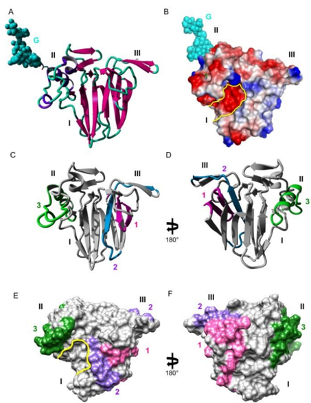
Figure 1 ( A ). Ribbon diagram of the Cup s 3 model showing the overall organization of the allergen in three distinct domains labeled I, II, and III, and the N - glycan chain (G) attached to residue Asn162 in domain II, represented in CPK colored cyan. ( B ). Molecular surface of Cup s 3 showing the electrostatic potentials patches. The electronegatively charged groove is outlined in yellow. ( C , D ). Mapping of the sequential IgE-binding epitopes 1 (colored pink), 2 (colored blue), and 3 (colored green) on the front face ( C ) and the back face ( D ) of the ribbon diagram of Cup s 3. ( E , F ). Mapping of the sequential IgE-binding epitopes 1 (colored pink), #2 (colored blue) and 3 (colored green) on the front face ( E ) and the back face ( F ) of the molecular surface of Cup s 3. Epitope 2 consists of two separate patches on the molecular surface of the TLP allergen. Note the coalescence of epitope 1 with both patches of epitope 2. Figure 1. ( A ). Ribbon diagram of the Cup s 3 model showing the overall organization of the allergen in three distinct domains labeled I, II, and III, and the N - glycan chain (G) attached to residue Asn162 in domain II, represented in CPK colored cyan. ( B ). Molecular surface of Cup s 3 showing the electrostatic potentials patches. The electronegatively charged groove is outlined in yellow. ( C , D ). Mapping of the sequential IgE-binding epitopes 1 (colored pink), 2 (colored blue), and 3 (colored green) on the front face ( C ) and the back face ( D ) of the ribbon diagram of Cup s 3. ( E , F ). Mapping of the sequential IgE-binding epitopes 1 (colored pink), #2 (colored blue) and 3 (colored green) on the front face ( E ) and the back face ( F ) of the molecular surface of Cup s 3. Epitope 2 consists of two separate patches on the molecular surface of the TLP allergen. Note the coalescence of epitope 1 with both patches of epitope 2.
Figure 1 ( A ). Ribbon diagram of the Cup s 3 model showing the overall organization of the allergen in three distinct domains labeled I, II, and III, and the N - glycan chain (G) attached to residue Asn162 in domain II, represented in CPK colored cyan. ( B ). Molecular surface of Cup s 3 showing the electrostatic potentials patches. The electronegatively charged groove is outlined in yellow. ( C , D ). Mapping of the sequential IgE-binding epitopes 1 (colored pink), 2 (colored blue), and 3 (colored green) on the front face ( C ) and the back face ( D ) of the ribbon diagram of Cup s 3. ( E , F ). Mapping of the sequential IgE-binding epitopes 1 (colored pink), #2 (colored blue) and 3 (colored green) on the front face ( E ) and the back face ( F ) of the molecular surface of Cup s 3. Epitope 2 consists of two separate patches on the molecular surface of the TLP allergen. Note the coalescence of epitope 1 with both patches of epitope 2. Figure 1. ( A ). Ribbon diagram of the Cup s 3 model showing the overall organization of the allergen in three distinct domains labeled I, II, and III, and the N - glycan chain (G) attached to residue Asn162 in domain II, represented in CPK colored cyan. ( B ). Molecular surface of Cup s 3 showing the electrostatic potentials patches. The electronegatively charged groove is outlined in yellow. ( C , D ). Mapping of the sequential IgE-binding epitopes 1 (colored pink), 2 (colored blue), and 3 (colored green) on the front face ( C ) and the back face ( D ) of the ribbon diagram of Cup s 3. ( E , F ). Mapping of the sequential IgE-binding epitopes 1 (colored pink), #2 (colored blue) and 3 (colored green) on the front face ( E ) and the back face ( F ) of the molecular surface of Cup s 3. Epitope 2 consists of two separate patches on the molecular surface of the TLP allergen. Note the coalescence of epitope 1 with both patches of epitope 2.CC-BY · From the PDF
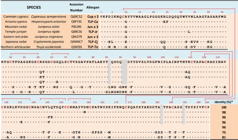
Table 2. Multiple amino-acid sequence alignments of seven thaumatin-like proteins from Cupressaceae pollen. The sequence of Jun v 3 is incomplete in the NCBI databank. Grey boxes correspond to the absence of insertion which are only present in TLP-Cj and TLP-To. Cystein residues are in red and red dotted lines indicate the eight disulfide bridges. Table 2. Multiple amino-acid sequence alignments of seven thaumatin-like proteins from Cupressaceae pollen. The sequence of Jun v 3 is incomplete in the NCBI databank. Grey boxes correspond to the absence of insertion which are only present in TLP-Cj and TLP-To. Cystein residues are in red and red dotted lines indicate the eight disulfide bridges.
Table 2. Multiple amino-acid sequence alignments of seven thaumatin-like proteins from Cupressaceae pollen. The sequence of Jun v 3 is incomplete in the NCBI databank. Grey boxes correspond to the absence of insertion which are only present in TLP-Cj and TLP-To. Cystein residues are in red and red dotted lines indicate the eight disulfide bridges. Table 2. Multiple amino-acid sequence alignments of seven thaumatin-like proteins from Cupressaceae pollen. The sequence of Jun v 3 is incomplete in the NCBI databank. Grey boxes correspond to the absence of insertion which are only present in TLP-Cj and TLP-To. Cystein residues are in red and red dotted lines indicate the eight disulfide bridges.CC-BY · From the PDF
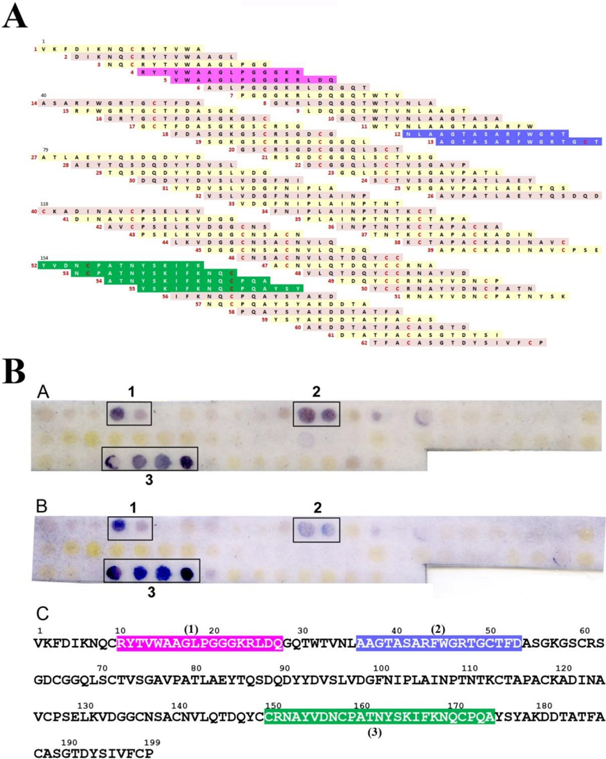
Figure 2 ( A ). Amino acid sequences of the 62 overlapping peptides used in the SPOT technique. Epitope 1 (pink background), 2 (purple background), and 3 (green background) are indicated. Cystein residues are in red. Peptide numbering in red. ( BA , BB ). Examples of SPOT results obtained with two different sera from cypress allergic patients used as a probe. ( BC ). Sequential IgE-binding epitopic stretches 1, 2, and 3 deduced from the stained spots are indicated in colored boxed white Figure 2. ( A ). Amino acid sequences of the 62 overlapping peptides used in the SPOT technique. Epitope 1 (pink background), 2 (purple background), and 3 (green background) are indicated. Cystein residues are in red. Peptide numbering in red. ( BA , BB ). Examples of SPOT results obtained with two different sera from cypress allergic patients used as a probe. ( BC ). Sequential IgE-binding epitopic stretches 1, 2, and 3 deduced from the stained spots are indicated in colored boxed white letters along the amino acid sequence of Cup s 3.
Figure 2 ( A ). Amino acid sequences of the 62 overlapping peptides used in the SPOT technique. Epitope 1 (pink background), 2 (purple background), and 3 (green background) are indicated. Cystein residues are in red. Peptide numbering in red. ( BA , BB ). Examples of SPOT results obtained with two different sera from cypress allergic patients used as a probe. ( BC ). Sequential IgE-binding epitopic stretches 1, 2, and 3 deduced from the stained spots are indicated in colored boxed white Figure 2. ( A ). Amino acid sequences of the 62 overlapping peptides used in the SPOT technique. Epitope 1 (pink background), 2 (purple background), and 3 (green background) are indicated. Cystein residues are in red. Peptide numbering in red. ( BA , BB ). Examples of SPOT results obtained with two different sera from cypress allergic patients used as a probe. ( BC ). Sequential IgE-binding epitopic stretches 1, 2, and 3 deduced from the stained spots are indicated in colored boxed white letters along the amino acid sequence of Cup s 3.CC-BY · From the PDF
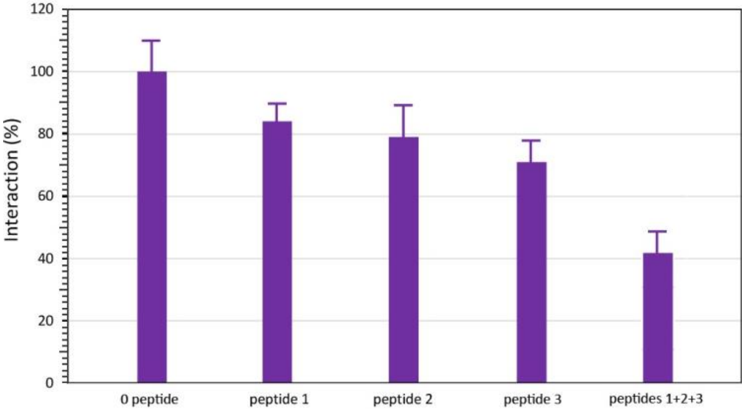
Figure 3 ELISA inhibition of the interaction between cypress pollen e in the presence of 2 mM of synthetic peptides corresponding 9PGGGKRLDQ27), epitope 2 (peptide 2: 45RFWGRTGIT53), Figure 3. ELISA inhibition of the interaction between cypress pollen extract and IgE from CPAPs, in the presence of 2 mM of synthetic peptides corresponding to epitope 1 (peptide 1: 9PGGGKRLDQ27), epitope 2 (peptide 2: 45RFWGRTGIT53), epitope 3 (peptide 3: 161TNYSKIFKN169), and an equimolar mixture of the three peptides 1, 2, and 3 (peptides 1 + 2 + 3). All values are the mean ± s.d. of three separate measurements performed in the same experimental conditions.
Figure 3 ELISA inhibition of the interaction between cypress pollen e in the presence of 2 mM of synthetic peptides corresponding 9PGGGKRLDQ27), epitope 2 (peptide 2: 45RFWGRTGIT53), Figure 3. ELISA inhibition of the interaction between cypress pollen extract and IgE from CPAPs, in the presence of 2 mM of synthetic peptides corresponding to epitope 1 (peptide 1: 9PGGGKRLDQ27), epitope 2 (peptide 2: 45RFWGRTGIT53), epitope 3 (peptide 3: 161TNYSKIFKN169), and an equimolar mixture of the three peptides 1, 2, and 3 (peptides 1 + 2 + 3). All values are the mean ± s.d. of three separate measurements performed in the same experimental conditions.CC-BY · From the PDF
Image 5
CC-BY · From the PDF
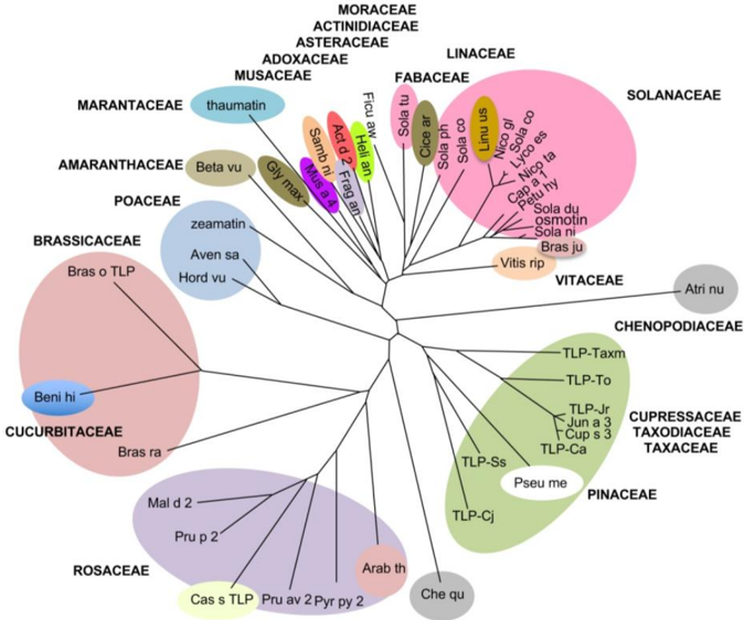
Figure 5 Unrooted phylogenetic tree built up from the multiple alignment of thaumatin-like proteins of Actinidia deliciosa (Act d 2), Arabidopsis thaliana (Arab th), Atriplex numularia (Atri nu), Avena sativa (Aven sa), Benincasa hispida (Beni hi), Beta vulgaris (Beta vu), Brassica juncea (Bras ju), Brassica oleracea (Bras ol), Brassica rapa (Bras ra), Capsicum annuum (Cap a 1), Castanea sativa (Cas s TLP), Chenopodium quinoa (Che qu), Cicer arietinum (Cice ar), Cryptomeria japonica (TLP-Cj), Cupressus arizonica (TLP-Ca), Cupressus sempervirens (Cup s 3), Fragaria x ananassa (Frag an), Ficus awkeotsang (Ficu aw), Glycine max (Glyc ma), Helianthus annuus (Heli an), Hordeum vulgare (Hord vu), Juniperus ashei (Jun a 3), Juniperus rigida (TLP-Jr), Linum usitatissimum (Linu us), Lycopersicum esculentum (Lyco es), Malus domestica (Mal d 2), Musa acuminata (Mus a 4), Nicotiana glauca (Nico gl), Nicotiana tabacum (osmotin), Petunia hybrida (Petu hy), Prunus avium (Pru av 2), Prunus persica (Pru p 2), Pseudotsuga menziesii (Pseu me), Pyrus pyriformis (Pyr py 2), Sambucus nigra (Samb ni), Sequoia sempervirens (TLPSs), Solanum commersonii (Sola co), Solanum dulcamara (Sola du), Solanum nigrum (Sola ni), Solanum phyreja (Sola ph), Solanum tuberosum (Sola tu), Taxodium distichum (TLP-Txm), Thaumatococcus daniellii (thaumatin), Thuja occidentalis (TLP-To), Vitis riparia (Viti ri), Vitis vinifera (Vit v TLP), Zea mays (zeamatin). Thaumatin-like proteins belonging to the same plant family are boxed and the different family boxes are labeled and differently colored. Figure 5. Unrooted phylogenetic tree built up from the multiple alignment of thaumatin-like proteins of Actinidia deliciosa (Act d 2), Arabidopsis thaliana (Arab th), Atriplex numularia (Atri nu), Avena sativa (Aven sa), Benincasa hispida (Beni hi), Beta vulgaris (Beta vu), Brassica juncea (Bras ju), Brassica oleracea (Bras ol), Brassica rapa (Bras ra), Capsicum annuum (Cap a 1), Castanea sativa (Cas s TLP), Chenopodium quinoa (Che qu), Cicer arietinum (Cice ar), Cryptomeria japonica (TLP-Cj), Cupressus arizonica (TLP-Ca), Cupressus sempervirens (Cup s 3), Fragaria × ananassa (Frag an), Ficus awkeotsang (Ficu aw), Glycine max (Glyc ma), Helianthus annuus (Heli an), Hordeum vulgare (Hord vu), Juniperus ashei (Jun a 3), Juniperus rigida (TLP-Jr), Linum usitatissimum (Linu us), Lycopersicum esculentum (Lyco es), Malus domestica (Mal d 2), Musa acuminata (Mus a 4), Nicotiana glauca (Nico gl), Nicotiana tabacum (osmotin), Petunia hybrida (Petu hy), Prunus avium (Pru av 2), Prunus persica (Pru p 2), Pseudotsuga menziesii (Pseu me), Pyrus pyriformis (Pyr py 2), Sambucus nigra (Samb ni), Sequoia sempervirens (TLP-Ss), Solanum commersonii (Sola co), Solanum dulcamara (Sola du), Solanum nigrum (Sola ni), Solanum phyreja (Sola ph), Solanum tuberosum (Sola tu), Taxodium distichum (TLP-Txm), Thaumatococcus daniellii (thaumatin), Thuja occidentalis (TLP-To), Vitis riparia (Viti ri), Vitis vinifera (Vit v TLP), Zea mays (zeamatin). Thaumatinlike proteins belonging to the same plant family are boxed and the different family boxes are labeled and differently colored.
Figure 5 Unrooted phylogenetic tree built up from the multiple alignment of thaumatin-like proteins of Actinidia deliciosa (Act d 2), Arabidopsis thaliana (Arab th), Atriplex numularia (Atri nu), Avena sativa (Aven sa), Benincasa hispida (Beni hi), Beta vulgaris (Beta vu), Brassica juncea (Bras ju), Brassica oleracea (Bras ol), Brassica rapa (Bras ra), Capsicum annuum (Cap a 1), Castanea sativa (Cas s TLP), Chenopodium quinoa (Che qu), Cicer arietinum (Cice ar), Cryptomeria japonica (TLP-Cj), Cupressus arizonica (TLP-Ca), Cupressus sempervirens (Cup s 3), Fragaria x ananassa (Frag an), Ficus awkeotsang (Ficu aw), Glycine max (Glyc ma), Helianthus annuus (Heli an), Hordeum vulgare (Hord vu), Juniperus ashei (Jun a 3), Juniperus rigida (TLP-Jr), Linum usitatissimum (Linu us), Lycopersicum esculentum (Lyco es), Malus domestica (Mal d 2), Musa acuminata (Mus a 4), Nicotiana glauca (Nico gl), Nicotiana tabacum (osmotin), Petunia hybrida (Petu hy), Prunus avium (Pru av 2), Prunus persica (Pru p 2), Pseudotsuga menziesii (Pseu me), Pyrus pyriformis (Pyr py 2), Sambucus nigra (Samb ni), Sequoia sempervirens (TLPSs), Solanum commersonii (Sola co), Solanum dulcamara (Sola du), Solanum nigrum (Sola ni), Solanum phyreja (Sola ph), Solanum tuberosum (Sola tu), Taxodium distichum (TLP-Txm), Thaumatococcus daniellii (thaumatin), Thuja occidentalis (TLP-To), Vitis riparia (Viti ri), Vitis vinifera (Vit v TLP), Zea mays (zeamatin). Thaumatin-like proteins belonging to the same plant family are boxed and the different family boxes are labeled and differently colored. Figure 5. Unrooted phylogenetic tree built up from the multiple alignment of thaumatin-like proteins of Actinidia deliciosa (Act d 2), Arabidopsis thaliana (Arab th), Atriplex numularia (Atri nu), Avena sativa (Aven sa), Benincasa hispida (Beni hi), Beta vulgaris (Beta vu), Brassica juncea (Bras ju), Brassica oleracea (Bras ol), Brassica rapa (Bras ra), Capsicum annuum (Cap a 1), Castanea sativa (Cas s TLP), Chenopodium quinoa (Che qu), Cicer arietinum (Cice ar), Cryptomeria japonica (TLP-Cj), Cupressus arizonica (TLP-Ca), Cupressus sempervirens (Cup s 3), Fragaria × ananassa (Frag an), Ficus awkeotsang (Ficu aw), Glycine max (Glyc ma), Helianthus annuus (Heli an), Hordeum vulgare (Hord vu), Juniperus ashei (Jun a 3), Juniperus rigida (TLP-Jr), Linum usitatissimum (Linu us), Lycopersicum esculentum (Lyco es), Malus domestica (Mal d 2), Musa acuminata (Mus a 4), Nicotiana glauca (Nico gl), Nicotiana tabacum (osmotin), Petunia hybrida (Petu hy), Prunus avium (Pru av 2), Prunus persica (Pru p 2), Pseudotsuga menziesii (Pseu me), Pyrus pyriformis (Pyr py 2), Sambucus nigra (Samb ni), Sequoia sempervirens (TLP-Ss), Solanum commersonii (Sola co), Solanum dulcamara (Sola du), Solanum nigrum (Sola ni), Solanum phyreja (Sola ph), Solanum tuberosum (Sola tu), Taxodium distichum (TLP-Txm), Thaumatococcus daniellii (thaumatin), Thuja occidentalis (TLP-To), Vitis riparia (Viti ri), Vitis vinifera (Vit v TLP), Zea mays (zeamatin). Thaumatinlike proteins belonging to the same plant family are boxed and the different family boxes are labeled and differently colored.CC-BY · From the PDF

Verification

Claims the paper makes

The claims and statistics of this paper haven’t been checked yet.

Run the check — free, no account needed. It reads the open-access text and checks the sources it cites.

A few free checks a day · papers already verified open instantly · results are public

Claims other papers make about this one

Each is checked against this paper’s own text: does it say what the citing paper says it does?

Self-check

Percentages, sums and reported differences re-computed from the paper’s own numbers.

The self-check needs the paper’s full text.

Citations

Cites

No references could be resolved yet.

Research works printed but not linked: 51

  1. Cecchi, 2007, Allergenic pollen and pollen allergy in Europe, Allergy, 62, 976, 10.1111/j.1398-9995.2007.01393.x · DOI · Paper page
  2. Bibi, 2002, Comparison of positive allergy skin tests among asthmatic children from rural and urban areas living within small geographic area, Ann. Allergy Asthma Immunol., 88, 416, 10.1016/S1081-1206(10)62374-5 · DOI
  3. Gonianakis, 2006, A 10-year aerobiological study (1994–2003) in the Mediterranean island of Crete, Greece: Trees, aerobiologic data, and botanical and clinical correlations, Allergy Asthma Proc., 27, 371, 10.2500/aap.2006.27.2935 · DOI
  4. Rapp, 2011, Aerobiological study in east-central Iberian Peninsula: Pollen diversity and dynamics for major taxa, Ann. Agric. Environ. Med., 18, 99
  5. Lamy, 2001, Ten years of cypress pollen counts in Marseille, Allergol. Immunopathol., 33, 103
  6. Kishikawa, 2017, Longitudinal monitoring of tree airborne pollen in japan, Arerugi, 66, 97
  7. Pham, 1994, Cypress pollen allergy. Identification of allergens and crossreactivity between divergent species, Clin. Exp. Allergy, 24, 558, 10.1111/j.1365-2222.1994.tb00953.x · DOI
  8. Mohanty, 2017, Molecular approaches for the analysis of airborne pollen: A case study of Juniperus pollen, Ann. Allergy Asthma Immunol., 118, 204, 10.1016/j.anai.2016.11.015 · DOI
  9. Guerin, 1996, Allergic Rhinitis to Thuja Pollen, Int. Arch. Allergy Immunol., 110, 91, 10.1159/000237317 · DOI
  10. Iacovacci, 1998, Juniperus oxycedrus: A new allergenic pollen from the Cupressaceae family, J. Allergy Clin. Immunol., 101, 755, 10.1016/S0091-6749(98)70304-5 · DOI
  11. Sneller, 1993, Pollen changes during five decades of urbanization in Tucson, Arizona, Ann. Allergy, 71, 519
  12. Charpin, 2013, Cypress pollen allergy, Rev. Mal. Respir., 30, 868, 10.1016/j.rmr.2013.09.014 · DOI
  13. Charpin, 2019, Cypress Pollinosis: From Tree to Clinic, Clin. Rev. Allergy Immunol., 56, 174, 10.1007/s12016-017-8602-y · DOI · Paper page
  14. Cortegano, 2004, Cloning and expression of a major allergen from Cupressus arizonica pollen, Cup a 3, a PR-5 protein expressed under polluted environment, Allergy, 59, 485, 10.1046/j.1398-9995.2003.00363.x · DOI
  15. Svoboda, 2018, A new allergen family involved in pollen food-associated syndrome: Snakin/gibberellin-regulated proteins, J. Allergy Clin. Immunol., 141, 411, 10.1016/j.jaci.2017.06.041 · DOI · Paper page
  16. Iizuka, 2021, Gibberellin-regulated protein sensitization in Japanese cedar (Cryptomeria japonica) pollen allergic Japanese cohorts, Allergy, 76, 2297, 10.1111/all.14816 · DOI
  17. Ehrenberg, 2020, Characterization of a 7 kDa pollen allergen belonging to the gibberellin-regulated protein family from three Cupressaceae species, Clin. Exp. Allergy, 50, 964, 10.1111/cea.13675 · DOI
  18. Keykhosravi, 2019, Pollen/Fruit Syndrome: Clinical Relevance of the Cypress Pollen Allergenic Gibberellin-Regulated Protein, Allergy Asthma Immunol. Res., 11, 143, 10.4168/aair.2019.11.1.143 · DOI
  19. Takei, 2022, Capsicum Allergy: Involvement of Cap a 7, a New Clinically Relevant Gibberellin-Regulated Protein Cross-Reactive With Cry j 7, the Gibberellin-Regulated Protein From Japanese Cedar Pollen, Allergy Asthma Immunol. Res., 14, 328, 10.4168/aair.2022.14.3.328 · DOI
  20. Panzani, 1986, Cross-reactivity between the pollens of Cupressus sempervirens (common cypress) and of Cryptomeria japonica (Japanese cedar), Ann. Allergy, 57, 26
  21. Breiteneder, 2004, Thaumatin-like proteins—A new family of pollen and fruit allergens, Allergy, 59, 479, 10.1046/j.1398-9995.2003.00421.x · DOI
  22. Ivanciuc, 2003, Detecting Potential IgE-Reactive Sites on Food Proteins Using a Sequence and Structure Database, SDAP-Food, J. Agric. Food Chem., 51, 4830, 10.1021/jf034218r · DOI
  23. Fujimura, 2007, Isolation and characterization of native Cry j 3 from Japanese cedar (Cryptomeria japonica) pollen, Allergy, 62, 547, 10.1111/j.1398-9995.2007.01331.x · DOI
  24. Palacin, A., Rivas, L.A., Gómez-Casado, C., Aguirre, J., Tordesillas, L., Bartra, J., Blanco, C., Carrillo, T., Cuesta-Herranz, J., and Bonny, J.A.C. (2012). The Involvement of Thaumatin-Like Proteins in Plant Food Cross-Reactivity: A Multicenter Study Using a Specific Protein Microarray. PLoS ONE, 7. · DOI · Paper page
  25. Goldblum, 2000, Variable Expression of Pathogenesis-Related Protein Allergen in Mountain Cedar (Juniperus ashei) Pollen, J. Immunol., 164, 2188, 10.4049/jimmunol.164.4.2188 · DOI
  26. Togawa, 2006, Identification of Italian cypress (Cupressus sempervirens) pollen allergen Cup s 3 using homology and cross-reactivity, Ann. Allergy Asthma Immunol., 97, 336, 10.1016/S1081-1206(10)60798-3 · DOI
  27. Smith, 1987, Measurement of protein using bicinchoninic acid, Anal. Biochem., 150, 76, 10.1016/0003-2697(85)90442-7 · DOI · Paper page
  28. Barre, 2000, Purification and structural analysis of an abundant thaumatin-like protein from ripe banana fruit, Planta, 211, 791, 10.1007/s004250000354 · DOI
  29. Tastet, 2003, A versatile electrophoresis system for the analysis of high- and low-molecular-weight proteins, Electrophoresis, 24, 1787, 10.1002/elps.200305400 · DOI
  30. Barre, 2022, Identification of Potential IgE-Binding Epitopes Contributing to the Cross-Reactivity of the Major Cupressaceae Pectate-Lyase Pollen Allergens (Group 1), Allergies, 2, 106, 10.3390/allergies2030010 · DOI
  31. Frank, 1992, Spot-synthesis: An easy technique for the positionally addressable paralel chemical synthesis on a membrane support, Tetrehedron, 48, 9217, 10.1016/S0040-4020(01)85612-X · DOI
  32. Laune, 2002, Application of the Spot method to the identification of peptides and amino acids from the antibody paratope that contribute to antigen binding, J. Immunol. Methods, 267, 53, 10.1016/S0022-1759(02)00140-0 · DOI
  33. Thompson, 1997, The CLUSTAL_X windows interface: Flexible strategies for multiple sequence alignment aided by quality analysis tools, Nucleic Acids Res., 25, 4876, 10.1093/nar/25.24.4876 · DOI
  34. Krieger, 2002, Increasing the precision of comparative models with YASARA NOVA—A self-parameterizing force field, Proteins, 47, 393, 10.1002/prot.10104 · DOI
  35. Leone, 2006, Resolution of the structure of the allergenic and antifungal banana fruit thaumatin-like protein at 1.7-Å, Biochimie, 88, 45, 10.1016/j.biochi.2005.07.001 · DOI
  36. Marangon, M., Van Sluyter, S.C., Waters, E.J., and Menz, R.I. (2014). Structure of Haze Forming Proteins in White Wines: Vitis vinifera Thaumatin-Like Proteins. PLoS ONE, 9. · DOI
  37. Batalia, 1996, The crystal structure of the antifungal protein zeamatin, a member of the thaumatin-like, PR-5 protein family, Nat. Struct. Biol., 3, 19, 10.1038/nsb0196-19 · DOI
  38. Laskowski, 1993, PROCHECK: A program to check the stereochemical quality of protein structures, J. Appl. Cryst., 26, 283, 10.1107/S0021889892009944 · DOI · Paper page
  39. Melo, 1998, Assessing protein structures with a non-local atomic interaction energy, J. Mol. Biol., 277, 1141, 10.1006/jmbi.1998.1665 · DOI
  40. Arnold, 2006, The SWISS-MODEL workspace: A web-based environment for protein structure homology modelling, Bioinformatics, 22, 195, 10.1093/bioinformatics/bti770 · DOI
  41. Benkert, 2011, Toward the estimation of the absolute quality of individual protein structure models, Bioinformatics, 27, 343, 10.1093/bioinformatics/btq662 · DOI
  42. Pettersen, 2004, UCSF Chimera—A visualization system for exploratory research and analysis, J. Comput. Chem., 25, 1605, 10.1002/jcc.20084 · DOI
  43. Vriet, 2003, A molecular basis for the endo-beta 1,3-glucanase activity of the thaumatin-like proteins from edible fruits, Biochimie, 85, 123, 10.1016/S0300-9084(03)00058-0 · DOI
  44. Bistoni, 2005, Biochemical and immunological characterization of pollen-derived beta-galactosidase reveals a new cross-reactive class of allergens among Mediterranean trees, Int. Arch. Allergy Immunol., 136, 123, 10.1159/000083319 · DOI
  45. Shahali, 2012, Differential IgE sensitization to cypress pollen associated to a basic allergen of 14 kDa, FEBS J., 279, 1445, 10.1111/j.1742-4658.2012.08536.x · DOI
  46. Soman, 2000, Homology Modeling and Characterization of IgE Binding Epitopes of Mountain Cedar Allergen Jun a 3, Biophys. J., 79, 161, 10.1016/S0006-3495(00)76410-1 · DOI
  47. Afferni, 1999, Role of carbohydrate moieties in IgE binding to allergenic components of Cupressus arizonica pollen extract, Clin. Exp. Allergy, 29, 1087, 10.1046/j.1365-2222.1999.00590.x · DOI
  48. Barletta, 1998, Arizona cypress (Cupressus arizonica) pollen allergens. Identification of crossreactive periodate-resistant and sensitive epitopes with monoclonal antibodies, Allergy, 53, 586, 10.1111/j.1398-9995.1998.tb03934.x · DOI
  49. Canini, 2004, Localisation of a carbohydrate epitope recognised by human IgE in pollen of Cupressaceae, J. Plant Res., 117, 147, 10.1007/s10265-003-0139-x · DOI
  50. Iacovacci, 2002, Comparison between the native glycosylated and the recombinant Cup a1 allergen: Role of carbohydrates in the histamine release from basophils, Clin. Exp. Allergy, 32, 1620, 10.1046/j.1365-2222.2002.01516.x · DOI
  51. Shahali, 2017, Identification of a polygalacturonase (Cup s 2) as the major CCD-bearing allergen in Cupressus sempervirens pollen, Allergy, 72, 1806, 10.1111/all.13191 · DOI

Cited by

Sorted by citations

Metadata

Authors
Barre, A., Sénéchal, H., Nguyen, C., Granier, C., Poncet, P., & Rougé, P.
Affiliations
  • UMR 152 PharmaDev, Research and Development Institute, Paul Sabatier University, 35 Chemin des Maraîchers, CEDEX 09, 31062 Toulouse, France
  • Allergy & Environment Research Team, Biochemistry Department, Armand Trousseau Children Hospital, AP-HP, 26 Avenue Dr Arnold Netter, 75571 Paris, France
  • Institut des Biomolécules Max Mousseron CNRS, Faculté de Pharmacie, 15 Avenue Charles Flahault, BP 14491, CEDEX 5, 34093 Montpellier, France
  • Sys2Diag UMR 9005 CNRS/ALCEDIAG Complex System Modeling and Engineering for Diagnosis, Cap Delta/Parc Euromédecine, 1682 rue de la Valsière CS 61003, CEDEX 4, 34184 Montpellier, France
Venue
Allergies
Publisher
MDPI AG
Published
2023-01-10
Volume
3
Issue
1
Pages
11-24
ISSN
2313-5786
Cited by
7
References
0
Language
en

About these checks

Registry, retraction, Cites and statistics results are computed, not judged, with one exception: a printed reference that no index lists is read once by a model before it counts as a citation error. Claims are extracted by a model and kept only when their quote appears verbatim in the text; each cited source is then checked, with a model’s help, for whether it says what the claim relies on. Those verdicts can be wrong — read the claim’s quote and the cited source before relying on one.

Statistics and arithmetic are recomputed, not judged.

Confidence bands are shown only for fields calibrated on our annotated test set. None is calibrated yet, so no band is shown.

What counts as a claim ›How we measure confidence ›How we check this ›

Are you an author, or do you see a problem with this page? Request a review