

The DOI matches its Crossref record; 2 of the 2 references it cites were found in a registry or an index.
Microscopy Methods for Biofilm Imaging: Focus on SEM and VP-SEM Pros and Cons
Abstract
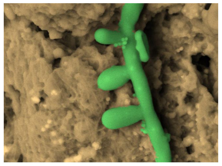
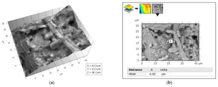
Several imaging methodologies have been used in biofilm studies, contributing to deepening the knowledge on their structure. This review illustrates the most widely used microscopy techniques in biofilm investigations, focusing on traditional and innovative scanning electron microscopy techniques such as scanning electron microscopy (SEM), variable pressure SEM (VP-SEM), environmental SEM (ESEM), and the more recent ambiental SEM (ASEM), ending with the cutting edge Cryo-SEM and focused ion beam SEM (FIB SEM), highlighting the pros and cons of several methods with particular emphasis on conventional SEM and VP-SEM. As each technique has its own advantages and disadvantages, the choice of the most appropriate method must be done carefully, based on the specific aim of the study. The evaluation of the drug effects on biofilm requires imaging methods that show the most detailed ultrastructural features of the biofilm. In this kind of research, the use of scanning electron microscopy with customized protocols such as osmium tetroxide (OsO4), ruthenium red (RR), tannic acid (TA) staining, and ionic liquid (IL) treatment is unrivalled for its image quality, magnification, resolution, minimal sample loss, and actual sample structure preservation. The combined use of innovative SEM protocols and 3-D image analysis software will allow for quantitative data from SEM images to be extracted; in this way, data from images of samples that have undergone different antibiofilm treatments can be compared.
Overview
The claims and statistics of this paper haven’t been checked yet.
Run the check — free, no account needed. It reads the open-access text and checks the sources it cites.
Integrity signals
Registry
Cites
Statistics
Figures
Verification
Claims the paper makes
The claims and statistics of this paper haven’t been checked yet.
Run the check — free, no account needed. It reads the open-access text and checks the sources it cites.
Claims other papers make about this one
Each is checked against this paper’s own text: does it say what the citing paper says it does?
In the citing paper
“Of various methods available for qualitative structural BFI detection on abiotic or biotic surfaces, scanning electron microscopy (SEM)-based detection of BFI structure is considered the gold standard.20-22”
From SEMTWIST Quantification of Biofilm Infection in Human Chronic Wound Using Scanning Electron Microscopy and Machine Learning (2025) · cites this paper as [21]
In the citing paper
“This resolution allows for in-depth examination of fine bacterial surface features, biofilm architecture, and nanoscale bacterial interactions.21”
From SEMTWIST Quantification of Biofilm Infection in Human Chronic Wound Using Scanning Electron Microscopy and Machine Learning (2025) · cites this paper as [21]
In the citing paper
“Conventional SEM and FESEM are the two rigorous methods for high magnification and high-resolution visualization of biofilm morphology.21”
From SEMTWIST Quantification of Biofilm Infection in Human Chronic Wound Using Scanning Electron Microscopy and Machine Learning (2025) · cites this paper as [21]
Self-check
Percentages, sums and reported differences re-computed from the paper’s own numbers.
The self-check needs the paper’s full text.
Citations
Cites
Oral Microbial Communities: Biofilms, Interactions, and Genetic Systems
Kolenbrander, P. E. (2000) · Annual Review of Microbiology · doi:10.1146/annurev.micro.54.1.413 · Cited by 894
Cabral, V., Znaidi, S., Walker, L. A., Martin-Yken, H., Dague, E., Legrand, M., Lee, K., Chauvel, M., Firon, A., Rossignol, T., et al. (2014) · PLoS Pathogens · doi:10.1371/journal.ppat.1004542 · Cited by 59
Research works printed but not linked: 109
- Costerton, 1978, How Bacteria Stick, Sci. Am., 238, 86, 10.1038/scientificamerican0178-86 · DOI
- Costerton, 1995, Microbial Biofilms, Annu. Rev. Microbiol., 49, 711, 10.1146/annurev.mi.49.100195.003431 · DOI
- Shapiro, 1998, Thinking about bacterial populations as multicellular organisms, Annu. Rev. Microbiol., 52, 81, 10.1146/annurev.micro.52.1.81 · DOI · Paper page
- 2018, Extracellular polymeric substances, a key element in understanding biofilm phenotype, AIMS Microbiol., 4, 274, 10.3934/microbiol.2018.2.274 · DOI
- Karygianni, 2020, Biofilm Matrixome: Extracellular Components in Structured Microbial Communities, Trends Microbiol., 28, 668, 10.1016/j.tim.2020.03.016 · DOI
- Yadav, M.K., and Singh, B.P. (2020). Chapter 1—Microbial biofilms and human disease: A concise review. New and Future Developments in Microbial-l Biotechnology and Bioengineering; Current Research and Future Trends in Microbial Biofilms, Elsevier. · DOI
- Suetens, 2018, Prevalence of healthcare-associated infections, estimated incidence and composite antimicrobial resistance index in acute care hospitals and long-term care facilities: Results from two European point prevalence surveys, 2016 to 2017, Eurosurveillance, 23, 1800516, 10.2807/1560-7917.ES.2018.23.46.1800516 · DOI
- Breidenstein, 2011, Pseudomonas aeruginosa: All roads lead to resistance, Trends Microbiol., 19, 419, 10.1016/j.tim.2011.04.005 · DOI
- Vestby, L.K., Grønseth, T., Simm, R., and Nesse, L.L. (2020). Bacterial Biofilm and its Role in the Pathogenesis of Disease. Antibiotics, 9. · DOI
- Li, 2020, Mechanisms and Control Measures of Mature Biofilm Resistance to Antimicrobial Agents in the Clinical Context, ACS Omega, 5, 22684, 10.1021/acsomega.0c02294 · DOI
- Papa, R., Garzoli, S., Vrenna, G., Sabatino, M., Sapienza, F., Relucenti, M., Donfrancesco, O., Fiscarelli, E.V., Artini, M., and Selan, L. (2020). Essential Oils Biofilm Modulation Activity, Chemical and Machine Learning Analysis. Application on Staphylococcus aureus Isolates from Cystic Fibrosis Patients. Int. J. Mol. Sci., 21. · DOI
- Hong, 2014, Hematoxylin and Eosin Staining for Detecting Biofilms: Practical and Cost-Effective Methods for Predicting Worse Outcomes After Endoscopic Sinus Surgery, Clin. Exp. Otorhinolaryngol., 7, 193, 10.3342/ceo.2014.7.3.193 · DOI
- Bulut, 2014, Effects of N-acetyl-cysteine and acetylsalicylic acid on the tonsil bacterial biofilm tissues by light and electron microscopy, Eur. Rev. Med. Pharmacol. Sci., 18, 3720
- 2007, Assessment of three-dimensional biofilm structure using an optical microscope, BioTechniques, 42, 616, 10.2144/000112403 · DOI
- Richardson, 2009, Microflora in teeth associated with apical periodontitis: A methodological observational study comparing two protocols and three microscopy techniques, Int. Endod. J., 42, 908, 10.1111/j.1365-2591.2009.01594.x · DOI
- Klausen, 2003, Biofilm formation by Pseudomonas aeruginosa wild type, flagella and type IV pili mutants, Mol. Microbiol., 48, 1511, 10.1046/j.1365-2958.2003.03525.x · DOI
- Bridier, 2010, The biofilm architecture of sixty opportunistic pathogens deciphered using a high throughput CLSM method, J. Microbiol. Methods, 82, 64, 10.1016/j.mimet.2010.04.006 · DOI
- Guilbaud, 2014, Exploring the Diversity of Listeria monocytogenes Biofilm Architecture by High-Throughput Confocal Laser Scanning Microscopy and the Predominance of the Honeycomb-Like Morphotype, Appl. Environ. Microbiol., 81, 1813, 10.1128/AEM.03173-14 · DOI
- Sun, L., Liao, K., and Wang, D. (2015). Effects of Magnolol and Honokiol on Adhesion, Yeast-Hyphal Transition, and Formation of Biofilm by Candida albicans. PLoS ONE, 10. · DOI
- Villacorte, 2015, Characterisation of algal organic matter produced by bloom-forming marine and freshwater algae, Water Res., 73, 216, 10.1016/j.watres.2015.01.028 · DOI
- Thornton, R.B., Rigby, P.J., Wiertsema, S.P., Filion, P., Langlands, J., Coates, H., Vijayasekaran, S., Keil, A.D., and Richmond, P.C. (2011). Multi-species bacterial biofilm and intracellular infection in otitis media. BMC Pediatr., 11. · DOI
- Bridier, 2014, A model-based approach to detect interspecific interactions during biofilm development, Biofouling, 30, 761, 10.1080/08927014.2014.923409 · DOI
- Bridier, A., Sanchez-Vizuete, M.D.P., Le Coq, D., Aymerich, S., Meylheuc, T., Maillard, J.-Y., Thomas, V., Dubois-Brissonnet, F., and Briandet, R. (2012). Biofilms of a Bacillus subtilis Hospital Isolate Protect Staphylococcus aureus from Biocide Action. PLoS ONE, 7. · DOI
- Doroshenko, 2014, Extracellular DNA Impedes the Transport of Vancomycin in Staphylococcus epidermidis Biofilms Preexposed to Subinhibitory Concentrations of Vancomycin, Antimicrob. Agents Chemother., 58, 7273, 10.1128/AAC.03132-14 · DOI
- Marchal, M., Briandet, R., Halter, D., Koechler, S., Dubow, M.S., Lett, M.-C., and Bertin, P.N. (2011). Subinhibitory Arsenite Concentrations Lead to Population Dispersal in Thiomonas sp.. PLoS ONE, 6. · DOI
- Verma, 2010, Structural changes induced by a lytic bacteriophage make ciprofloxacin effective against older biofilm ofKlebsiella pneumoniae, Biofouling, 26, 729, 10.1080/08927014.2010.511196 · DOI
- Hope, 2002, Determining the spatial distribution of viable and nonviable bacteria in hydrated microcosm dental plaques by viability profiling, J. Appl. Microbiol., 93, 448, 10.1046/j.1365-2672.2002.01703.x · DOI
- Persat, 2020, Mechanomicrobiology: How bacteria sense and respond to forces, Nat. Rev. Microbiol., 18, 227, 10.1038/s41579-019-0314-2 · DOI
- Li, 2014, Residence-time dependent cell wall deformation of different Staphylococcus aureus strains on gold measured using surface-enhanced-fluorescence, Soft Matter, 10, 7638, 10.1039/C4SM00584H · DOI
- Perozo, 2002, Physical principles underlying the transduction of bilayer defor-mation forces during mechanosensitive channel gating, Nat. Struct. Biol., 9, 696, 10.1038/nsb827 · DOI
- Baro, A.M., and Reifenberger, R.G. (2012). Atomic Force Microscopy in Liquid: Biological Applications, Wiley-VCH. · DOI
- Beaussart, 2014, Quantifying the forces guiding microbial cell adhesion using single-cell force spectroscopy, Nat. Protoc., 9, 1049, 10.1038/nprot.2014.066 · DOI
- Peterson, 2015, Viscoelasticity of biofilms and their recalcitrance to mechanical and chemical challenges, FEMS Microbiol. Rev., 39, 234, 10.1093/femsre/fuu008 · DOI · Paper page
- Zeng, 2014, Single-Cell Force Spectroscopy of Bacteria Enabled by Naturally Derived Proteins, Langmuir, 30, 4019, 10.1021/la404673q · DOI
- Tarafdar, A., Lee, J.-U., Jeong, J.-E., Lee, H., Jung, Y., Oh, H.-B., Woo, H.Y., and Kwon, J.-H. (2020). Biofilm development of Bacillus siamensis ATKU1 on pristine short chain low-density polyethylene: A case study on microbe-microplastics interaction. J. Hazard. Mater., 124516. · DOI
- Boyd, 2014, Structural Features of the Pseudomonas fluorescens Biofilm Adhesin LapA Required for LapG-Dependent Cleavage, Biofilm Formation, and Cell Surface Localization, J. Bacteriol., 196, 2775, 10.1128/JB.01629-14 · DOI
- Lim, 2011, Studying the effect of alginate overproduction on Pseudomonas aeruginosa biofilm by atomic force microscopy, J. Nanosci. Nanotechnol., 11, 5676, 10.1166/jnn.2011.4491 · DOI
- Ovchinnikova, 2013, Surface Thermodynamic and Adhesion Force Evaluation of the Role of Chitin-Binding Protein in the Physical Interaction between Pseudomonas aeruginosaand Candida albicans, Langmuir, 29, 4823, 10.1021/la400554g · DOI
- Potthoff, 2015, Bacterial adhesion force quantification by fluidic force microscopy, Nanoscale, 7, 4070, 10.1039/C4NR06495J · DOI
- Ansari, 2013, Effect of Jujube Honey on Candida albicans Growth and Biofilm Formation, Arch. Med. Res., 44, 352, 10.1016/j.arcmed.2013.06.003 · DOI
- Chatterjee, 2014, Atomic force microscopy in biofilm study, Microscopy (Oxf. Engl.), 63, 269
- Li, 2014, The importance of the viable but non-culturable state in human bacterial pathogens, Front. Microbiol., 5, 258, 10.3389/fmicb.2014.00258 · DOI
- Danin, 2014, Éric; Girou, E.; Legrand, P.; Louis, B.; Fodil, R.; Christov, C.; Devaquet, J.; Isabey, D.; Brochard, L. Description and Microbiology of Endotracheal Tube Biofilm in Mechanically Ventilated Subjects, Respir. Care, 60, 21, 10.4187/respcare.02722 · DOI
- Korber, 2008, Differential Adaptive Response and Survival of Salmonella enterica Serovar Enteritidis Planktonic and Biofilm Cells Exposed to Benzalkonium Chloride, Antimicrob. Agents Chemother., 52, 3669, 10.1128/AAC.00073-08 · DOI
- Nandakumar, 2004, In vitro laser ablation of laboratory developed biofilms using an Nd:YAG laser of 532 nm wavelength, Biotechnol. Bioeng., 86, 729, 10.1002/bit.10829 · DOI
- Qin, 2009, Organic compounds inhibiting S. epidermidis adhesion and biofilm formation, Ultramicroscopy, 109, 881, 10.1016/j.ultramic.2009.03.040 · DOI
- Sharma, 2010, Nanocharacterization in Dentistry, Int. J. Mol. Sci., 11, 2523, 10.3390/ijms11062523 · DOI
- Bossù, M., Selan, L., Artini, M., Relucenti, M., Familiari, G., Papa, R., Vrenna, G., Spigaglia, P., Barbanti, F., and Salucci, A. (2020). Characterization of Scardovia wiggsiae Biofilm by Original Scanning Electron Microscopy Protocol. Microorganisms, 8. · DOI · Paper page
- Hung, 2013, Escherichia coli Biofilms Have an Organized and Complex Extracellular Matrix Structure, mBio, 4, e00645-13, 10.1128/mBio.00645-13 · DOI
- Rodrigues, 2013, Effect of magnetic hyperthermia on the structure of biofilm and cellular viability of a food spoilage bacterium, Biofouling, 29, 1225, 10.1080/08927014.2013.834893 · DOI
- Piccolomini, 2008, Influence of temperature on biofilm formation by Listeria monocytogenes on various food-contact surfaces: Relationship with motility and cell surface hydrophobicity, J. Appl. Microbiol., 104, 1552, 10.1111/j.1365-2672.2007.03688.x · DOI
- Pompilio, 2006, Biofilm Formation by the Emerging Fungal Pathogen Trichosporon asahii: Development, Architecture, and Antifungal Resistance, Antimicrob. Agents Chemother., 50, 3269, 10.1128/AAC.00556-06 · DOI
- Hasan, S., Danishuddin, M., and Khan, A.U. (2015). Inhibitory effect of zingiber officinale towards Streptococcus mutans virulence and caries development: In vitro and in vivo studies. BMC Microbiol., 15. · DOI
- Sun, 2015, Inhibited biofilm formation and improved antibacterial activity of a novel nanoemulsion against cariogenic Streptococcus mutans in vitro and in vivo, Int. J. Nanomed., 10, 447, 10.2147/IJN.S72920 · DOI
- Patel, 2013, Use of a stainless steel washer platform to study Acinetobacter baumannii adhesion and biofilm formation on abiotic surfaces, Microbiology (Read. Engl.), 159, 2594, 10.1099/mic.0.068825-0 · DOI
- Samaranayake, Y.H., Cheung, B.P.K., Yau, J.Y.Y., Yeung, S.K.W., and Samaranayake, Y.H. (2013). Human Serum Promotes Candida albicans Biofilm Growth and Virulence Gene Expression on Silicone Biomaterial. PLoS ONE, 8. · DOI
- Chen, 2015, Sublethal Concentrations of Carbapenems Alter Cell Morphology and Genomic Expression of Klebsiella pneumoniae Biofilms, Antimicrob. Agents Chemother., 59, 1707, 10.1128/AAC.04581-14 · DOI
- Bressan, 2014, Effect of Chlorhexidine in Preventing Plaque Biofilm on Healing Abutment: A crossover controlled study, Implant. Dent., 23, 64, 10.1097/ID.0000000000000018 · DOI
- Ceresa, 2015, Inhibition of Candida albicans adhesion on medical-grade silicone by a Lactobacillus -derived biosurfactant, J. Appl. Microbiol., 118, 1116, 10.1111/jam.12760 · DOI
- Garcez, 2013, Effects of Photodynamic Therapy on Gram-Positive and Gram-Negative Bacterial Biofilms by Bioluminescence Imaging and Scanning Electron Microscopic Analysis, Photomed. Laser Surg., 31, 519, 10.1089/pho.2012.3341 · DOI
- Nishitani, 2015, Quantifying the natural history of biofilm formation in vivo during the establishment of chronic implant-associated Staphylococcus aureus osteomyelitis in mice to identify critical pathogen and host factors, J. Orthop. Res., 33, 1311, 10.1002/jor.22907 · DOI
- Alhede, 2012, Combination of microscopic techniques reveals a comprehensive visual impression of biofilm structure and composition, FEMS Immunol. Med. Microbiol., 65, 335, 10.1111/j.1574-695X.2012.00956.x · DOI
- Timp, 2008, Chapter 14 Electron Microscopy of Hydrated Samples, Methods Cell Biol., 89, 391, 10.1016/S0091-679X(08)00614-6 · DOI
- Oliveira, 2003, Comparison of hexamethyldisilazane and critical point drying treatments for SEM analysis of anaerobic biofilms and granular sludge, QJM Int. J. Med., 52, 429
- Karcz, 2011, Application of lyophilization to prepare the nitrifying bacterial biofilm for imaging with scanning electron microscopy, Scanning, 34, 26, 10.1002/sca.20275 · DOI
- Bossù, M., Matassa, R., Relucenti, M., Iaculli, F., Salucci, A., Di Giorgio, G., Familiari, G., Polimeni, A., and Di Carlo, S. (2020). Morpho-Chemical Observations of Human Deciduous Teeth Enamel in Response to Biomimetic Toothpastes Treatment. Materials, 13. · DOI
- Saccucci, 2019, Enamel remineralization and repair results of Biomimetic Hydroxyapatite toothpaste on deciduous teeth: An effective option to fluoride toothpaste, J. Nanobiotechnol., 17, 17, 10.1186/s12951-019-0454-6 · DOI
- Redler, 2019, Ultrastructural Assessment of the Anterolateral Ligament, Orthop. J. Sports Med., 7, 2325967119887920, 10.1177/2325967119887920 · DOI
- Priester, 2007, Enhanced visualization of microbial biofilms by staining and environmental scanning electron microscopy, J. Microbiol. Methods, 68, 577, 10.1016/j.mimet.2006.10.018 · DOI
- Weber, 2013, Comparison of SEM and VPSEM imaging techniques with respect toStreptococcus mutansbiofilm topography, FEMS Microbiol. Lett., 350, 175, 10.1111/1574-6968.12334 · DOI
- Ishii, 2004, Two Morphological Types of Cell Appendages on a Strongly Adhesive Bacterium, Acinetobacter sp. Strain Tol 5, Appl. Environ. Microbiol., 70, 5026, 10.1128/AEM.70.8.5026-5029.2004 · DOI
- Weimer, 2006, Studies of the Extracellular Glycocalyx of the Anaerobic Cellulolytic Bacterium Ruminococcus albus 7, Appl. Environ. Microbiol., 72, 7559, 10.1128/AEM.01632-06 · DOI
- Joubert, 2017, Visualization of Aspergillus fumigatus biofilms with Scanning Electron Microscopy and Variable Pressure-Scanning Electron Microscopy: A comparison of processing techniques, J. Microbiol. Methods, 132, 46, 10.1016/j.mimet.2016.11.002 · DOI
- Fassel, 1999, Ruthenium Red and the Bacterial Glycocalyx, Biotech. Histochem., 74, 194, 10.3109/10520299909047974 · DOI
- Sutherland, 2001, Biofilm exopolysaccharides: A strong and sticky framework, Microbiology, 147, 3, 10.1099/00221287-147-1-3 · DOI
- Erlandsen, 2004, High-resolution Visualization of the Microbial Glycocalyx with Low-voltage Scanning Electron Microscopy: Dependence on Cationic Dyes, J. Histochem. Cytochem., 52, 1427, 10.1369/jhc.4A6428.2004 · DOI
- Reichhardt, 2015, Analysis of the Aspergillus fumigatus Biofilm Extracellular Matrix by Solid-State Nuclear Magnetic Resonance Spectroscopy, Eukaryot. Cell, 14, 1064, 10.1128/EC.00050-15 · DOI
- Trent, 1983, Ruthenium tetraoxide staining of polymers for electron micros-copy, Macromolecules, 16, 589, 10.1021/ma00238a021 · DOI
- Behnke, 1970, Preservation of intercellular substances by the cationic dye alcian blue in preparative procedures for electron microscopy, J. Ultrastruct. Res., 31, 424, 10.1016/S0022-5320(70)90159-0 · DOI
- Fassel, 1997, Paraformaldehyde effect on ruthenium red and ly-sine preservation and staining of the staphylococcal glycocalyx, Microsc. Res. Tech., 36, 422, 10.1002/(SICI)1097-0029(19970301)36:5<422::AID-JEMT12>3.0.CO;2-U · DOI
- Forte, 2020, Pharmacological restoration of autophagy reduces hypertension-related stroke occurrence, Autophagy, 16, 1468, 10.1080/15548627.2019.1687215 · DOI
- Grimaldi, 2019, Microglia-Derived Microvesicles Affect Microglia Phenotype in Glioma, Front. Cell. Neurosci., 13, 41, 10.3389/fncel.2019.00041 · DOI
- Nardoni, M., Della Valle, E., Liberti, M., Relucenti, M., Casadei, M.A., Paolicelli, P., Apollonio, F., and Petralito, S. (2018). Can Pulsed Electromagnetic Fields Trigger On-Demand Drug Release from High-Tm Magnetoliposomes?. Nanomaterials, 8. · DOI
- Hayat, M.A. (1993). Stains and Cytochemical Methods, Plenum Press.
- Tapia, 2012, High-contrast en bloc staining of neuronal tissue for field emission scanning electron microscopy, Nat. Protoc., 7, 193, 10.1038/nprot.2011.439 · DOI · Paper page
- Murakami, 1974, A revised tannin-osmium method for non-coated SEM specimens, Arch. Histol. Jpn., 36, 189, 10.1679/aohc1950.36.189 · DOI
- Murakami, 1973, A metal impregnation method of biological specimens for SEM, Arch. Histol. Jpn., 35, 323, 10.1679/aohc1950.35.323 · DOI
- Murakami, 1976, Puncture perfusion of small tissue pieces for SEM, Arch. Histol. Jpn., 39, 99, 10.1679/aohc1950.39.99 · DOI
- Murakami, 1977, Modified tannin osmium conductive staining method for non-coated SEM specimens. Its application to microdissection SEM of the spleen, Arch. Histol. Jpn., 40, 35, 10.1679/aohc1950.40.35 · DOI
- Gunji, 1980, Conductive staining in SEM with especial reference to tissue transparency, Scanning, 3, 227, 10.1002/sca.4950030317 · DOI
- Arimoto, 2008, Development of new techniques for scanning electron microscope observation using ionic liquid, Electrochim. Acta, 53, 6228, 10.1016/j.electacta.2008.01.001 · DOI
- Tsuda, 2011, SEM Observation of Wet Biological Specimens Pretreated with Room-Temperature Ionic Liquid, ChemBioChem, 12, 2547, 10.1002/cbic.201100476 · DOI
- Brodusch, 2014, Ionic liquid-based observation technique for nonconductive materials in the scanning electron microscope: Application to the characterization of a rare earth ore, Microsc. Res. Tech., 77, 225, 10.1002/jemt.22333 · DOI
- Imashuku, 2012, Possibility of Scanning Electron Microscope Observation and Energy Dispersive X-Ray Analysis in Microscale Region of Insulating Samples Using Diluted Ionic Liquid, Microsc. Microanal., 18, 365, 10.1017/S1431927611012669 · DOI
- Torto, 2018, The Effect of Postmastectomy Radiation Therapy on Breast Implants: Material Analysis on Silicone and Polyurethane Prosthesis, Ann. Plast. Surg., 81, 228, 10.1097/SAP.0000000000001461 · DOI
- Relucenti, 2020, SEM BSE 3D Image Analysis of Human Incus Bone Affected by Cholesteatoma Ascribes to Osteoclasts the Bone Erosion and VpSEM dEDX Analysis Reveals New Bone Formation, Scanning, 2020, 1, 10.1155/2020/9371516 · DOI
- Relucenti, 2019, Ciliated cell observation by SEM on the surface of human incudo-malleolar-joint articular cartilage: Are they a new chondrocyte phenotype?, Acta Oto-Laryngol., 139, 439, 10.1080/00016489.2019.1575520 · DOI
- Fulcher, 2001, Demonstration of biofilm in infectious crystalline keratopathy using ruthenium red and electron microscopy, Ophthalmology, 108, 1088, 10.1016/S0161-6420(01)00561-9 · DOI
- Luft, 1964, Electron microscopy of cell extraneous coats as revealed by ruthenium red staining, J. Cell Biol., 23, 54A
- Fassel, 1992, Comparison of Alcian blue and ruthenium red effects on preservation of outer envelope ultrastructure in methanotrophic bacteria, Microsc. Res. Tech., 20, 87, 10.1002/jemt.1070200109 · DOI
- Bleck, 2009, Comparison of different methods for thin section EM analysis of Mycobacterium smegmatis, J. Microsc., 237, 23, 10.1111/j.1365-2818.2009.03299.x · DOI
- Hrubanova, 2018, The innovation of cryo-SEM freeze-fracturing methodology demonstrated on high pressure frozen biofilm, Micron, 110, 28, 10.1016/j.micron.2018.04.006 · DOI
- Nishiyama, 2014, Atmospheric scanning electron microscope system with an open sample chamber: Configuration and applications, Ultramicroscopy, 147, 86, 10.1016/j.ultramic.2014.06.001 · DOI
- Maruyama, 2012, Immuno EM–OM correlative microscopy in solution by atmospheric scanning electron microscopy (ASEM), J. Struct. Biol., 180, 259, 10.1016/j.jsb.2012.08.006 · DOI
- Hirano, 2014, Electron microscopy of primary cell cultures in solution and correlative optical microscopy using ASEM, Ultramicroscopy, 143, 52, 10.1016/j.ultramic.2013.10.010 · DOI
- Sato, 2012, Rapid imaging of mycoplasma in solution using Atmospheric Scanning Electron Microscopy (ASEM), Biochem. Biophys. Res. Commun., 417, 1213, 10.1016/j.bbrc.2011.12.111 · DOI
- Sugimoto, 2016, Imaging of bacterial multicellular behaviour in biofilms in liquid by atmospheric scanning electron microscopy, Sci. Rep., 6, 25889, 10.1038/srep25889 · DOI
- Gu, 2017, Substrate effects on cell-envelope deformation during early-stage Staphylococcus aureus biofilm formation, Soft Matter, 13, 2967, 10.1039/C6SM02815B · DOI
- Cao, 2018, Nanostructured titanium surfaces exhibit recalcitrance towards Staphylococcus epidermidis biofilm formation, Sci. Rep., 8, 1071, 10.1038/s41598-018-19484-x · DOI
Cited by
Sorted by citations
Cheah, Y. T., & Chan, D. J. C. (2022) · Journal of Applied Microbiology · doi:10.1111/jam.15455 · Cited by 108
Singh, S., Muniz De Oliveira, F., Wang, C., Kumar, M., Xuan, Y., DeMazumder, D., Sen, C. K., & Roy, S. (2025) · Advances in Wound Care · doi:10.1089/wound.2024.0291 · Cited by 6
Metadata
- Authors
- Relucenti, M., Familiari, G., Donfrancesco, O., Taurino, M., Li, X., Chen, R., Artini, M., Papa, R., & Selan, L.
- Venue
- Biology
- Publisher
- MDPI AG
- Published
- 2021-01-12
- Volume
- 10
- Issue
- 1
- Pages
- 51
- ISSN
- 2079-7737
- PMC
- PMC7828176
- Cited by
- 228
- References
- 2
- Language
- en
- Funders
- Grant from the Italian Ministry of Foreign Affairs and International Cooperation PGR01023. (PGR01023)
About these checks
Registry, retraction, Cites and statistics results are computed, not judged, with one exception: a printed reference that no index lists is read once by a model before it counts as a citation error. Claims are extracted by a model and kept only when their quote appears verbatim in the text; each cited source is then checked, with a model’s help, for whether it says what the claim relies on. Those verdicts can be wrong — read the claim’s quote and the cited source before relying on one.
Statistics and arithmetic are recomputed, not judged.
Confidence bands are shown only for fields calibrated on our annotated test set. None is calibrated yet, so no band is shown.
What counts as a claim ›How we measure confidence ›How we check this ›
Are you an author, or do you see a problem with this page? Request a review
How to cite
Cite CoChat for the checks on this page, and the paper for its own findings.
- This page
- CoChat. (2026). Citation check: Microscopy Methods for Biofilm Imaging: Focus on SEM and VP-SEM Pros and Cons. https://app.cochat.ai/papers/10.3390/biology10010051
- The paper
- Relucenti, M., Familiari, G., Donfrancesco, O., Taurino, M., Li, X., Chen, R., Artini, M., Papa, R., & Selan, L. (2021). Microscopy Methods for Biofilm Imaging: Focus on SEM and VP-SEM Pros and Cons. Biology. https://doi.org/10.3390/biology10010051
