

The DOI matches its Crossref record; 40 of the 40 references it cites were found in a registry or an index.
Curcumin Regulates Anti-Inflammatory Responses by JAK/STAT/SOCS Signaling Pathway in BV-2 Microglial Cells
Open accessAbstract
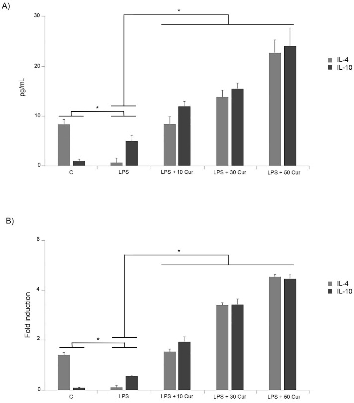
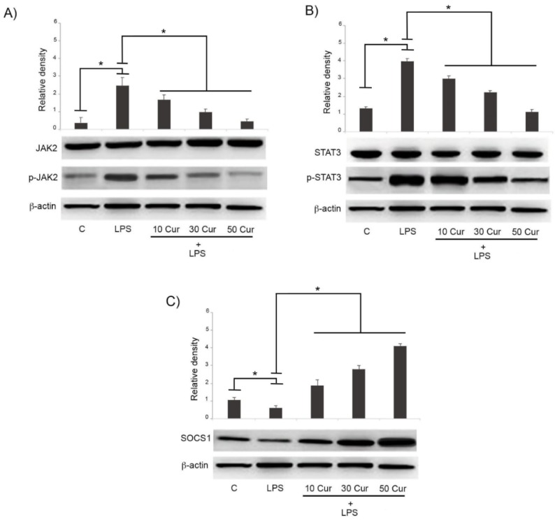
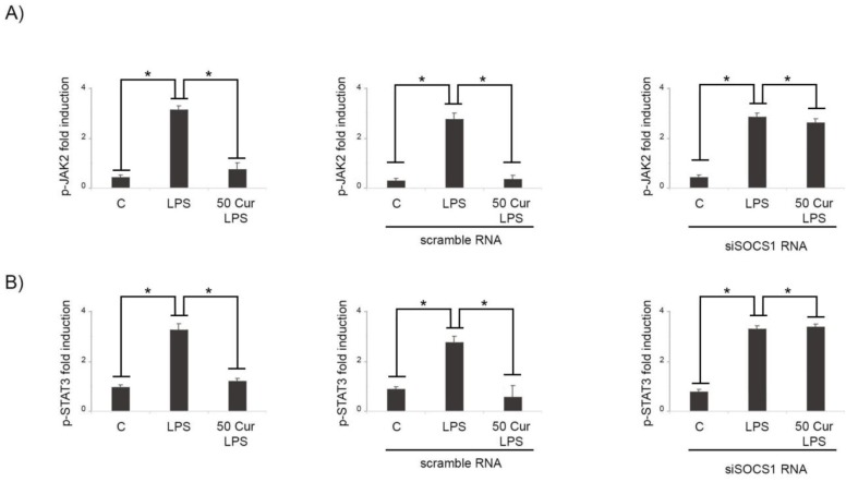
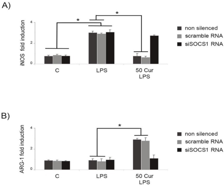
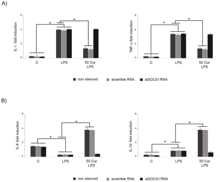
Microglia play important physiological roles in central nervous system (CNS) homeostasis and in the pathogenesis of inflammatory brain diseases. Inflammation stimulates microglia to secrete cytokines and chemokines that guide immune cells to sites of injury/inflammation. Neuroinflammation is also strongly implicated in the pathogenesis of a number of neurodegenerative diseases, including Alzheimer's disease and Parkinson's disease, for which nutritional intervention could represent a benefit due to a lack of clinically efficacious drugs. To this end, the anti-inflammatory mechanisms of several phytochemicals, including curcumin, have been extensively studied. The present experiments show that the administration of curcumin is able to increase the production of the anti-inflammatory cytokines, IL-4 and IL-10, in murine BV-2 microglial cells treated with lipopolysaccharide (LPS). Consistent with these data, curcumin stimulation upregulates the expression of Suppressors of cytokine signaling (SOCS)-1, whereas phosphorylation of the JAK2 and STAT3 was reduced. Taken together, these results provide evidence that curcumin is able to regulate neuroinflammatory reactions by eliciting anti-inflammatory responses in microglia through JAK/STAT/SOCS signaling pathway modulation.
Overview
The claims and statistics of this paper haven’t been checked yet.
Run the check — free, no account needed. It reads the open-access text and checks the sources it cites.
Integrity signals
Registry
Cites
Statistics
Figures
Verification
Claims the paper makes
The claims and statistics of this paper haven’t been checked yet.
Run the check — free, no account needed. It reads the open-access text and checks the sources it cites.
Claims other papers make about this one
Each is checked against this paper’s own text: does it say what the citing paper says it does?
In the citing paper
“curcumin has been demonstrated to inhibit NF-κB and MAPK activation in LPS-stimulated microglia (Porro et al., 2019; Zhang et al., 2019)”
From Exploring the Therapeutic Potential of Phytochemicals in Alzheimer’s Disease: Focus on Polyphenols and Monoterpenes (2022) · cites this paper as Porro et al., 2019
Self-check
Percentages, sums and reported differences re-computed from the paper’s own numbers.
The self-check needs the paper’s full text.
Citations
Cites
The IL-4 receptor: signaling mechanisms and biologic functions
Nelms, K., Keegan, A. D., Zamorano, J., Ryan, J. J., & Paul, W. E. (1999) · Annual Review of Immunology · doi:10.1146/annurev.immunol.17.1.701 · Cited by 1,545 · cited 1×
“IL-4 serves as an immunosuppressive cytokine that may inhibit the synthesis of several pro-inflammatory cytokines, as previously reported [24].”
ResultRegulation of JAK–STAT signalling in the immune system
Shuai, K., & Liu, B. (2003) · Nature reviews. Immunology · doi:10.1038/nri1226 · Cited by 1,410
Aggarwal, B. B., & Sung, B. (2009) · Trends in Pharmacological Sciences · doi:10.1016/j.tips.2008.11.002 · Cited by 1,140
Safety and Anti-Inflammatory Activity of Curcumin: A Component of Tumeric ( Curcuma longa )
Chainani-Wu, N. (2003) · The Journal of Alternative and Complementary Medicine · doi:10.1089/107555303321223035 · Cited by 1,106
Amalraj, A., Pius, A., Gopi, S., & Gopi, S. (2016) · Journal of Traditional and Complementary Medicine · doi:10.1016/j.jtcme.2016.05.005 · Cited by 976
Butovsky, O., Ziv, Y., Schwartz, A., Landa, G., Talpalar, A. E., Pluchino, S., Martino, G., & Schwartz, M. (2006) · Molecular and Cellular Neuroscience · doi:10.1016/j.mcn.2005.10.006 · Cited by 923
SOCS1/JAB Is a Negative Regulator of LPS-Induced Macrophage Activation
Kinjyo, I., Hanada, T., Inagaki-Ohara, K., Mori, H., Aki, D., Ohishi, M., Yoshida, H., Kubo, M., & Yoshimura, A. (2002) · Immunity · doi:10.1016/s1074-7613(02)00446-6 · Cited by 630
Factors regulating microglia activation
Kierdorf, K., & Prinz, M. (2013) · Frontiers in Cellular Neuroscience · doi:10.3389/fncel.2013.00044 · Cited by 360
Biological and therapeutic activities, and anticancer properties of curcumin
PERRONE, D., Ardito, F., Giannatempo, G., Dioguardi, M., Troiano, G., Russo, L. L., LILLO, A. D., Laino, L., & Muzio, L. L. (2015) · Experimental and Therapeutic Medicine · doi:10.3892/etm.2015.2749 · Cited by 351
IL-10 inhibits transcription of cytokine genes in human peripheral blood mononuclear cells.
Wang, P., Wu, P., Siegel, M. I., Egan, R. W., & Billah, M. M. (1994) · The Journal of Immunology · doi:10.4049/jimmunol.153.2.811 · Cited by 329
Cited by
Sorted by citations
The JAK/STAT signaling pathway: from bench to clinic
Hu, X., li, J., Fu, M., Zhao, X., & Wang, W. (2021) · Signal Transduction and Targeted Therapy · doi:10.1038/s41392-021-00791-1 · Cited by 2,633
Microglia polarization from M1 to M2 in neurodegenerative diseases
Guo, S., Wang, H., & Yin, Y. (2022) · Frontiers in Aging Neuroscience · doi:10.3389/fnagi.2022.815347 · Cited by 920
Microglia: Agents of the CNS Pro-Inflammatory Response
Rodríguez-Gómez, J. A., Kavanagh, E., Engskog-Vlachos, P., Engskog, M. K. R., Herrera, A. J., Espinosa-Oliva, A. M., Joseph, B., Hajji, N., Venero, J. L., & Burguillos, M. A. (2020) · Cells · doi:10.3390/cells9071717 · Cited by 365
Tang, X., Yu, H., Bui, B., Wang, L., Xing, C., Wang, S., Chen, M., Hu, Z., & Chen, W. (2020) · Bioactive Materials · doi:10.1016/j.bioactmat.2020.11.006 · Cited by 269
Curcumin’s Nanomedicine Formulations for Therapeutic Application in Neurological Diseases
Salehi, B., Călina, D. C., Docea, A. O., Koirala, N., Aryal, S., Lombardo, D., Pasqua, L., Taheri, Y., Castillo, C. M. S., Martorell, M., et al. (2020) · Journal of Clinical Medicine · doi:10.3390/jcm9020430 · Cited by 199
Mamun, A. A., Chauhan, A., Qi, S., Ngwa, C., Xu, Y., Sharmeen, R., Hazen, A. L., Li, J., Aronowski, J. A., McCullough, L. D., et al. (2019) · Proceedings of the National Academy of Sciences · doi:10.1073/pnas.1914742117 · Cited by 188
Neuroprotective Activities of Curcumin in Parkinson’s Disease: A Review of the Literature
Nebrisi, E. E. (2021) · International Journal of Molecular Sciences · doi:10.3390/ijms222011248 · Cited by 165
Signal pathways in the treatment of Alzheimer’s disease with traditional Chinese medicine
Ding, M.-R., Qu, Y.-J., Hu, B., & An, H. (2022) · Biomedicine & Pharmacotherapy · doi:10.1016/j.biopha.2022.113208 · Cited by 145
Rusek, M., Smith, J., El-Khatib, K., Aikins, K., Czuczwar, S. J., & Pluta, R. M. (2023) · International Journal of Molecular Sciences · doi:10.3390/ijms24010864 · Cited by 151
Basheer, A. S., Abas, F., Othman, I., & Naidu, R. (2021) · Cancers · doi:10.3390/cancers13164226 · Cited by 120
Ruganzu, J. B., Zheng, Q., Wu, X., He, Y., Peng, X., Jin, H., Zhou, J., Ma, R., Ji, S., Ma, Y., et al. (2020) · Experimental Neurology · doi:10.1016/j.expneurol.2020.113506 · Cited by 119
Potential therapeutic effects of curcumin mediated by JAK/STAT signaling pathway: A review
Ashrafizadeh, M., Rafiei, H., Mohammadinejad, R., Afshar, E. G., Farkhondeh, T., & Samarghandian, S. (2020) · Phytotherapy Research · doi:10.1002/ptr.6642 · Cited by 121
Inflammation and Alzheimer’s Disease: Mechanisms and Therapeutic Implications by Natural Products
Rather, M. A., Khan, A., Alshahrani, S., Rashid, H., Qadri, M. M., Rashid, S., Alsaffar, R. M., Kamal, M. A., & Rehman, M. U. (2021) · Mediators of Inflammation · doi:10.1155/2021/9982954 · Cited by 114
Promising drug targets and associated therapeutic interventions in Parkinson’s disease
Rai, S. N., Singh, P., Varshney, R., Chaturvedi, V. K., Vamanu, E., Singh, M. P., & Singh, S. P. (2021) · Neural Regeneration Research · doi:10.4103/1673-5374.306066 · Cited by 109
Gao, Y., Zhuang, Z., Lu, Y., Tao, T., Zhou, Y., Liu, G., Wang, H., Zhang, D., Wu, L., Dai, H., et al. (2019) · Frontiers in Neuroscience · doi:10.3389/fnins.2019.01223 · Cited by 105
Curcumin as Prospective Anti-Aging Natural Compound: Focus on Brain
Benameur, T., Soleti, R., Panaro, M. A., Torre, M. E. L., Monda, V., Messina, G., & Porro, C. (2021) · Molecules · doi:10.3390/molecules26164794 · Cited by 105
New Promising Therapeutic Avenues of Curcumin in Brain Diseases
Benameur, T., Giacomucci, G., Panaro, M. A., Ruggiero, M., Trotta, T., Monda, V., Pizzolorusso, I., Lofrumento, D. D., Porro, C., & Messina, G. (2021) · Molecules · doi:10.3390/molecules27010236 · Cited by 108
Action Mechanisms of Curcumin in Alzheimer’s Disease and Its Brain Targeted Delivery
Ege, D. (2021) · Materials · doi:10.3390/ma14123332 · Cited by 97
Sun, W., Li, H., Shen, Y., & Xiao, H. (2021) · Pathology - Research and Practice · doi:10.1016/j.prp.2021.153576 · Cited by 90
Li, J., Shui, X., Sun, R., Wan, L., Zhang, B., Xiao, B., & Luo, Z. (2021) · Frontiers in Cellular Neuroscience · doi:10.3389/fncel.2021.736310 · Cited by 100
Alzheimer’s disease: natural products as inhibitors of neuroinflammation
Olajide, O. A., & Sarker, S. D. (2020) · Inflammopharmacology · doi:10.1007/s10787-020-00751-1 · Cited by 92
Molecular Interactions for the Curcumin-Polymer Complex with Enhanced Anti-Inflammatory Effects
He, Y., Liu, H., Bian, W., Liu, Y., Liu, X., Ma, S., Zheng, X., Du, Z., Zhang, K., & Ouyang, D. (2019) · Pharmaceutics · doi:10.3390/pharmaceutics11090442 · Cited by 83
Challenges and Future Trends in Atopic Dermatitis
Gatmaitan, J. G., & Lee, J. H. (2023) · International Journal of Molecular Sciences · doi:10.3390/ijms241411380 · Cited by 94
Mechanism underlying treatment of ischemic stroke using acupuncture: transmission and regulation
Cao, B.-Q., Tan, F., Zhan, J., & Lai, P.-H. (2020) · Neural Regeneration Research · doi:10.4103/1673-5374.297061 · Cited by 86
Benameur, T., Soleti, R., & Porro, C. (2021) · Nutrients · doi:10.3390/nu13041318 · Cited by 85
Microglial polarization in TBI: Signaling pathways and influencing pharmaceuticals
Li, Y., Ren, X., Zhang, L., Wang, Y., & Chen, T. (2022) · Frontiers in Aging Neuroscience · doi:10.3389/fnagi.2022.901117 · Cited by 77
Piccialli, I., Tedeschi, V., Caputo, L., D’Errico, S., Ciccone, R., De Feo, V., Secondo, A., & Pannaccione, A. (2022) · Frontiers in Pharmacology · doi:10.3389/fphar.2022.876614 · Cited by 73
Levy, E. H., DELVIN, E. E., Marcil, V., & Spahis, S. (2020) · American Journal of Physiology-Endocrinology and Metabolism · doi:10.1152/ajpendo.00298.2020 · Cited by 73
Sintov, A. C. (2020) · International Journal of Pharmaceutics · doi:10.1016/j.ijpharm.2020.119725 · Cited by 67
Kooshki, L., Zarneshan, S. N., Fakhri, S., Moradi, S. Z., & Echeverría, J. (2023) · Phytomedicine · doi:10.1016/j.phymed.2023.154686 · Cited by 66
Antiaging effects of dietary supplements and natural products
Gao, L., Liu, X., Luo, X., Lou, X., Li, P., Li, X., & Liu, X. (2023) · Frontiers in Pharmacology · doi:10.3389/fphar.2023.1192714 · Cited by 66
JAK2/STAT3 as a new potential target to manage neurodegenerative diseases: An interactive review
Panda, S. P., Kesharwani, A., Datta, S., Prasanth, D., Panda, S. K., & Guru, A. (2024) · European Journal of Pharmacology · doi:10.1016/j.ejphar.2024.176490 · Cited by 61
Bacterial resistance to antimicrobial photodynamic therapy: A critical update
Surur, A. K., de Oliveira, A. B., de Annunzio, S. R., Ferrisse, T. M., & Fontana, C. R. (2024) · Journal of Photochemistry and Photobiology B Biology · doi:10.1016/j.jphotobiol.2024.112905 · Cited by 62
Inflammaging and Brain: Curcumin and Its Beneficial Potential as Regulator of Microglia Activation
Cianciulli, A., Calvello, R., Ruggiero, M., & Panaro, M. A. (2022) · Molecules · doi:10.3390/molecules27020341 · Cited by 60
Akter, K., Gul, K., & Mumtaz, S. (2025) · Current Issues in Molecular Biology · doi:10.3390/cimb47090716 · Cited by 67
Extracellular Vesicles miRNA Cargo for Microglia Polarization in Traumatic Brain Injury
Panaro, M. A., Benameur, T., & Porro, C. (2020) · Biomolecules · doi:10.3390/biom10060901 · Cited by 56
Zheng, Y., Zhang, X., Zhang, R., Wang, Z., Gan, J., Gao, Q., Yang, L., Xu, P., & Jiang, X. (2023) · International Journal of Molecular Medicine · doi:10.3892/ijmm.2023.5314 · Cited by 52
Rainha, J., Gomes, D., Rodrigues, L. R., & Rodrigues, J. L. (2020) · Life · doi:10.3390/life10050056 · Cited by 49
Hypothalamic Neuropeptide Brain Protection: Focus on Oxytocin
Panaro, M. A., Benameur, T., & Porro, C. (2020) · Journal of Clinical Medicine · doi:10.3390/jcm9051534 · Cited by 50
Biomaterials-based anti-inflammatory treatment strategies for Alzheimer’s disease
Chu, J., Zhang, W., Liu, Y., Gong, B., Ji, W., Yin, T., Gao, C., Liangwen, D., Hao, M., Chen, C., et al. (2023) · Neural Regeneration Research · doi:10.4103/1673-5374.374137 · Cited by 48
Kopalli, S. R., Prakash, A. V., & Koppula, S. (2022) · Molecules · doi:10.3390/molecules27144660 · Cited by 45
Mechanistic Insight into the Effects of Curcumin on Neuroinflammation-Driven Chronic Pain
Hasriadi, H., Wasana, P. W. D., Vajragupta, O., Rojsitthisak, P., & Towiwat, P. (2021) · Pharmaceuticals · doi:10.3390/ph14080777 · Cited by 42
Joshi, P., Bisht, A., Joshi, S., Semwal, D. K., Nema, N. K., Dwivedi, J., & Sharma, S. K. (2022) · Phytotherapy Research · doi:10.1002/ptr.7522 · Cited by 39
Barbalho, S. M., Boaro, B. L., Oliveira, J. D. S. C., Patočka, J., Lamas, C. B., Tanaka, M., & Laurindo, L. F. (2025) · Pharmaceuticals · doi:10.3390/ph18010133 · Cited by 46
Modulating Neuroinflammation as a Prospective Therapeutic Target in Alzheimer’s Disease
Lee, E., & Chang, Y. (2025) · Cells · doi:10.3390/cells14030168 · Cited by 39
Yan, M., Sun, Z., Zhang, S., Yang, G., Jiang, X. Y., Wang, G., Li, R., Wang, Q., & Tian, X. (2024) · Brain Research Bulletin · doi:10.1016/j.brainresbull.2024.110988 · Cited by 38
Anti-Neuroinflammatory Potential of Natural Products in the Treatment of Alzheimer’s Disease
Deng, M., Yan, W., Gu, Z., Li, Y., Chen, L., & He, B. (2023) · Molecules · doi:10.3390/molecules28031486 · Cited by 38
The Role of Natural Compounds and their Nanocarriers in the Treatment of CNS Inflammation
Khadka, B., Lee, J., Park, D. H., Kim, K.-T., & Bae, J. (2020) · Biomolecules · doi:10.3390/biom10101401 · Cited by 34
Wang, X., Cao, Z., Su, J., Ma, Y., Zhang, S., Shao, Z., Ge, X., Cheng, X., & Zhou, Z. (2024) · International Journal of Biological Macromolecules · doi:10.1016/j.ijbiomac.2024.139392 · Cited by 32
Das, S. S., Gopal, P. M., Thomas, J. V., Mohan, M. C., Thomas, S. C., Maliakel, B. P., Krishnakumar, I. M., & Chakrapani, P. S. B. (2023) · Frontiers in Dementia · doi:10.3389/frdem.2023.1222708 · Cited by 35
Wen, X., & Hu, J. (2024) · International Immunopharmacology · doi:10.1016/j.intimp.2024.112936 · Cited by 32
Liu, J., Jiao, L., Zhong, X., Yao, W., Du, K., Lu, S., Wu, Y., Ma, T., Tong, J., Xu, M., et al. (2022) · Frontiers in Aging Neuroscience · doi:10.3389/fnagi.2022.856628 · Cited by 31
Sun, M., Liu, N., Sun, J., Li, X., Wang, H., Zhang, W., Xie, Q., & Wang, M. (2022) · Neuroscience Letters · doi:10.1016/j.neulet.2022.136821 · Cited by 33
Almutary, A. G., Begum, M. Y., Kyada, A., Gupta, S. K., Jyothi, S. R., Chaudhary, K., Sharma, S., Sinha, A., Abomughaid, M. M., Imran, M., et al. (2024) · Ageing Research Reviews · doi:10.1016/j.arr.2024.102548 · Cited by 28
Calvello, R., Cianciulli, A., Porro, C., Moda, P., Nuccio, F. D., Nicolardi, G., Giannotti, L., Panaro, M. A., & Lofrumento, D. D. (2021) · Nutrients · doi:10.3390/nu13051418 · Cited by 26
Inflammation, Autoimmunity and Neurodegenerative Diseases, Therapeutics and Beyond
Garmendia, J. V., Sanctis, C. V. D., Das, V., Annadurai, N., Hajdúch, M., & Sanctis, J. B. D. (2023) · Current Neuropharmacology · doi:10.2174/1570159x22666231017141636 · Cited by 27
Tong, Q., & Wu, Z.-Q. (2023) · Clinical anatomy (New York, N.Y. Print) · doi:10.1002/ca.24057 · Cited by 19
Qu, Y.-J., Zhen, R.-R., Zhang, L., Gu, C., Chen, L., Peng, X., Hu, B., & An, H. (2020) · BMC Complementary Medicine and Therapies · doi:10.1186/s12906-020-02951-2 · Cited by 23
The Potential Role of Nutrition in Lung Cancer Establishment and Progression
Porro, C., Torre, M. E. L., Tartaglia, N., Benameur, T., Santini, M., Ambrosi, A., Messina, G., Cibelli, G., Fiorelli, A., Polito, R., et al. (2022) · Life · doi:10.3390/life12020270 · Cited by 26
Lagoa, R., Logesh, R., Violante, C., Babiaka, S. B., Marques‐da‐Silva, D., Kapoor, B., Reis, F., & Atanasov, A. G. (2025) · Biotechnology Advances · doi:10.1016/j.biotechadv.2025.108568 · Cited by 24
Qiao, J., Wang, C., Chen, Y., Yu, S., Liu, Y., Yu, S., Jiang, L., Jin, C., Wang, X., Zhang, P., et al. (2023) · Antioxidants · doi:10.3390/antiox12040920 · Cited by 23
Hemshekhar, M., Anaparti, V., El-Gabalawy, H. S., & Mookherjee, N. (2021) · Arthritis Research & Therapy · doi:10.1186/s13075-021-02423-z · Cited by 22
Jithavech, P., Suwattananuruk, P., Hasriadi, H., Muangnoi, C., Thitikornpong, W., Towiwat, P., Vajragupta, O., & Rojsitthisak, P. (2022) · PLoS ONE · doi:10.1371/journal.pone.0265689 · Cited by 20
Nawaz, A., Latif, M. S., Shah, M. K. A., Elsayed, T. M. A., Ahmad, S., & Khan, H. A. (2023) · Gels · doi:10.3390/gels9030201 · Cited by 20
Targeting the JAK/STAT3/SOCS signaling pathway in Alzheimer’s disease
Al-Kuraishy, H. M., Sulaiman, G. M., Mohammed, H. A., Saad, . M., Al-Gareeb, A. I., & Albuhadily, A. K. (2025) · Inflammopharmacology · doi:10.1007/s10787-025-01796-w · Cited by 20
Comparative Analysis of Multiple Neurodegenerative Diseases Based on Advanced Epigenetic Aging Brain
Shi, F., He, Y., Chen, Y., Yin, X., Sha, X., & Wang, Y. (2021) · Frontiers in Genetics · doi:10.3389/fgene.2021.657636 · Cited by 15
Development of a Slow-Degrading Polymerized Curcumin Coating for Intracortical Microelectrodes
Ziemba, A. M., Woodson, M. C. C., Funnell, J. L., Wich, D. M., Balouch, B., Rende, D., Amato, D. N., Bao, J., Oprea, I., Cao, D., et al. (2023) · ACS Applied Bio Materials · doi:10.1021/acsabm.2c00969 · Cited by 19
IRF5 Signaling in Phagocytes Is Detrimental to Neonatal Hypoxic Ischemic Encephalopathy
Mamun, A. A., Yu, H., Sharmeen, R., McCullough, L. D., & Liu, F. (2020) · Translational Stroke Research · doi:10.1007/s12975-020-00832-x · Cited by 17
Domínguez-Avila, J. A., Salazar‐López, N. J., Montiel‐Herrera, M., Martı́nez-Martı́nez, A., Villegas‐Ochoa, M. A., & González-Aguilar, G. A. (2022) · Journal of Food Biochemistry · doi:10.1111/jfbc.14260 · Cited by 16
Gholami, A. A., Tahmasebi, F., Haghir, H., & Babaloo, H. (2025) · Inflammopharmacology · doi:10.1007/s10787-025-01884-x · Cited by 15
Wang, R., Yu, H., Chen, P., Yuan, T., & Zhang, J. (2023) · International Journal of Molecular Sciences · doi:10.3390/ijms241512479 · Cited by 15
Retinasamy, T., Lee, A. L. Y., Lee, H. S., Lee, V. L. L., Shaikh, M. F., & Yeong, K. Y. (2024) · ACS Chemical Neuroscience · doi:10.1021/acschemneuro.4c00205 · Cited by 16
Park, J., Lee, C.-H., & Kim, Y. T. (2023) · Pharmaceuticals · doi:10.3390/ph16070941 · Cited by 17
Oxley, R. A., & Peart, D. J. (2023) · Nutrition and Health · doi:10.1177/02601060231186439 · Cited by 17
Yu, L., Zhang, C., Gu, L., Chen, H., Huo, Y., Wang, S., Tao, J., Xu, C. L., Zhang, Q., Ma, M., et al. (2024) · Molecular Neurobiology · doi:10.1007/s12035-023-03896-6 · Cited by 14
Neuroinflammation and Disease: Pathways and Opportunities
Hoffman, O., Varvel, N. H., Roopra, A., & Dingledine, R. J. (2025) · The Annual Review of Pharmacology and Toxicology · doi:10.1146/annurev-pharmtox-062124-043519 · Cited by 14
Cross-kingdom regulation of ginseng miRNA156 on immunity and metabolism
Wang, J., Li, C., Ruan, J., Yang, C., Tian, Y., Lu, B., & Wang, Y. (2024) · International Immunopharmacology · doi:10.1016/j.intimp.2024.112577 · Cited by 12
Ortiz-Islas, E., Montes, P., Rodríguez-Pérez, C. E., Ruiz-Sánchez, E., Sánchez-Barbosa, T., Pichardo-Rojas, D., Zavala‐Tecuapetla, C., Carvajal, K., & Campos‐Peña, V. (2025) · Pharmaceutics · doi:10.3390/pharmaceutics17010128 · Cited by 14
Ji, X., Chai, J., Zhao, S., & Zhao, Y. (2025) · Frontiers in Pharmacology · doi:10.3389/fphar.2025.1605027 · Cited by 16
Corneal Application of R9-SOCS1-KIR Peptide Alleviates Endotoxin-Induced Uveitis
Ahmed, C. M. I., Patel, A. P., Ildefonso, C. J., Johnson, H. M., & Lewin, A. S. (2021) · Translational Vision Science & Technology · doi:10.1167/tvst.10.3.25 · Cited by 12
Autophagy Modulation and Its Implications on Glioblastoma Treatment
Chen, J., Salinas, A., Morales, M. A., & Fang, X. (2023) · Current Issues in Molecular Biology · doi:10.3390/cimb45110546 · Cited by 13
Li, Q., Ding, Y., Ou, Y., Li, M., Jithavech, P., Buranasudja, V., Sritularak, B., Xu, Y., Rojsitthisak, P., & Han, J. (2023) · Current Pharmaceutical Design · doi:10.2174/0113816128263974231029180947 · Cited by 11
Xian, L., Cheng, S., Chen, W., Zhong, C., Hu, Z., & Deng, X. (2024) · Frontiers in Medicine · doi:10.3389/fmed.2024.1320811 · Cited by 9
Gharat, S., Basudkar, V., & Momin, M. M. (2024) · Immunological Investigations · doi:10.1080/08820139.2024.2301997 · Cited by 9
Tylek, K., Trojan, E., Leśkiewicz, M., Francavilla, F., Lacivita, E., Leopoldo, M., & Basta‐Kaim, A. (2023) · ACS Chemical Neuroscience · doi:10.1021/acschemneuro.3c00525 · Cited by 8
The Anti-Vitiligo Effects of Feshurin In Vitro from Ferula samarcandica and the Mechanism of Action
Nueraihemaiti, M., Deng, Z., Kamoldinov, K., Niu, C., Habasi, M., & Aisa, H. A. (2024) · Pharmaceuticals · doi:10.3390/ph17091252 · Cited by 7
Barangi, S., Hosseinzadeh, P., Karimi, G., Najaran, Z. T., & Mehri, S. (2023) · PubMed Central · doi:10.22038/ijbms.2023.68292.14905 · Cited by 7
Covid-19 pandemic: What is the truth?
Blaylock, R. L. (2021) · Surgical Neurology International · doi:10.25259/sni_1008_2021 · Cited by 7
Su, A.-L., Zhao, S., Hong-Na, Z. H. U., Ying, Q. I. A. O., & Zhang, T. (2024) · Biomedical Research · doi:10.2220/biomedres.45.13 · Cited by 6
Wang, Y., Liu, J., Jiang, Q., Mi, Y., Meng, Q., Mu, D., & Hou, Y. (2024) · Journal of Drug Delivery Science and Technology · doi:10.1016/j.jddst.2024.105418 · Cited by 6
Bioactive Natural Compounds for Therapeutic and Nutraceutical Applications in Neurodegeneration
Panaro, M. A., & Porro, C. (2022) · Nutrients · doi:10.3390/nu14112216 · Cited by 6
Astaneh, M. E., & Fereydouni, N. (2024) · Regenerative Therapy · doi:10.1016/j.reth.2024.11.016 · Cited by 5
Gui, S., Ni, J., Mizutani, S., SHIGEMATSU, N., Nakanishi, H., Kashiwazaki, H., & Wu, Z. (2024) · Food Science & Nutrition · doi:10.1002/fsn3.4045 · Cited by 5
Curcumin for protecting mitochondria and downregulating inflammation
Salimi, A., Jamali, Z., & Shirmard, L. R. (2022) · Elsevier eBooks · doi:10.1016/b978-0-323-90256-4.00014-x · Cited by 5
Fang, F., Zhang, S., Chen, C., Wang, X., Luo, C., & Wei, Q. L. (2023) · Optical Materials · doi:10.1016/j.optmat.2023.113800 · Cited by 5
Probing curcumin reactive conformers in keto-enol tautomerization enhanced by clustering with t-SNE
Rusydi, F., Susanti, E. D., Puspitasari, I. P. I., Fadilla, R. N., Madinah, R., & Mark-Lee, W. F. (2026) · Journal of Molecular Modeling · doi:10.1007/s00894-025-06621-3 · Cited by 4
Exosomes and their Cargo as a New Avenue for Brain and Treatment of CNS-Related Diseases
Benameur, T., Panaro, M. A., & Porro, C. (2022) · The Open Neurology Journal · doi:10.2174/1874205x-v16-e2201190 · Cited by 4
Kuru, G., Üner, G., & Bedіr, E. (2022) · Phytochemistry Reviews · doi:10.1007/s11101-022-09829-w · Cited by 4
Ji, Q., Han, L., Zhang, T., Xia, X., & Xiang, X. (2023) · Arabian Journal of Chemistry · doi:10.1016/j.arabjc.2023.104931 · Cited by 4
Opportunities and challenges of foodborne polyphenols applied to anti-aging health foods
Zhuang, H., Zhang, X., Wu, S., Pang, Y., & Yan, H. (2024) · Food Science and Biotechnology · doi:10.1007/s10068-024-01686-x · Cited by 5
Metadata
- Authors
- Porro, C., Cianciulli, A., Trotta, T., Lofrumento, D. D., & Panaro, M. A.
- Affiliations
- University of Foggia (IT)
- University of Bari Aldo Moro (IT)
- University of Salento (IT)
- Corresponding author
- Panaro, Maria Antonietta
- ORCID
- Venue
- Biology
- Publisher
- MDPI AG
- Published
- 2019-06-27
- Volume
- 8
- Issue
- 3
- Pages
- 51
- ISSN
- 2079-7737 (ISSN-L)
- PMC
- PMC6784227
- OpenAlex
- W2953720445
- Cited by
- 124
- References
- 40
- Citations by year
- 2026: 10 · 2025: 14 · 2024: 19 · 2023: 28 · 2022: 17 · 2021: 18 · 2020: 15 · 2019: 3
- FWCI
- 4.58
- Type
- journal article
- Language
- en
- Open access
- gold
- dx.doi.org
- Licence
- Locations
- Biology (publishedVersion, cc-by)
- Europe PMC (PubMed Central) (publishedVersion, cc-by)
- PubMed Central (publishedVersion, cc-by)
- PubMed (publishedVersion)
- DOAJ (DOAJ: Directory of Open Access Journals) (submittedVersion, cc-by-sa)
- Institutional Research Information System University of Ferrara (University of Ferrara) (submittedVersion, public-domain)
- AIRUS (submittedVersion)
- CINECA IRIS Institutional Research Information System (University of Bari Aldo Moro) (submittedVersion, cc-by-nc-nd)
- Funders
- Università degli Studi di Foggia
- Topics
- Life Sciences › Biochemistry, Genetics and Molecular Biology › Molecular Medicine › Curcumin's Biomedical Applications
- Life Sciences › Neuroscience › Neurology › Neuroinflammation and Neurodegeneration Mechanisms
- Life Sciences › Immunology and Microbiology › Immunology › Cytokine Signaling Pathways and Interactions
- Keywords
- curcumin, microglia, neuroinflammation, SOCS1, STAT3 phosphorylation, JAK2 phosphorylation, IL-4, IL-10, anti-inflammatory cytokines, lipopolysaccharide, BV-2 cells, neurodegenerative diseases, phytochemicals
About these checks
Registry, retraction, Cites and statistics results are computed, not judged, with one exception: a printed reference that no index lists is read once by a model before it counts as a citation error. Claims are extracted by a model and kept only when their quote appears verbatim in the text; each cited source is then checked, with a model’s help, for whether it says what the claim relies on. Those verdicts can be wrong — read the claim’s quote and the cited source before relying on one.
Statistics and arithmetic are recomputed, not judged.
Confidence bands are shown only for fields calibrated on our annotated test set. None is calibrated yet, so no band is shown.
What counts as a claim ›How we measure confidence ›How we check this ›
Are you an author, or do you see a problem with this page? Request a review
