

The DOI matches its Crossref record; 92 of the 95 references it cites were found in a registry or an index.
Calcium Overload and Mitochondrial Metabolism
Open accessAbstract
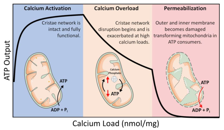
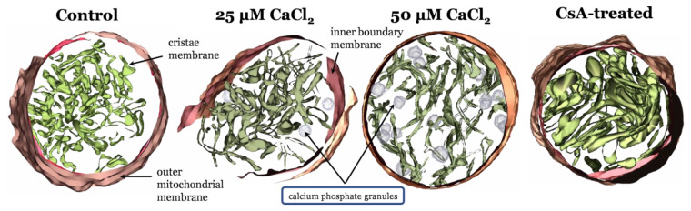
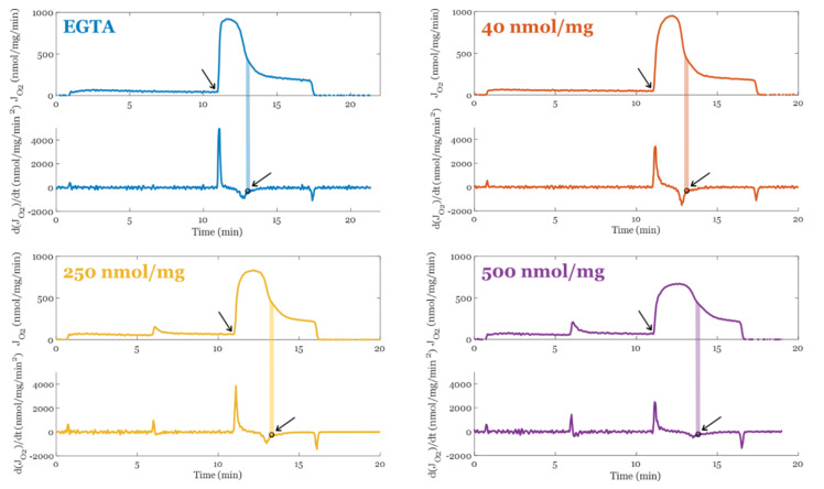
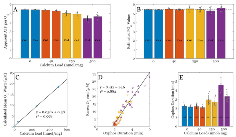
Mitochondria calcium is a double-edged sword. While low levels of calcium are essential to maintain optimal rates of ATP production, extreme levels of calcium overcoming the mitochondrial calcium retention capacity leads to loss of mitochondrial function. In moderate amounts, however, ATP synthesis rates are inhibited in a calcium-titratable manner. While the consequences of extreme calcium overload are well-known, the effects on mitochondrial function in the moderately loaded range remain enigmatic. These observations are associated with changes in the mitochondria ultrastructure and cristae network. The present mini review/perspective follows up on previous studies using well-established cryo–electron microscopy and poses an explanation for the observable depressed ATP synthesis rates in mitochondria during calcium-overloaded states. The results presented herein suggest that the inhibition of oxidative phosphorylation is not caused by a direct decoupling of energy metabolism via the opening of a calcium-sensitive, proteinaceous pore but rather a separate but related calcium-dependent phenomenon. Such inhibition during calcium-overloaded states points towards mitochondrial ultrastructural modifications, enzyme activity changes, or an interplay between both events.
Overview
The claims and statistics of this paper haven’t been checked yet.
Run the check — free, no account needed. It reads the open-access text and checks the sources it cites.
Integrity signals
Registry
Cites
Statistics
Figures
Verification
Claims the paper makes
The claims and statistics of this paper haven’t been checked yet.
Run the check — free, no account needed. It reads the open-access text and checks the sources it cites.
Claims other papers make about this one
Each is checked against this paper’s own text: does it say what the citing paper says it does?
The source backs part of the claim, not all of it — or backs it for a different population, condition or comparison.
In the citing paper
“Ca2+ activates mitochondrial metabolism and stimulates ATP synthesis”
From CYFIP1 governs the development of cortical axons by modulating calcium availability (2025) · cites this paper as [104]
Experimental — accuracy not yet measuredThe cited source says what the claim attributes to it.
In the citing paper
“mitochondria also buffer cytosolic calcium51,103,104.”
From CYFIP1 governs the development of cortical axons by modulating calcium availability (2025) · cites this paper as [104]
Self-check
Percentages, sums and reported differences re-computed from the paper’s own numbers.
The self-check needs the paper’s full text.
Citations
Cites
Computer Visualization of Three-Dimensional Image Data Using IMOD
Kremer, J. R., Mastronarde, D. N., & McIntosh, J. R. (1996) · Journal of Structural Biology · doi:10.1006/jsbi.1996.0013 · Cited by 5,530
Mitochondrial Transport of Cations: Channels, Exchangers, and Permeability Transition
Bernardi, P. (1999) · Physiological Reviews · doi:10.1152/physrev.1999.79.4.1127 · Cited by 1,546
Annexins: linking Ca2+ signalling to membrane dynamics
Gerke, V., Creutz, C. E., & Moss, S. E. (2005) · Nature Reviews Molecular Cell Biology · doi:10.1038/nrm1661 · Cited by 1,412
The mitochondrial calcium uniporter is a highly selective ion channel
Kirichok, Y., Krapivinsky, G., & Clapham, D. E. (2004) · Nature · doi:10.1038/nature02246 · Cited by 1,409
Mitochondria and calcium: from cell signalling to cell death
Duchen, M. R. (2000) · The Journal of Physiology · doi:10.1111/j.1469-7793.2000.00057.x · Cited by 1,261
Microdomains of Intracellular Ca2+: Molecular Determinants and Functional Consequences
Rizzuto, R., & Pozzan, T. (2005) · Physiological Reviews · doi:10.1152/physrev.00004.2005 · Cited by 1,234
What is the mitochondrial permeability transition pore?
Halestrap, A. P. (2009) · Journal of Molecular and Cellular Cardiology · doi:10.1016/j.yjmcc.2009.02.021 · Cited by 966
Role of Mitochondrial Ca2+ in the Regulation of Cellular Energetics
Glancy, B. C., & Balaban, R. S. (2012) · Biochemistry · doi:10.1021/bi2018909 · Cited by 649
Annexins – unique membrane binding proteins with diverse functions
Rescher, U., & Gerke, V. (2004) · Journal of Cell Science · doi:10.1242/jcs.01245 · Cited by 595
MICU1 Controls Both the Threshold and Cooperative Activation of the Mitochondrial Ca2+ Uniporter
Csordás, G., Golenár, T., Seifert, E. L., Kamer, K. J., Sancak, Y., Perocchi, F., Moffat, C., Weaver, D., de la Fuente, S., Bogorad, R. L., et al. (2013) · Cell Metabolism · doi:10.1016/j.cmet.2013.04.020 · Cited by 499
Research works printed but not linked: 1
- Andrade, L.O. (2019). Chapter Three—Annexins and Plasma Membrane Repai. Plasma Membrane Repair, Elsevier.
Cited by
Sorted by citations
Liu, T., Li, X., Zhou, X., Chen, W. J., Wen, A., Liu, M., & Ding, Y. (2024) · Neural Regeneration Research · doi:10.4103/nrr.nrr-d-24-00568 · Cited by 123
Li, F., Guan, Z., Gao, Y., Bai, Y., Zhan, X., Ji, X., Xu, J., Zhou, H., & Rao, Z. (2024) · Hepatology Communications · doi:10.1097/hc9.0000000000000399 · Cited by 62
Wu, J., Yi, S., Cao, Y., Zu, M., Li, B., Yang, W. J., Shahbazi, M., Wan, Y., Reis, R. L., Kundu, S. C., et al. (2023) · Biomaterials · doi:10.1016/j.biomaterials.2023.122332 · Cited by 59
Rahi, V., & Kaundal, R. K. (2024) · Life Sciences · doi:10.1016/j.lfs.2024.122651 · Cited by 57
Role of hydrogen sulfide in health and disease
Jin, Y., Yuan, H., Liu, Y., Zhu, Y., Wang, Y., Liang, X., Gao, W., Ren, Z., Ji, X., & Wu, D. (2024) · MedComm · doi:10.1002/mco2.661 · Cited by 54
Mitochondria as intracellular signalling organelles. An update
Popov, L. (2023) · Cellular Signalling · doi:10.1016/j.cellsig.2023.110794 · Cited by 57
Zhou, H., Wu, C., Jin, Y., Wu, O., Chen, L., Guo, Z., Xinzhou, W., Chen, Q., Kwan, K. Y.-H., Li, Y. M., et al. (2024) · Journal of Orthopaedic Translation · doi:10.1016/j.jot.2024.08.016 · Cited by 60
Li, B., Huo, S., Du, J., Zhang, X., Zhang, J., Wang, Q., Song, M., & Li, Y. (2023) · The Science of The Total Environment · doi:10.1016/j.scitotenv.2023.164620 · Cited by 43
Jiang, T., Ruan, N., Luo, P., Wang, Q., Wei, X., Li, Y., Dai, Y., Li, L., Lv, J., Liu, Y., et al. (2024) · Ageing Research Reviews · doi:10.1016/j.arr.2024.102320 · Cited by 43
Anthracycline-induced cardiotoxicity: An overview from cellular structural perspective
Li, H., Wang, M., & Huang, Y. (2024) · Biomedicine & Pharmacotherapy · doi:10.1016/j.biopha.2024.117312 · Cited by 39
Sirtuins in mitophagy: key gatekeepers of mitochondrial quality
Lagunas‐Rangel, F. A. (2025) · Molecular and Cellular Biochemistry · doi:10.1007/s11010-025-05358-0 · Cited by 42
The pathogenesis and therapeutic strategies of heat stroke-induced myocardial injury
Xia, R., Sun, M., Li, Y., Yin, J., Liu, H., Yang, J., Liu, J., He, Y., Wu, B., Yang, G., et al. (2024) · Frontiers in Pharmacology · doi:10.3389/fphar.2023.1286556 · Cited by 30
Gou, F., Lin, Q., Tu, X., Zhu, J., Li, X., Chen, S., & Hu, C. (2024) · Journal of Agricultural and Food Chemistry · doi:10.1021/acs.jafc.4c02265 · Cited by 26
Calcium signaling hypothesis: A non-negligible pathogenesis in Alzheimer’s disease
Wang, M., Zhang, H., Liang, J., Huang, J., Wu, T., & Chen, N. (2025) · Journal of Advanced Research · doi:10.1016/j.jare.2025.01.007 · Cited by 29
“Abraxane‐Like” Radiosensitizer for In Situ Oral Cancer Therapy
Gong, Z., Fu, Y., Gao, Y., Jiao, F., Su, Q., Sang, X., Chen, B., Deng, X., & Liu, X. (2024) · Advanced Science · doi:10.1002/advs.202309569 · Cited by 23
Jian, Y., Cheng, L., Hong, Y., Li, Z., Li, C., Ban, X., Zhu, L., & Gu, Z. (2023) · Nutrients · doi:10.3390/nu15245080 · Cited by 24
Mitochondria-targeting therapeutic strategies for chronic kidney disease
Sun, A., Pollock, C. A., & Huang, C. (2024) · Biochemical Pharmacology · doi:10.1016/j.bcp.2024.116669 · Cited by 22
Abdullaev, I., Gayibov, U., Omonturdiev, S., Fotima, S., Gayibova, S., & Aripov, T. (2025) · Journal of Biomedical Research · doi:10.7555/jbr.38.20240387 · Cited by 24
Mitochondrial Transplantation/Transfer: Promising Therapeutic Strategies for Spinal Cord Injury
Xiong, X., Zhou, C., Yu, Y., Xie, Q., Xia, L., Li, Q., Lin, H., Zhang, S., & Liang, W. (2025) · Journal of Orthopaedic Translation · doi:10.1016/j.jot.2025.04.017 · Cited by 21
Javadpour, P., Abbaszadeh, F., Ahmadiani, A., Rezaei, M., & Ghasemi, R. (2024) · Molecular Neurobiology · doi:10.1007/s12035-024-04009-7 · Cited by 21
Cox7a1 controls skeletal muscle physiology and heart regeneration through complex IV dimerization
García‐Poyatos, C., Arora, P., Calvo, E., Marques, I. J., Kirschke, N., Galardi‐Castilla, M., Lembke, C., Meer, M., Fernández-Montes, P., Ernst, A., et al. (2024) · Developmental Cell · doi:10.1016/j.devcel.2024.04.012 · Cited by 21
Buja, L. M. (2024) · Experimental and Molecular Pathology · doi:10.1016/j.yexmp.2024.104944 · Cited by 21
Importance of proteins and mitochondrial changes as freshness indicators in fish muscle post‐mortem
Hematyar, N., Policar, T., & Rustad, T. (2024) · Journal of the Science of Food and Agriculture · doi:10.1002/jsfa.14044 · Cited by 19
Liu, H., Jiang, Y., Cong, L., Zhang, X., Zhou, Y., Pan, X., Liu, S., Wang, R., & Cao, X. (2025) · Free Radical Biology and Medicine · doi:10.1016/j.freeradbiomed.2025.01.006 · Cited by 18
Advances in nanotherapeutics for tumor treatment by targeting calcium overload
Wang, C., Peng, J., Xiao, Y., Zhang, Z., Yang, X., Liang, X., Yang, J., Zhou, X., & Li, C. (2024) · Colloids and Surfaces B Biointerfaces · doi:10.1016/j.colsurfb.2024.114190 · Cited by 18
Zhou, Z., Bai, X., Zhou, Z.-Y., Lin, Z., Liao, B., Gong, F., Wang, M.-Q., Chen, X.-B., Liu, Y., Chen, H., et al. (2025) · Journal of Controlled Release · doi:10.1016/j.jconrel.2025.113728 · Cited by 17
Zhang, Y., Cui, Y., Sun, C., Guo, J., & Li, M. (2025) · Cellular Signalling · doi:10.1016/j.cellsig.2025.111713 · Cited by 16
From oxidative stress to metabolic dysfunction: The role of TRPM2
Li, Y., Ren, H.-C., Li, H., Xing, M., & Cao, J. (2024) · International Journal of Biological Macromolecules · doi:10.1016/j.ijbiomac.2024.138081 · Cited by 16
Artificial Mitochondrial Nanorobots Deliver Energy In Vivo by Oral Administration
Li, N., Zhou, M., Wu, Z., Chen, Y., Duan, Y., Zhang, Z.-Q., Wu, Z., Xia, X., Shen, J., Mao, C., et al. (2025) · Advanced Materials · doi:10.1002/adma.202500495 · Cited by 17
Chen, Z., Chen, P., Li, J., Landao‐Bassonga, E., Papadimitriou, J. M., Gao, J., Liu, D., Tai, A. C. P., Ma, J., Lloyd, D. G., et al. (2025) · Science Advances · doi:10.1126/sciadv.ads6132 · Cited by 17
Ghosh, S., Debnath, I., Bhunia, S., Nandi, S., Ashique, S., Nayak, A., Mallick, S., & Basak, S. (2025) · Chinese Herbal Medicines · doi:10.1016/j.chmed.2025.09.005 · Cited by 16
Electro-metabolic coupling in atrial fibrillation: A deeper understanding of the metabolic driver
Qin, X., Jin, L., Gong, H., & Zheng, Q. (2024) · Biomedicine & Pharmacotherapy · doi:10.1016/j.biopha.2024.117536 · Cited by 14
Wang, Y., Che, H., Qu, L., Lu, X., Dong, M., Sun, B. H., & Guan, H. (2024) · iScience · doi:10.1016/j.isci.2024.111373 · Cited by 14
Zaher, A., & Stephens, S. B. (2025) · Cells · doi:10.3390/cells14060399 · Cited by 14
Bartmański, M., Pawłowski, Ł., Knabe, A., Mania, S., Banach‐Kopeć, A., Sakowicz‐Burkiewicz, M., & Ronowska, A. (2024) · ACS Applied Materials & Interfaces · doi:10.1021/acsami.4c13529 · Cited by 13
The Role of N6-Methyladenosine in Mitochondrial Dysfunction and Pathology
Yan, W., Saqirile, Li, K., Li, K., & Wang, C. (2025) · International Journal of Molecular Sciences · doi:10.3390/ijms26083624 · Cited by 13
Troise, D. E., Infante, B., Mercuri, S., Lindholm, B., Kublickiene, K. R., & Stallone, G. (2025) · Cells · doi:10.3390/cells14181443 · Cited by 12
Liu, J., Xie, Y., Ma, J., & Chu, H. (2024) · Chemico-Biological Interactions · doi:10.1016/j.cbi.2024.110948 · Cited by 12
Guo, J., Wang, Y., Shi, C., Zhang, D., Zhang, Q., Wang, L., & Gong, Z. (2024) · Cellular Signalling · doi:10.1016/j.cellsig.2024.111284 · Cited by 13
Grepper, D., Tabasso, C., Zanou, N., Aguettaz, A. K. F., Castro‐Sepúlveda, M., Ziegler, D. V., Lagarrigue, S., Arribat, Y., Martinotti, A., Ebrahimi, A., et al. (2024) · iScience · doi:10.1016/j.isci.2024.110510 · Cited by 12
Wang, C., Ji, X., Wang, X., Song, Y., Pan, C., Qian, M., & Jin, Y. (2025) · Environmental Pollution · doi:10.1016/j.envpol.2025.126014 · Cited by 14
Xiong, Z., Li, L., Ouyang, K., Qu, M., & Qiu, Q. (2025) · Agriculture · doi:10.3390/agriculture15141477 · Cited by 17
Zhao, Y., Wang, X., He, M., Zeng, G., Xu, Z., Zhang, L., Kang, Y., & Xue, P. (2023) · Small · doi:10.1002/smll.202307404 · Cited by 11
Shapiro, I. M., Risbud, M. V., & Landis, W. J. (2024) · Bone · doi:10.1016/j.bone.2024.117112 · Cited by 12
Gu, Y., Zhao, J., Wang, J., Jiang, Y., Jing, Y., Xu, K., & Su, J. (2024) · Medicine Plus · doi:10.1016/j.medp.2024.100033 · Cited by 11
Xing, Y., Shi, H., Gao, X., Zhu, X., Zhang, D., Fang, L., Wang, J., Liu, C., Wu, D., Wang, X., et al. (2024) · Journal of Agricultural and Food Chemistry · doi:10.1021/acs.jafc.4c01201 · Cited by 12
Huber, T., Horioka-Duplix, M., Chen, Y., Saca, V. R., Ceraudo, E., Chen, Y., & Sakmar, T. P. (2024) · Science Signaling · doi:10.1126/scisignal.adp3967 · Cited by 12
Xu, Y., Qu, X., Liang, M., Huang, D., Jin, M., Sun, L., Chen, X., Liu, F., & Qiu, Z. (2024) · Frontiers in Medicine · doi:10.3389/fmed.2024.1457882 · Cited by 17
The Role of Autophagy in Excitotoxicity, Synaptic Mitochondrial Stress and Neurodegeneration
Chu, C. T. (2025) · Autophagy Reports · doi:10.1080/27694127.2025.2464376 · Cited by 12
Li, Z., Xiao, F., Fan, J., Zhang, W., Liu, J., Liu, B., & Zhang, H. (2023) · Nanomedicine · doi:10.2217/nnm-2023-0200 · Cited by 11
A deadly taste: linking bitter taste receptors and apoptosis
Miller, Z. A., Carey, R. M., & Lee, R. J. (2025) · APOPTOSIS · doi:10.1007/s10495-025-02091-3 · Cited by 10
Shishkova, D. K., Markova, V. E., Markova, Y. O., Sinitsky, M. Y., Sinitskaya, А. V., Матвеева, В. Г., Torgunakova, E., Lazebnaya, A. A., Stepanov, A. D., & Кутихин, А. Г. (2025) · Biochemistry (Moscow) · doi:10.1134/s0006297924604064 · Cited by 10
Xiao, L., Chen, X.-J., Feng, J.-K., Li, W.-N., Yuan, S., & Hu, Y. (2023) · Fitoterapia · doi:10.1016/j.fitote.2023.105600 · Cited by 11
Yu, H., Huang, Y., Cai, Z., Huang, K., Yu, T., Lan, H., Zhang, Q., Wu, L., & Luo, H. (2024) · ACS Applied Materials & Interfaces · doi:10.1021/acsami.3c14865 · Cited by 9
Pang, Y., Hu, H., Xu, K., Cao, T., Wang, Z., Nie, J., Zheng, H., Luo, H., Wang, F., Xiong, C., et al. (2024) · Investigative Ophthalmology & Visual Science · doi:10.1167/iovs.65.5.36 · Cited by 10
Evaluating the safety and efficiency of nanomaterials: A focus on mitochondrial health
Siquan, L., Weilin, C., Chen, X., Meiyan, Z., Weihong, G., & Xiaoli, F. (2024) · Biomedicine & Pharmacotherapy · doi:10.1016/j.biopha.2024.117484 · Cited by 10
Kong, L., Yang, H., Yang, J., Jiang, L., Xu, B., Yang, T., & Liu, W. (2025) · Ecotoxicology and Environmental Safety · doi:10.1016/j.ecoenv.2025.117835 · Cited by 9
Exploring Calcium Channels as Potential Therapeutic Targets in Blast Traumatic Brain Injury
Wachtler, N., O’Brien, R., Ehrlich, B. E., & McGuone, D. (2025) · Pharmaceuticals · doi:10.3390/ph18020223 · Cited by 12
Szewczyk, A., Rembiałkowska, N., Saczko, J., Daczewska, M., Novickij, V., & Kulbacka, J. (2025) · Biological Research · doi:10.1186/s40659-025-00591-9 · Cited by 9
The Calci-Inflammatory Network: A Paradigm Shift in Understanding Milk Fever
Ametaj, B. N. (2025) · Dairy · doi:10.3390/dairy6030022 · Cited by 9
Li, D., Liu, L., Liu, D.-Q., Zhang, L.-Q., Zhou, Y., & Mei, W. (2025) · International Immunopharmacology · doi:10.1016/j.intimp.2025.114863 · Cited by 12
Pei, W., Dai, L., Li, M., Cao, S., Xiao, Y., Yang, Y., Ma, M., Deng, M., Mo, Y., & Liu, M. (2025) · Biomolecules · doi:10.3390/biom15070970 · Cited by 10
Zhang, W., Zhang, X., Lei, M., Zhang, D., Qin, G., Zhou, J., Ji, L., & Chen, L. (2024) · Antioxidants · doi:10.3390/antiox13060725 · Cited by 8
Yelisyeyeva, O. P., Kaminskyy, D. V., Semen, M. O., Chelpanova, I. V., & Semen, K. O. (2025) · Biomedicines · doi:10.3390/biomedicines13010161 · Cited by 9
Mitochondrial Transplantation: A Paradigm Shift in Osteoporosis Therapy
Shen, J., Lai, Y., Peng, Q., Lin, X., Chen, S., Guo, L., Xu, M., Lu, Y., Zhu, J., Lin, X., et al. (2025) · International Journal of Nanomedicine · doi:10.2147/ijn.s537166 · Cited by 10
Sidorenko, D. A., Galkin, G. V., Bokov, R. O., Tyrina, E. A., Vilchinskaya, N. A., Lvova, I. D., Tyganov, S. A., Shenkman, B. S., & Sharlo, K. A. (2024) · Free Radical Biology and Medicine · doi:10.1016/j.freeradbiomed.2024.10.285 · Cited by 7
Zhao, X., Dilixiati, A., Zhang, L., Aihemaiti, A., Song, Y., Zhao, G., Fu, X., Wang, X., & Wusiman, A. (2024) · Animals · doi:10.3390/ani14010152 · Cited by 7
Zhang, Y., Chen, B., Wang, M., Liu, H., Chen, M., Zhu, J., Zhang, Y., Wang, X., Wu, Y., Liu, D., et al. (2024) · Biochimica et Biophysica Acta (BBA) - Molecular Basis of Disease · doi:10.1016/j.bbadis.2024.167274 · Cited by 7
The mitochondrial intermembrane space – a permanently proteostasis-challenged compartment
Weith, M., Weiss, K., Stobbe, D., & Riemer, J. (2025) · Biological Chemistry · doi:10.1515/hsz-2025-0108 · Cited by 10
Cellot, G., & Ballerini, L. (2025) · Life · doi:10.3390/life15111776 · Cited by 10
Endoplasmic reticulum, mitochondria, and their crosstalk in retinal ganglion cell degeneration
Li, B., Shi, Y., Liu, M., Cao, W., Zhang, F., & Kang, Z. (2025) · Biomedicine & Pharmacotherapy · doi:10.1016/j.biopha.2025.118835 · Cited by 7
Next Generation Calcium Nanomaterials: Disrupting Tumor Ca2⁺ Homeostasis for Enhanced Cancer Therapy
Gangadhar, L., Sana, S. S., Venkatesan, R., Bhandare, R. R., Kim, S.-C., & Yasin, H. K. A. (2026) · International Journal of Nanomedicine · doi:10.2147/ijn.s557935 · Cited by 7
Jeyamogan, S., Leventhal, J. R., Mathew, J. M., & Zhang, Z. J. (2023) · Frontiers in Immunology · doi:10.3389/fimmu.2023.1270300 · Cited by 7
Li, J., Bai, J., Cui, S., Han, Z., Liu, T., Liu, Y., Guan, C., Zheng, J., & Wang, H. (2025) · Archives of Biochemistry and Biophysics · doi:10.1016/j.abb.2025.110566 · Cited by 6
Sarcoplasmic reticulum-mitochondrial calcium communication: A new target for cardiovascular therapy
Wang, J., Li, S., Yu, C., Wang, Y., Xu, S., Wang, Y., Zhao, L., & Zhang, J. (2025) · Biomedicine & Pharmacotherapy · doi:10.1016/j.biopha.2025.118424 · Cited by 6
Sidorenko, D. A., Bokov, R. O., Galkin, G. V., Vilchinskaya, N. A., Tyganov, S. A., Lvova, I. D., Shenkman, B. S., Mirzoev, T. M., & Sharlo, K. A. (2025) · Pflügers Archiv - European Journal of Physiology · doi:10.1007/s00424-025-03108-1 · Cited by 6
Jing, Q., Zhao, M., Tang, Y., Chen, T., Sun, M., Mi, D., Zou, L., Wang, R., Lu, J., & Shi, S. (2025) · Acta Pharmaceutica Sinica B · doi:10.1016/j.apsb.2025.10.046 · Cited by 6
An adverse outcome pathway for chemical-induced Parkinson’s disease: Calcium is key
Meerman, J. J., Legler, J., Piersma, A. H., Westerink, R. H. S., & Heusinkveld, H. J. (2023) · NeuroToxicology · doi:10.1016/j.neuro.2023.11.001 · Cited by 8
Shen, D., Qi, C., Hu, P., Li, J., & Shen, Y. (2025) · Reproductive Sciences · doi:10.1007/s43032-025-01827-5 · Cited by 6
Happ, K., Barawi, S., Niederer, D., Schwiete, C., Heinrich, C., Franz, A., Wahl, P., & Behringer, M. (2025) · Scientific Reports · doi:10.1038/s41598-025-11654-y · Cited by 5
Toosky, M., & Kheradvar, A. (2025) · Mitochondrion · doi:10.1016/j.mito.2025.102098 · Cited by 5
Mitochondrial regulation of obesity by POMC neurons
Luo, X.-D., Tang, S., Luo, X., Quzhen, L., Xia, R.-H., & Wang, X.-W. (2025) · Biochimica et Biophysica Acta (BBA) - Molecular Basis of Disease · doi:10.1016/j.bbadis.2025.167682 · Cited by 5
Sun, Z., Nie, M., Wang, X., Jiao, B., Fu, J., & Du, A. (2025) · European journal of medical research · doi:10.1186/s40001-025-02630-3 · Cited by 5
Effects of Calcium Ion Dyshomeostasis and Calcium Ion-Induced Excitotoxicity in Parkinson’s Disease
Nam, D., Kim, H. J., Han, S. J., Son, I., & Ho, D. H. (2024) · Journal of Molecular Pathology · doi:10.3390/jmp5040037 · Cited by 7
Paolini, E., Longo, M., Meroni, M., Podini, P., Maggioni, M., Quattrini, A., Fracanzani, A. L., & Dongiovanni, P. (2025) · Antioxidants · doi:10.3390/antiox14060618 · Cited by 6
Candida and Long Covid: Mannan Not from HeavenPreprint
Chambers, P. W. (2024) · Qeios · doi:10.32388/je31eo.5 · Cited by 5
CD38-NAD(+) Axis: New Insights into Glaucoma Therapy
Zhang, W., Zheng, H., Pang, Y., Wang, Y., & Zhang, X. (2025) · Molecular Neurobiology · doi:10.1007/s12035-025-05243-3 · Cited by 6
Shi, R., Zhang, Y., Chen, G., Zhang, J., Liu, J., Zhu, H., Sun, M., & Duan, Y. (2025) · Cell Proliferation · doi:10.1111/cpr.70120 · Cited by 4
Wang, L., Hsiao, A., Carli, J., Raza, A., Lin, Z., Arnaud, D., Davies, J. M., Franklin‐Tong, V. E., Smirnoff, N., & Bosch, M. (2026) · The Plant Cell · doi:10.1093/plcell/koag031 · Cited by 5
Mechanisms and therapeutic potential of mitochondrial-targeted therapies in bone repair
Xu, N., Ma, W. H., Liu, G., & Yang, F. (2026) · Annals of Medicine · doi:10.1080/07853890.2026.2641277 · Cited by 4
Du, W., Yang, Y., Liu, J., Zhu, Y., Shen, T., Chen, Q., & Miyazaki, T. (2024) · ACS Biomaterials Science & Engineering · doi:10.1021/acsbiomaterials.4c00306 · Cited by 4
TBBPA exposure causes cartilage cell damage in both in vitro and in vivo models
Yin, B., Liu, C., Sun, H. Y., & Zhang, W. (2024) · Ecotoxicology and Environmental Safety · doi:10.1016/j.ecoenv.2024.117271 · Cited by 4
Tao, X., Wang, Y., Jin, J., Yan, H., Yang, H., Wan, X., Li, P., Xiao, Y., Yu, Q., Liu, L., et al. (2025) · Journal of Advanced Research · doi:10.1016/j.jare.2025.05.031 · Cited by 4
Overmeyer, C., Jorgensen, K., & Vohra, B. P. S. (2023) · Molecular and Cellular Neuroscience · doi:10.1016/j.mcn.2023.103853 · Cited by 4
Chen, Y., Li, B., Xu, H., & Wu, L. (2024) · Frontiers in Pharmacology · doi:10.3389/fphar.2024.1439816 · Cited by 4
Gil-Martins, E., Cagide, F., Borer, A., Barbosa, D. J., Fernandes, C., Chavarria, D., Remião, F., Borges, F., & Silva, R. (2025) · Chemico-Biological Interactions · doi:10.1016/j.cbi.2025.111425 · Cited by 4
Protective Effect of Vitamin K2 (MK-7) on Acute Lung Injury Induced by Lipopolysaccharide in Mice
Yang, W., Wang, Y., Liu, L., Liu, L., Li, S., & Li, Y. (2024) · Current Issues in Molecular Biology · doi:10.3390/cimb46030110 · Cited by 4
Shin, S., & Cho, S. K. (2025) · Life Sciences · doi:10.1016/j.lfs.2025.123444 · Cited by 3
Xu, L., Jiang, J., Wang, Y., Wei, F., & Zhu, H. (2025) · Biochemical and Biophysical Research Communications · doi:10.1016/j.bbrc.2025.151857 · Cited by 3
Tang, Y., Huang, J., Cui, C., Yang, F., Li, K., Gao, B., Fu, S., & Yang, X. (2025) · International Journal of Nanomedicine · doi:10.2147/ijn.s523621 · Cited by 3
Metadata
- Authors
- Walkon, L. L., Strubbe-Rivera, J. O., & Bazil, J. N.
- Affiliations
- Michigan State University (US)
- Department of Pharmacology and Toxicology, Michigan State University, East Lansing, MI 48824, USA
- Venue
- Biomolecules
- Publisher
- MDPI AG
- Published
- 2022-12-17
- Volume
- 12
- Issue
- 12
- Pages
- 1891
- ISSN
- 2218-273X (ISSN-L)
- PMC
- PMC9775684
- OpenAlex
- W4313236994
- Cited by
- 187
- References
- 95
- Citations by year
- 2026: 50 · 2025: 75 · 2024: 50 · 2023: 12
- FWCI
- 3.57
- Type
- journal article
- Language
- en
- Open access
- gold
- doi.org
- Licence
- Locations
- Biomolecules (publishedVersion, cc-by)
- PubMed Central (publishedVersion, cc-by)
- PubMed (publishedVersion)
- DOAJ (DOAJ: Directory of Open Access Journals) (submittedVersion, cc-by-sa)
- Funders
- National Institutes of Health (F31HL152623)
- Topics
- Life Sciences › Biochemistry, Genetics and Molecular Biology › Molecular Biology › Mitochondrial Function and Pathology
- Life Sciences › Biochemistry, Genetics and Molecular Biology › Molecular Biology › ATP Synthase and ATPases Research
- Life Sciences › Biochemistry, Genetics and Molecular Biology › Cell Biology › Autophagy in Disease and Therapy
- Keywords
- mitochondrial calcium overload, mitochondrial calcium retention capacity, ATP synthesis inhibition, oxidative phosphorylation, mitochondrial ultrastructure, cryo-electron microscopy, mitochondrial function
- MeSH terms
- Adenosine Triphosphate, Calcium, Energy Metabolism, Mitochondria, Cryoelectron Microscopy
About these checks
Registry, retraction, Cites and statistics results are computed, not judged, with one exception: a printed reference that no index lists is read once by a model before it counts as a citation error. Claims are extracted by a model and kept only when their quote appears verbatim in the text; each cited source is then checked, with a model’s help, for whether it says what the claim relies on. Those verdicts can be wrong — read the claim’s quote and the cited source before relying on one.
Statistics and arithmetic are recomputed, not judged.
Confidence bands are shown only for fields calibrated on our annotated test set. None is calibrated yet, so no band is shown.
What counts as a claim ›How we measure confidence ›How we check this ›
Are you an author, or do you see a problem with this page? Request a review
