

The DOI matches its Crossref record.
Nanoparticle Activation Methods in Cancer Treatment
Abstract
In this review, we intend to highlight the progress which has been made in recent years around different types of smart activation nanosystems for cancer treatment. Conventional treatment methods, such as chemotherapy or radiotherapy, suffer from a lack of specific targeting and consequent off-target effects. This has led to the development of smart nanosystems which can effect specific regional and temporal activation. In this review, we will discuss the different methodologies which have been designed to permit activation at the tumour site. These can be divided into mechanisms which take advantage of the differences between healthy cells and cancer cells to trigger activation, and those which activate by a mechanism extrinsic to the cell or tumour environment.
Overview
The claims and statistics of this paper haven’t been checked yet.
Run the check — free, no account needed. It reads the open-access text and checks the sources it cites.
Integrity signals
Registry
Cites
Statistics
Figures
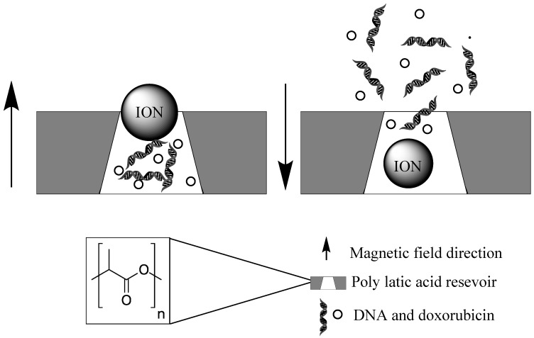
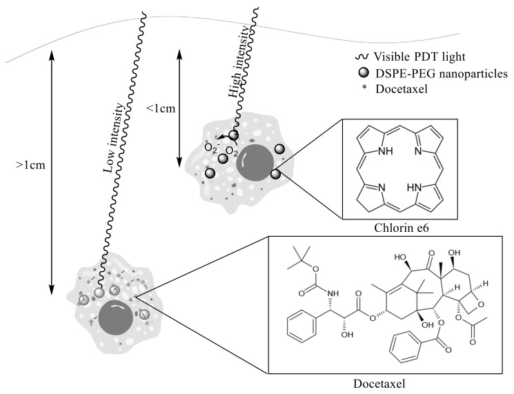
Verification
Claims the paper makes
The claims and statistics of this paper haven’t been checked yet.
Run the check — free, no account needed. It reads the open-access text and checks the sources it cites.
Claims other papers make about this one
Each is checked against this paper’s own text: does it say what the citing paper says it does?
In the citing paper
“Nanocarriers for cancer treatment are aimed to deliver the loaded-molecules to and around the targeted pathologic tissue, killing cancer cells without harming the normal ones.141”
From Current Advances in Chitosan Nanoparticles Based Oral Drug Delivery for Colorectal Cancer Treatment (2022) · cites this paper as [141]
Self-check
Percentages, sums and reported differences re-computed from the paper’s own numbers.
The self-check needs the paper’s full text.
Citations
Cites
No references could be resolved yet.
Research works printed but not linked: 133
- Boron, 2004, Regulation of intracellular pH, Adv. Physiol. Educ., 28, 160, 10.1152/advan.00045.2004 · DOI · Paper page
- Kato, 2013, Acidic extracellular microenvironment and cancer, Cancer Cell Int., 13, 89, 10.1186/1475-2867-13-89 · DOI
- Lin, 2005, pH-sensitive polyelectrolyte complex gel microspheres composed of chitosan/sodium tripolyphosphate/dextran sulfate: Swelling kinetics and drug delivery properties, Colloids Surf. B Biointerfaces, 44, 143, 10.1016/j.colsurfb.2005.06.010 · DOI
- Mura, 2013, Stimuli-responsive nanocarriers for drug delivery, Nat. Mater., 12, 991, 10.1038/nmat3776 · DOI · Paper page
- Yang, 2011, pH-Sensitive Micelles Self-Assembled from Amphiphilic Copolymer Brush for Delivery of Poorly Water-Soluble Drugs, Biomacromolecules, 12, 116, 10.1021/bm101058w · DOI
- Gao, 2010, pH-Responsive Nanoparticles for Drug Delivery, Mol. Pharm., 7, 1913, 10.1021/mp100253e · DOI
- Bradley, 1988, Mechanism of multidrug resistance, Biochim. Biophys. Acta - Rev. Cancer, 948, 87, 10.1016/0304-419X(88)90006-6 · DOI
- Wang, 2018, Mechanisms of drug release in pH-sensitive micelles for tumour targeted drug delivery system: A review, Int. J. Pharm., 535, 253, 10.1016/j.ijpharm.2017.11.003 · DOI
- Dawidar, 2011, Biodegradable pH-responsive alginate-poly (lactic-co-glycolic acid) nano/micro hydrogel matrices for oral delivery of silymarin, Carbohydr. Polym., 83, 1345, 10.1016/j.carbpol.2010.09.055 · DOI
- Tian, 2017, pH-responsive poly (acrylic acid)-gated mesoporous silica and its application in oral colon targeted drug delivery for doxorubicin, Colloids Surf. B Biointerfaces, 154, 287, 10.1016/j.colsurfb.2017.03.024 · DOI
- Hsu, C.-H., and Cheng, A.-L. (2007). Clinical studies with Curcumin. The Molecular Targets and Therapeutic Uses of Curcumin in Health and Disease, Springer. · DOI
- Cui, 2009, Enhancement of oral absorption of curcumin by self-microemulsifying drug delivery systems, Int. J. Pharm., 371, 148, 10.1016/j.ijpharm.2008.12.009 · DOI
- Raghunand, 1999, Enhancement of chemotherapy by manipulation of tumour pH, Br. J. Cancer, 80, 1005, 10.1038/sj.bjc.6690455 · DOI
- Peppicelli, 2017, The acidic microenvironment as a possible niche of dormant tumor cells, Cell. Mol. Life Sci., 74, 2761, 10.1007/s00018-017-2496-y · DOI
- Jo, 2017, In vivo quantitative imaging of tumor pH by nanosonophore assisted multispectral photoacoustic imaging, Nat. Commun., 8, 471, 10.1038/s41467-017-00598-1 · DOI
- Ray, 2019, PEG-b-poly (carbonate)-derived nanocarrier platform with pH-responsive properties for pancreatic cancer combination therapy, Colloids Surf. B Biointerfaces, 174, 126, 10.1016/j.colsurfb.2018.10.069 · DOI
- Qian, 2019, Tumor-microenvironment controlled nanomicelles with AIE property for boosting cancer therapy and apoptosis monitoring, Biomaterials, 188, 96, 10.1016/j.biomaterials.2018.10.003 · DOI
- Glunde, 2003, Extracellular acidification alters lysosomal trafficking in human breast cancer cells, Neoplasia, 5, 533, 10.1016/S1476-5586(03)80037-4 · DOI
- Griset, 2009, Expansile Nanoparticles: Synthesis, Characterization, and in Vivo Efficacy of an Acid-Responsive Polymeric Drug Delivery System, J. Am. Chem. Soc., 131, 2469, 10.1021/ja807416t · DOI
- Xu, 2016, Ultra-pH-Responsive and Tumor-Penetrating Nanoplatform for Targeted siRNA Delivery with Robust Anti-Cancer Efficacy, Angew. Chemie Int. Ed., 55, 7091, 10.1002/anie.201601273 · DOI
- Hwang, 2015, Functional Nanovalves on Protein-Coated Nanoparticles for In vitro and In vivo Controlled Drug Delivery, Small, 11, 319, 10.1002/smll.201400765 · DOI
- Muniswamy, 2019, ‘Dendrimer-Cationized-Albumin’ encrusted polymeric nanoparticle improves BBB penetration and anticancer activity of doxorubicin, Int. J. Pharm., 555, 77, 10.1016/j.ijpharm.2018.11.035 · DOI
- Dai, 2018, Dual-stimuli-responsive TiO x /DOX nanodrug system for lung cancer synergistic therapy, RSC Adv., 8, 21975, 10.1039/C8RA02899K · DOI
- Yue, 2011, Iron Oxide Nanotubes for Magnetically Guided Delivery and pH-Activated Release of Insoluble Anticancer Drugs, Adv. Funct. Mater., 21, 3446, 10.1002/adfm.201100510 · DOI
- Mi, 2016, A pH-activatable nanoparticle with signal-amplification capabilities for non-invasive imaging of tumour malignancy, Nat. Nanotechnol., 11, 724, 10.1038/nnano.2016.72 · DOI
- Dong, 2016, CaCO3 nanoparticles as an ultra-sensitive tumor-pH-responsive nanoplatform enabling real-time drug release monitoring and cancer combination therapy, Biomaterials, 110, 60, 10.1016/j.biomaterials.2016.09.025 · DOI
- Choi, 2016, PEGylated lipid bilayer-supported mesoporous silica nanoparticle composite for synergistic co-delivery of axitinib and celastrol in multi-targeted cancer therapy, Acta Biomater., 39, 94, 10.1016/j.actbio.2016.05.012 · DOI
- Schomburg, 2013, BRENDA in 2013: integrated reactions, kinetic data, enzyme function data, improved disease classification: new options and contents in BRENDA, Nucleic Acids Res., 41, D764, 10.1093/nar/gks1049 · DOI
- Li, 2017, Cancer-Associated, Stimuli-Driven, Turn on Theranostics for Multimodality Imaging and Therapy, Adv. Mater., 29, 1606857, 10.1002/adma.201606857 · DOI
- Shin, 2016, Cancer Targeted Enzymatic Theranostic Prodrug: Precise Diagnosis and Chemotherapy, Bioconjug. Chem., 27, 1419, 10.1021/acs.bioconjchem.6b00184 · DOI
- Tarro, 2005, Early diagnosis of lung cancer by detection of tumor liberated protein, J. Cell. Physiol., 203, 1, 10.1002/jcp.20195 · DOI
- Chan, 2017, Protease-activated nanomaterials for targeted cancer theranostics, Nanomedicine, 12, 2153, 10.2217/nnm-2017-0068 · DOI
- Han, 2017, Dual Enzymatic Reaction-Assisted Gemcitabine Delivery Systems for Programmed Pancreatic Cancer Therapy, ACS Nano, 11, 1281, 10.1021/acsnano.6b05541 · DOI
- Nairon, 2018, Abstract 4664: Enzymatically-responsive tumor-targeted mesoporous silica nanoparticle for identification of pancreatic cancer, Cancer Res., 78, 4664, 10.1158/1538-7445.AM2018-4664 · DOI
- Liu, 2018, Crosslinked self-assembled nanoparticles for chemo-sonodynamic combination therapy favoring antitumor, antimetastasis management and immune responses, J. Control. Release, 290, 150, 10.1016/j.jconrel.2018.10.007 · DOI
- Dzamukova, 2015, Enzyme-activated intracellular drug delivery with tubule clay nanoformulation, Sci. Rep., 5, 10560, 10.1038/srep10560 · DOI
- Talalay, 2010, NAD(P)H:quinone acceptor oxidoreductase 1 (NQO1), a multifunctional antioxidant enzyme and exceptionally versatile cytoprotector, Arch. Biochem. Biophys., 501, 116, 10.1016/j.abb.2010.03.019 · DOI
- Huang, 2014, Self-assembled hybrid nanoparticles for targeted co-delivery of two drugs into cancer cells, Chem. Commun., 50, 3103, 10.1039/c3cc49003c · DOI
- Shi, 2018, Molecular imaging of telomerase and the enzyme activity-triggered drug release by using a conformation-switchable nanoprobe in cancerous cells, Sci. Rep., 8, 16341, 10.1038/s41598-018-34670-7 · DOI
- Mannaris, 2018, Gas-Stabilizing Gold Nanocones for Acoustically Mediated Drug Delivery, Adv. Healthc. Mater., 7, 1800184, 10.1002/adhm.201800184 · DOI
- Decock, 2008, Cathepsin B, cathepsin H, cathepsin X and cystatin C in sera of patients with early-stage and inflammatory breast cancer, Int. J. Biol. Mark., 23, 161, 10.1177/172460080802300305 · DOI
- Pardo, 2018, Gold Nanorod-Based Nanohybrids for Combinatorial Therapeutics, ACS Omega, 3, 12633, 10.1021/acsomega.8b01591 · DOI
- Yildiz, 2018, Doxorubicin-loaded protease-activated near-infrared fluorescent polymeric nanoparticles for imaging and therapy of cancer, Int. J. Nanomed., 13, 6961, 10.2147/IJN.S174068 · DOI
- Coussens, 2002, Matrix metalloproteinase inhibitors and cancer: Trials and tribulations, Science, 295, 2387, 10.1126/science.1067100 · DOI · Paper page
- Somerville, 2003, Matrix metalloproteinases: Old dogs with new tricks, Genome Biol., 4, 216, 10.1186/gb-2003-4-6-216 · DOI
- Davies, 1995, Structures and mechanisms of glycosyl hydrolases, Structure, 3, 853, 10.1016/S0969-2126(01)00220-9 · DOI
- Shin, 2013, Oncogenic role of protein tyrosine kinase 7 in esophageal squamous cell carcinoma, Cancer Sci., 104, 1120, 10.1111/cas.12194 · DOI
- Yang, 2018, Stepwise Degradable Nanocarriers Enabled Cascade Delivery for Synergistic Cancer Therapy, Adv. Funct. Mater., 28, 1800706, 10.1002/adfm.201800706 · DOI
- Lian, 2018, Enzyme-MOF Nanoreactor Activates Nontoxic Paracetamol for Cancer Therapy, Angew. Chemie Int. Ed., 57, 5725, 10.1002/anie.201801378 · DOI
- Davis, 2010, Evidence of RNAi in humans from systemically administered siRNA via targeted nanoparticles, Nature, 464, 1067, 10.1038/nature08956 · DOI
- Wang, 2016, Self-assembled IR780-loaded transferrin nanoparticles as an imaging, targeting and PDT/PTT agent for cancer therapy, Sci. Rep., 6, 27421, 10.1038/srep27421 · DOI
- Dixit, 2015, Transferrin receptor-targeted theranostic gold nanoparticles for photosensitizer delivery in brain tumors, Nanoscale, 7, 1782, 10.1039/C4NR04853A · DOI
- Ciechanover, 1983, pH and the recycling of transferrin during receptor-mediated endocytosis, Proc. Natl. Acad. Sci. USA, 80, 2258, 10.1073/pnas.80.8.2258 · DOI
- Sutherland, 1981, Ubiquitous cell-surface glycoprotein on tumor cells is proliferation-associated receptor for transferrin, Proc. Natl. Acad. Sci. USA, 78, 4515, 10.1073/pnas.78.7.4515 · DOI · Paper page
- Zhang, Q., Wang, N., Ma, M., Luo, Y., and Chen, H. (2019). Transferrin Receptor-Mediated Sequential Intercellular Nanoparticles Relay for Tumor Deep Penetration and Sonodynamic Therapy. Adv. Ther., 1800152. · DOI
- Wosikowski, 1997, Altered gene expression in drug-resistant human breast cancer cells, Clin. Cancer Res., 3, 2405
- Akimoto, 1999, Inverse relationship between epidermal growth factor receptor expression and radiocurability of murine carcinomas, Clin. Cancer Res., 5, 2884
- Galizia, 2006, Prognostic Significance of Epidermal Growth Factor Receptor Expression in Colon Cancer Patients Undergoing Curative Surgery, Ann. Surg. Oncol., 13, 823, 10.1245/ASO.2006.05.052 · DOI
- Henriksen, L., Grandal, M.V., Knudsen, S.L.J., van Deurs, B., and Grøvdal, L.M. (2013). Internalization Mechanisms of the Epidermal Growth Factor Receptor after Activation with Different Ligands. PLoS ONE, 8. · DOI
- Merten, 2015, Antibody–Drug Conjugates for Tumor Targeting—Novel Conjugation Chemistries and the Promise of non-IgG Binding Proteins, Bioconjug. Chem., 26, 2176, 10.1021/acs.bioconjchem.5b00260 · DOI
- Ducry, 2010, Antibody−Drug Conjugates: Linking Cytotoxic Payloads to Monoclonal Antibodies, Bioconjug. Chem., 21, 5, 10.1021/bc9002019 · DOI
- Roncato, 2018, Improvement and extension of anti-EGFR targeting in breast cancer therapy by integration with the Avidin-Nucleic-Acid-Nano-Assemblies, Nat. Commun., 9, 4070, 10.1038/s41467-018-06602-6 · DOI
- Zhang, 2007, Prostate-specific membrane antigen (PSMA) protein expression in normal and neoplastic tissues and its sensitivity and specificity in prostate adenocarcinoma: an immunohistochemical study using mutiple tumour tissue microarray technique, Histopathology, 50, 472, 10.1111/j.1365-2559.2007.02635.x · DOI
- Rauscher, 2016, 68Ga-PSMA ligand PET/CT in patients with prostate cancer: How we review and report, Cancer Imaging, 16, 14, 10.1186/s40644-016-0072-6 · DOI
- Liu, 1998, Constitutive and antibody-induced internalization of prostate-specific membrane antigen, Cancer Res., 58, 4055
- Mangadlao, 2018, Prostate-Specific Membrane Antigen Targeted Gold Nanoparticles for Theranostics of Prostate Cancer, ACS Nano, 12, 3714, 10.1021/acsnano.8b00940 · DOI
- Brown, 1998, The unique physiology of solid tumors: Opportunities (and problems) for cancer therapy, Cancer Res., 58, 1408
- Feng, 2017, Theranostic Liposomes with Hypoxia-Activated Prodrug to Effectively Destruct Hypoxic Tumors Post-Photodynamic Therapy, ACS Nano, 11, 927, 10.1021/acsnano.6b07525 · DOI
- Yang, 2016, Structure-Dependent and Glutathione-Responsive Biodegradable Dendritic Mesoporous Organosilica Nanoparticles for Safe Protein Delivery, Chem. Mater., 28, 9008, 10.1021/acs.chemmater.6b03896 · DOI
- Robinson, R.K., Batt, C.A., and Patel, P.D. (2000). Encyclopedia of Food Microbiology, Academic Press.
- Mullin, 2013, Nanoparticle delivery enhancement with acoustically activated microbubbles, IEEE Trans. Ultrason. Ferroelectr. Freq. Control, 60, 65, 10.1109/TUFFC.2013.2538 · DOI
- Deng, 2004, Ultrasound-induced cell membrane porosity, Ultrasound Med. Biol., 30, 519, 10.1016/j.ultrasmedbio.2004.01.005 · DOI
- Dewitte, 2011, mRNA-Lipoplex loaded microbubble contrast agents for ultrasound-assisted transfection of dendritic cells, Biomaterials, 32, 9128, 10.1016/j.biomaterials.2011.08.024 · DOI
- Burke, 2014, Ultrasound-activated agents comprised of 5FU-bearing nanoparticles bonded to microbubbles inhibit solid tumor growth and improve survival, Mol. Ther., 22, 321, 10.1038/mt.2013.259 · DOI
- Kwan, 2015, Ultrasound-Propelled Nanocups for Drug Delivery, Small, 11, 5305, 10.1002/smll.201501322 · DOI
- Burke, 2011, Covalently linking poly(lactic-co-glycolic acid) nanoparticles to microbubbles before intravenous injection improves their ultrasound-targeted delivery to skeletal muscle, Small, 7, 1227, 10.1002/smll.201001934 · DOI
- Chappell, 2008, Targeted delivery of nanoparticles bearing fibroblast growth factor-2 by ultrasonic microbubble destruction for therapeutic arteriogenesis, Small, 4, 1769, 10.1002/smll.200800806 · DOI
- Price, 1998, Delivery of colloidal particles and red blood cells to tissue through microvessel ruptures created by targeted microbubble destruction with ultrasound, Circulation, 98, 1264, 10.1161/01.CIR.98.13.1264 · DOI
- Cochran, 2011, Doxorubicin and paclitaxel loaded microbubbles for ultrasound triggered drug delivery, Int. J. Pharm., 414, 161, 10.1016/j.ijpharm.2011.05.030 · DOI
- Yuan, 2018, MRI tracing non-invasive TiO2-based nanoparticles activated by ultrasound for multi-mechanism therapy of prostatic cancer, Nanotechnology, 29, 125101, 10.1088/1361-6528/aaa92a · DOI
- Lee, 2016, Theranostic gas-generating nanoparticles for targeted ultrasound imaging and treatment of neuroblastoma, J. Control. Release, 223, 197, 10.1016/j.jconrel.2015.12.051 · DOI
- Marshalek, 2016, Intracellular delivery and ultrasonic activation of folate receptor-targeted phase-change contrast agents in breast cancer cells in vitro, J. Control. Release, 243, 69, 10.1016/j.jconrel.2016.09.010 · DOI
- Wang, 2007, Direct-Current Nanogenerator Driven by Ultrasonic Waves, Science, 316, 102, 10.1126/science.1139366 · DOI
- Cucullo, 2005, Very low intensity alternating current decreases cell proliferation, Glia, 51, 65, 10.1002/glia.20188 · DOI
- Marino, 2018, Ultrasound-Activated Piezoelectric Nanoparticles Inhibit Proliferation of Breast Cancer Cells, Sci. Rep., 8, 6257, 10.1038/s41598-018-24697-1 · DOI
- Pearce, 2013, Magnetic Heating of Nanoparticles: The Importance of Particle Clustering to Achieve Therapeutic Temperatures, J. Nanotechnol. Eng. Med., 4, 011005, 10.1115/1.4024904 · DOI
- Shah, 2015, Impact of magnetic field parameters and iron oxide nanoparticle properties on heat generation for use in magnetic hyperthermia, J. Magn. Magn. Mater., 387, 96, 10.1016/j.jmmm.2015.03.085 · DOI
- Noto, 1989, Effects of sodium ascorbate (vitamin C) and 2-methyl-1,4-naphthoquinone (vitamin K3) treatment on human tumor cell growth in vitro. I. Synergism of combined vitamin C and K3 action, Cancer, 63, 901, 10.1002/1097-0142(19890301)63:5<901::AID-CNCR2820630518>3.0.CO;2-G · DOI
- Johannsen, 2005, Clinical hyperthermia of prostate cancer using magnetic nanoparticles: Presentation of a new interstitial technique, Int. J. Hyperth., 21, 637, 10.1080/02656730500158360 · DOI
- Teijeiro, 2013, Magnetic nanoparticle-based hyperthermia for cancer treatment, Rep. Pract. Oncol. Radiother., 18, 397, 10.1016/j.rpor.2013.09.011 · DOI
- Vedova, 2015, Gold Nanoparticle-Based Sensors Activated by External Radio Frequency Fields, Small, 11, 248, 10.1002/smll.201401187 · DOI
- Raoof, 2012, Stability of antibody-conjugated gold nanoparticles in the endolysosomal nanoenvironment: implications for noninvasive radiofrequency-based cancer therapy, Nanomed. Nanotechnol. Biol. Med., 8, 1096, 10.1016/j.nano.2012.02.001 · DOI
- Mironava, 2017, Gold Nanoparticles and Radio Frequency Field Interactions: Effects of Nanoparticle Size, Charge, Aggregation, Radio Frequency, and Ionic Background, Langmuir, 33, 13114, 10.1021/acs.langmuir.7b03210 · DOI
- Katagiri, 2011, Magnetoresponsive On-Demand Release of Hybrid Liposomes Formed from Fe3O4 Nanoparticles and Thermosensitive Block Copolymers, Small, 7, 1683, 10.1002/smll.201002180 · DOI
- Pradhan, 2010, Targeted temperature sensitive magnetic liposomes for thermo-chemotherapy, J. Control. Release, 142, 108, 10.1016/j.jconrel.2009.10.002 · DOI
- Plassat, 2011, Anti-Estrogen-Loaded Superparamagnetic Liposomes for Intracellular Magnetic Targeting and Treatment of Breast Cancer Tumors, Adv. Funct. Mater., 21, 83, 10.1002/adfm.201001450 · DOI
- Hu, 2012, Multifunctional Nanocapsules for Simultaneous Encapsulation of Hydrophilic and Hydrophobic Compounds and On-Demand Release, ACS Nano, 6, 2558, 10.1021/nn205023w · DOI
- Hua, 2011, The effectiveness of a magnetic nanoparticle-based delivery system for BCNU in the treatment of gliomas, Biomaterials, 32, 516, 10.1016/j.biomaterials.2010.09.065 · DOI
- Yang, 2011, Self-protecting core-shell magnetic nanoparticles for targeted, traceable, long half-life delivery of BCNU to gliomas, Biomaterials, 32, 6523, 10.1016/j.biomaterials.2011.05.047 · DOI
- Nigam, 2017, Dendrimer-conjugated iron oxide nanoparticles as stimuli-responsive drug carriers for thermally-activated chemotherapy of cancer, Colloids Surf. B Biointerfaces, 155, 182, 10.1016/j.colsurfb.2017.04.025 · DOI
- Stanley, 2012, Radio-Wave Heating of Iron Oxide Nanoparticles Can Regulate Plasma Glucose in Mice, Science, 336, 604, 10.1126/science.1216753 · DOI
- Huang, 2012, Self-assembling PVA-F127 thermosensitive nanocarriers with highly sensitive magnetically-triggered drug release for epilepsy therapy in vivo, J. Mater. Chem., 22, 8566, 10.1039/c2jm00032f · DOI
- Satarkar, 2008, Hydrogel nanocomposites as remote-controlled biomaterials, Acta Biomater., 4, 11, 10.1016/j.actbio.2007.07.009 · DOI
- Hu, 2008, Surfactant-Free, Self-Assembled PVA-Iron Oxide/Silica Core-Shell Nanocarriers for Highly Sensitive, Magnetically Controlled Drug Release and Ultrahigh Cancer Cell Uptake Efficiency, Adv. Funct. Mater., 18, 2946, 10.1002/adfm.200800428 · DOI
- Hu, 2008, Core/Single-Crystal-Shell Nanospheres for Controlled Drug Release via a Magnetically Triggered Rupturing Mechanism, Adv. Mater., 20, 2690, 10.1002/adma.200800193 · DOI
- Bringas, 2012, Triggered release in lipid bilayer-capped mesoporous silica nanoparticles containing SPION using an alternating magnetic field, Chem. Commun., 48, 5647, 10.1039/c2cc31563g · DOI
- Cai, 2009, Magnetically Triggered Reversible Controlled Drug Delivery from Microfabricated Polymeric Multireservoir Devices, Adv. Mater., 21, 4045, 10.1002/adma.200900593 · DOI
- Thomas, 2010, Noninvasive Remote-Controlled Release of Drug Molecules in Vitro Using Magnetic Actuation of Mechanized Nanoparticles, J. Am. Chem. Soc., 132, 10623, 10.1021/ja1022267 · DOI
- Walker, 2017, Radio-frequency actuated polymer-based phononic meta-materials for control of ultrasonic waves, NPG Asia Mater., 9, e350, 10.1038/am.2016.209 · DOI
- Park, 2011, Clustered magnetite nanocrystals cross-linked with PEI for efficient siRNA delivery, Biomacromolecules, 12, 457, 10.1021/bm101244j · DOI
- Allison, 2010, Oncologic photodynamic therapy photosensitizers: A clinical review, Photodiagn. Photodyn. Ther., 7, 61, 10.1016/j.pdpdt.2010.02.001 · DOI
- Wang, 2011, Upconversion nanoparticles: Synthesis, surface modification and biological applications, Nanomedicine, 7, 710, 10.1016/j.nano.2011.02.013 · DOI
- Haynes, W.M. (2009). CRC Handbook of Chemistry and Physics: A Ready-Reference Book of Chemical and Physical Data, CRC Press.
- Meng, 2018, Light-Activated Core–Shell Nanoparticles for Spatiotemporally Specific Treatment of Metastatic Triple-Negative Breast Cancer, ACS Nano, 12, 2789, 10.1021/acsnano.7b09210 · DOI
- Lucky, 2015, Titania Coated Upconversion Nanoparticles for Near-Infrared Light Triggered Photodynamic Therapy, ACS Nano, 9, 191, 10.1021/nn503450t · DOI
- Dai, 2017, 808 nm near-infrared light controlled dual-drug release and cancer therapy in vivo by upconversion mesoporous silica nanostructures, J. Mater. Chem. B, 5, 2086, 10.1039/C7TB00224F · DOI
- Huang, 2016, Ultralow-Power Near Infrared Lamp Light Operable Targeted Organic Nanoparticle Photodynamic Therapy, J. Am. Chem. Soc., 138, 14586, 10.1021/jacs.6b05390 · DOI
- Vankayala, 2015, Nucleus-Targeting Gold Nanoclusters for Simultaneous In Vivo Fluorescence Imaging, Gene Delivery, and NIR-Light Activated Photodynamic Therapy, Adv. Funct. Mater., 25, 5934, 10.1002/adfm.201502650 · DOI
- Qiu, 2018, Novel concept of the smart NIR-light-controlled drug release of black phosphorus nanostructure for cancer therapy, Proc. Natl. Acad. Sci. USA, 115, 501, 10.1073/pnas.1714421115 · DOI
- Liu, 2015, Gold Nanorods Coated with Mesoporous Silica Shell as Drug Delivery System for Remote Near Infrared Light-Activated Release and Potential Phototherapy, Small, 11, 2323, 10.1002/smll.201402145 · DOI
- He, 2015, Self-Assembled Core–Shell Nanoparticles for Combined Chemotherapy and Photodynamic Therapy of Resistant Head and Neck Cancers, ACS Nano, 9, 991, 10.1021/nn506963h · DOI
- Qian, 2016, Light-Activated Hypoxia-Responsive Nanocarriers for Enhanced Anticancer Therapy, Adv. Mater., 28, 3313, 10.1002/adma.201505869 · DOI
- Guan, 2015, Multifunctional upconversion–nanoparticles–trismethylpyridylporphyrin–fullerene nanocomposite: A near-infrared light-triggered theranostic platform for imaging-guided photodynamic therapy, NPG Asia Mater., 7, e205, 10.1038/am.2015.82 · DOI
- Yue, 2016, ROS-Responsive Mitochondria-Targeting Blended Nanoparticles: Chemo- and Photodynamic Synergistic Therapy for Lung Cancer with On-Demand Drug Release upon Irradiation with a Single Light Source, Theranostics, 6, 2352, 10.7150/thno.15433 · DOI
- Lu, 2015, A Chlorin-Based Nanoscale Metal–Organic Framework for Photodynamic Therapy of Colon Cancers, J. Am. Chem. Soc., 137, 7600, 10.1021/jacs.5b04069 · DOI
- Solís-López, M., and Ramírez-Zamora, R.M. (2019, January 24). Fenton Reaction—An Overview | ScienceDirect Topics. Available online: https://www.sciencedirect.com/topics/chemistry/fenton-reaction.
- Kim, 2017, Continuous O2-Evolving MnFe2O4 Nanoparticle-Anchored Mesoporous Silica Nanoparticles for Efficient Photodynamic Therapy in Hypoxic Cancer, J. Am. Chem. Soc., 139, 10992, 10.1021/jacs.7b05559 · DOI
- Espinosa, 2016, Duality of Iron Oxide Nanoparticles in Cancer Therapy: Amplification of Heating Efficiency by Magnetic Hyperthermia and Photothermal Bimodal Treatment, ACS Nano, 10, 2436, 10.1021/acsnano.5b07249 · DOI
- Goodman, 2017, Near-infrared remotely triggered drug-release strategies for cancer treatment, Proc. Natl. Acad. Sci. USA, 114, 12419, 10.1073/pnas.1713137114 · DOI
- Yang, 2016, Near-infrared light-activated cancer cell targeting and drug delivery with aptamer-modified nanostructures, Nano Res., 9, 139, 10.1007/s12274-015-0898-4 · DOI
- Chen, 2015, Nanoscintillator-Mediated X-ray Inducible Photodynamic Therapy for In Vivo Cancer Treatment, Nano Lett., 15, 2249, 10.1021/nl504044p · DOI
- Kotagiri, 2018, Radionuclides transform chemotherapeutics into phototherapeutics for precise treatment of disseminated cancer, Nat. Commun., 9, 275, 10.1038/s41467-017-02758-9 · DOI
- Stolik, 2000, Measurement of the penetration depths of red and near infrared light in human “ex vivo” tissues, J. Photochem. Photobiol. B Biol., 57, 90, 10.1016/S1011-1344(00)00082-8 · DOI
Cited by
Sorted by citations
Current Advances in Chitosan Nanoparticles Based Oral Drug Delivery for Colorectal Cancer Treatment
Choukaife, H., Seyam, S., Alallam, B., Doolaanea, A. A., & Alfatama, M. (2022) · International Journal of Nanomedicine · doi:10.2147/ijn.s375229 · Cited by 127
Metadata
- Authors
- White, B. D., Duan, C., & Townley, H. E.
- Venue
- Biomolecules
- Publisher
- MDPI AG
- Published
- 2019-05-24
- Volume
- 9
- Issue
- 5
- Pages
- 202
- ISSN
- 2218-273X
- PMC
- PMC6572460
- Cited by
- 32
- References
- 0
- Language
- en
About these checks
Registry, retraction, Cites and statistics results are computed, not judged, with one exception: a printed reference that no index lists is read once by a model before it counts as a citation error. Claims are extracted by a model and kept only when their quote appears verbatim in the text; each cited source is then checked, with a model’s help, for whether it says what the claim relies on. Those verdicts can be wrong — read the claim’s quote and the cited source before relying on one.
Statistics and arithmetic are recomputed, not judged.
Confidence bands are shown only for fields calibrated on our annotated test set. None is calibrated yet, so no band is shown.
What counts as a claim ›How we measure confidence ›How we check this ›
Are you an author, or do you see a problem with this page? Request a review
