

The DOI matches its Crossref record.
Noninvasive Estimation of Tumor Interstitial Fluid Pressure from Subharmonic Scattering of Ultrasound Contrast Microbubbles
Abstract
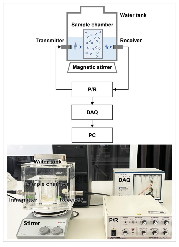
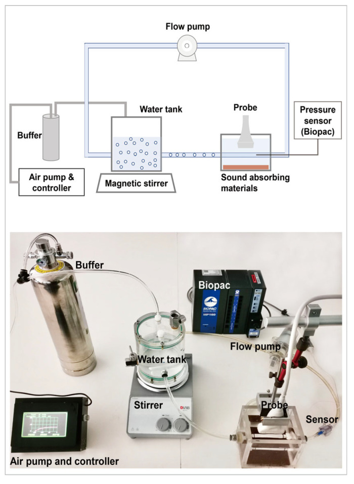
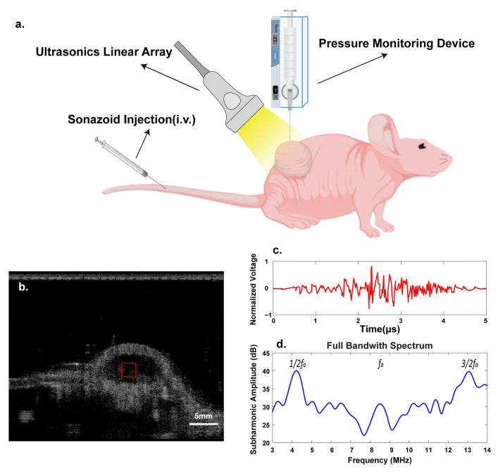
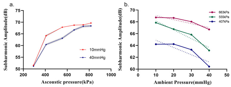
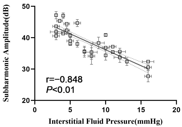
The noninvasive estimation of interstitial fluid pressure (IFP) using ultrasound contrast agent (UCA) microbubbles as pressure sensors will provide tumor treatments and efficacy assessments with a promising tool. This study aimed to verify the efficacy of the optimal acoustic pressure in vitro in the prediction of tumor IFPs based on UCA microbubbles’ subharmonic scattering. A customized ultrasound scanner was used to generate subharmonic signals from microbubbles’ nonlinear oscillations, and the optimal acoustic pressure was determined in vitro when the subharmonic amplitude reached the most sensitive to hydrostatic pressure changes. This optimal acoustic pressure was then applied to predict IFPs in tumor-bearing mouse models, which were further compared with the reference IFPs measured using a standard tissue fluid pressure monitor. An inverse linear relationship and good correlation (r = −0.853, p < 0.001) existed between the subharmonic amplitude and tumor IFPs at the optimal acoustic pressure of 555 kPa, and pressure sensitivity was 1.019 dB/mmHg. No statistical differences were found between the pressures measured by the standard device and those estimated via the subharmonic amplitude, as confirmed by cross-validation (mean absolute errors from 2.00 to 3.09 mmHg, p > 0.05). Our findings demonstrated that in vitro optimized acoustic parameters for UCA microbubbles’ subharmonic scattering can be applied for the noninvasive estimation of tumor IFPs.
Overview
The claims and statistics of this paper haven’t been checked yet.
Run the check — free, no account needed. It reads the open-access text and checks the sources it cites.
Integrity signals
Registry
Cites
Statistics
Figures
Verification
Claims the paper makes
The claims and statistics of this paper haven’t been checked yet.
Run the check — free, no account needed. It reads the open-access text and checks the sources it cites.
Claims other papers make about this one
Each is checked against this paper’s own text: does it say what the citing paper says it does?
The source backs part of the claim, not all of it — or backs it for a different population, condition or comparison.
In the citing paper
“In solid tumors, IFP is markedly elevated (typically 10–40 mmHg), due to leaky vasculature and dysfunctional lymphatic drainage.”
From Overcoming tumor microenvironment barriers: transformable and bioinspired nanomedicine strategies for deep tumor penetration (2026) · cites this paper as [56]
Experimental — accuracy not yet measuredIn the citing paper
“This reduces the interstitial pressure gradient that drives convection, creating a hydrodynamic barrier to nanoparticle extravasation and tissue diffusion [56].”
From Overcoming tumor microenvironment barriers: transformable and bioinspired nanomedicine strategies for deep tumor penetration (2026) · cites this paper as [56]
Self-check
Percentages, sums and reported differences re-computed from the paper’s own numbers.
The self-check needs the paper’s full text.
Citations
Cites
No references could be resolved yet.
Research works printed but not linked: 36
- Salavati, H., Debbaut, C., Pullens, P., and Ceelen, W. (2022). Interstitial fluid pressure as an emerging biomarker in solid tumors. Biochim. Biophys. Acta. Rev. Cancer, 1877. · DOI
- Boucher, 1992, Microvascular pressure is the principal driving force for interstitial hypertension in solid tumors: Implications for vascular collapse, Cancer Res., 52, 5110
- Haider, 2020, Recent advances in tumor microenvironment associated therapeutic strategies and evaluation models, Mater. Sci. Eng. C Mater. Biol. Appl., 116, 111229, 10.1016/j.msec.2020.111229 · DOI
- Heldin, 2004, High interstitial fluid pressure—An obstacle in cancer therapy, Nat. Rev. Cancer, 4, 806, 10.1038/nrc1456 · DOI · Paper page
- Schumacher, 2019, Targeting tumor interstitial fluid pressure: Will it yield novel successful therapies for solid tumors?, Expert Opin. Ther. Targets, 23, 1005, 10.1080/14728222.2019.1702974 · DOI
- Ozerdem, 2005, A simple method for measuring interstitial fluid pressure in cancer tissues, Microvasc. Res., 70, 116, 10.1016/j.mvr.2005.07.003 · DOI
- Khalifa, 2014, Models and methods for analyzing DCE-MRI: A review, Med. Phys., 41, 124301, 10.1118/1.4898202 · DOI
- Chong, 2018, Imaging with ultrasound contrast agents: Current status and future, Abdom. Radiol., 43, 762, 10.1007/s00261-018-1516-1 · DOI
- Forsberg, 2000, Subharmonic imaging of contrast agents, Ultrasonics, 38, 93, 10.1016/S0041-624X(99)00148-1 · DOI
- Versluis, 2020, Ultrasound Contrast Agent Modeling: A Review, Ultrasound Med. Biol., 46, 2117, 10.1016/j.ultrasmedbio.2020.04.014 · DOI
- Helfield, 2019, A Review of Phospholipid Encapsulated Ultrasound Contrast Agent Microbubble Physics, Ultrasound Med. Biol., 45, 282, 10.1016/j.ultrasmedbio.2018.09.020 · DOI
- Eisenbrey, J.R., Sridharan, A., Liu, J.B., and Forsberg, F. (2015). Recent Experiences and Advances in Contrast-Enhanced Subharmonic Ultrasound. BioMed. Res. Int., 2015. · DOI
- Dahibawkar, 2015, High and low frequency subharmonic imaging of angiogenesis in a murine breast cancer model, Ultrasonics, 62, 50, 10.1016/j.ultras.2015.04.012 · DOI
- Jones, 2018, Three-dimensional transcranial microbubble imaging for guiding volumetric ultrasound-mediated blood-brain barrier opening, Theranostics, 8, 2909, 10.7150/thno.24911 · DOI
- Sridharan, 2020, Characterizing Breast Lesions Using Quantitative Parametric 3D Subharmonic Imaging: A Multicenter Study, Acad. Radiol., 27, 1065, 10.1016/j.acra.2019.10.029 · DOI
- Shi, 1999, Pressure dependence of subharmonic signals from contrast microbubbles, Ultrasound Med. Biol., 25, 275, 10.1016/S0301-5629(98)00163-X · DOI
- Halldorsdottir, 2011, Subharmonic contrast microbubble signals for noninvasive pressure estimation under static and dynamic flow conditions, Ultrason. Imaging, 33, 153, 10.1177/016173461103300301 · DOI
- Andersen, 2010, Impact of acoustic pressure on ambient pressure estimation using ultrasound contrast agent, Ultrasonics, 50, 294, 10.1016/j.ultras.2009.09.016 · DOI
- Li, 2012, Simulation of noninvasive blood pressure estimation using ultrasound contrast agent microbubbles, IEEE Trans. Ultrason. Ferroelectr. Freq. Control., 59, 715, 10.1109/TUFFC.2012.2249 · DOI
- Nio, 2020, Optimal Control of SonoVue Microbubbles to Estimate Hydrostatic Pressure, IEEE Trans. Ultrason. Ferroelectr. Freq. Control, 67, 557, 10.1109/TUFFC.2019.2948759 · DOI
- Li, F., Li, D., and Yan, F. (2018). Improvement of Detection Sensitivity of Microbubbles as Sensors to Detect Ambient Pressure. Sensors, 18. · DOI
- Xu, 2021, Subharmonic Scattering of SonoVue Microbubbles Within 10–40-mmHg Overpressures In Vitro, IEEE Trans. Ultrason. Ferroelectr. Freq. Control, 68, 3583, 10.1109/TUFFC.2021.3101694 · DOI
- Zheng, 2022, Noninvasive Assessment of Intracranial Pressure Using Subharmonic-Aided Pressure Estimation: An Experimental Study in Canines, J. Trauma Acute Care Surg., 93, 882, 10.1097/TA.0000000000003720 · DOI
- Dave, 2012, Noninvasive LV pressure estimation using subharmonic emissions from microbubbles, JACC Cardiovasc. Imaging, 5, 87, 10.1016/j.jcmg.2011.08.017 · DOI
- Dave, 2012, Investigating the efficacy of subharmonic aided pressure estimation for portal vein pressures and portal hypertension monitoring, Ultrasound Med. Biol., 38, 1784, 10.1016/j.ultrasmedbio.2012.06.013 · DOI
- Dave, 2013, On the implementation of an automated acoustic output optimization algorithm for subharmonic aided pressure estimation, Ultrasonics, 53, 880, 10.1016/j.ultras.2012.12.010 · DOI
- Eisenbrey, 2013, Chronic liver disease: Noninvasive subharmonic aided pressure estimation of hepatic venous pressure gradient, Radiology, 268, 581, 10.1148/radiol.13121769 · DOI
- Gupta, 2021, Diagnosing Portal Hypertension with Noninvasive Subharmonic Pressure Estimates from a US Contrast Agent, Radiology, 298, 104, 10.1148/radiol.2020202677 · DOI
- Dave, 2017, Non-Invasive Intra-cardiac Pressure Measurements Using Subharmonic-Aided Pressure Estimation: Proof of Concept in Humans, Ultrasound Med. Biol., 43, 2718, 10.1016/j.ultrasmedbio.2017.07.009 · DOI
- Esposito, 2023, Noninvasive Evaluation of Cardiac Chamber Pressures Using Subharmonic-Aided Pressure Estimation With Definity Microbubbles, JACC Cardiovasc. Imaging, 16, 224, 10.1016/j.jcmg.2022.09.013 · DOI
- Halldorsdottir, 2014, Subharmonic aided pressure estimation for monitoring interstitial fluid pressure in tumours--in vitro and in vivo proof of concept, Ultrasonics, 54, 1938, 10.1016/j.ultras.2014.04.022 · DOI
- Halldorsdottir, 2017, Subharmonic-Aided Pressure Estimation for Monitoring Interstitial Fluid Pressure in Tumors: Calibration and Treatment with Paclitaxel in Breast Cancer Xenografts, Ultrasound Med. Biol., 43, 1401, 10.1016/j.ultrasmedbio.2017.02.011 · DOI
- Hansem, 2019, Intratumor Heterogeneity in Interstitial Fluid Pressure in Cervical and Pancreatic Carcinoma Xenografts, Transl. Oncol., 12, 1079, 10.1016/j.tranon.2019.05.012 · DOI
- Lu, 2023, Correlation Between Portal Vein Pressure and Subharmonic Scattering Signals From SonoVue Microbubbles in Canines, Ultrasound Med. Biol., 49, 203, 10.1016/j.ultrasmedbio.2022.08.014 · DOI
- Mulvana, 2011, Temperature-dependent differences in the nonlinear acoustic behavior of ultrasound contrast agents revealed by high-speed imaging and bulk acoustics, Ultrasound Med. Biol., 37, 1509, 10.1016/j.ultrasmedbio.2011.05.020 · DOI
- Chen, 2022, Acoustic characterization of tissue-mimicking materials for ultrasound perfusion imaging research, Ultrasound Med. Biol., 48, 124, 10.1016/j.ultrasmedbio.2021.09.004 · DOI
Cited by
Sorted by citations
Sheng, J., Yuan, W., Zhang, M., Chen, Z., Qi, Y., Zhao, Y., & Zhang, S. (2026) · Journal of Nanobiotechnology · doi:10.1186/s12951-026-04028-7 · Cited by 35
Metadata
- Authors
- Wang, Y., Lu, H., Huang, L., Li, D., Qiu, W., Li, L., Xu, G., Su, M., Zhou, J., & Li, F.
- Venue
- Biosensors
- Publisher
- MDPI AG
- Published
- 2023-05-08
- Volume
- 13
- Issue
- 5
- Pages
- 528
- ISSN
- 2079-6374
- PMC
- PMC10216054
- Cited by
- 13
- References
- 0
- Language
- en
- Funders
- National Natural Science Foundation of China (81671698, 81927808, 12074402, 12004411, JCYJ20200109114825064, ZDSYS201802061806314, GJHZ20210705141404012, 2022M712262)
- Shenzhen Basic Research Program (81671698, 81927808, 12074402, 12004411, JCYJ20200109114825064, ZDSYS201802061806314, GJHZ20210705141404012, 2022M712262)
- Shenzhen Key Laboratory of Ultrasound Imaging and Therapy (81671698, 81927808, 12074402, 12004411, JCYJ20200109114825064, ZDSYS201802061806314, GJHZ20210705141404012, 2022M712262)
- Shenzhen Science and Technology Program (81671698, 81927808, 12074402, 12004411, JCYJ20200109114825064, ZDSYS201802061806314, GJHZ20210705141404012, 2022M712262)
- China Postdoctoral Science Foundation (81671698, 81927808, 12074402, 12004411, JCYJ20200109114825064, ZDSYS201802061806314, GJHZ20210705141404012, 2022M712262)
About these checks
Registry, retraction, Cites and statistics results are computed, not judged, with one exception: a printed reference that no index lists is read once by a model before it counts as a citation error. Claims are extracted by a model and kept only when their quote appears verbatim in the text; each cited source is then checked, with a model’s help, for whether it says what the claim relies on. Those verdicts can be wrong — read the claim’s quote and the cited source before relying on one.
Statistics and arithmetic are recomputed, not judged.
Confidence bands are shown only for fields calibrated on our annotated test set. None is calibrated yet, so no band is shown.
What counts as a claim ›How we measure confidence ›How we check this ›
Are you an author, or do you see a problem with this page? Request a review
How to cite
Cite CoChat for the checks on this page, and the paper for its own findings.
- This page
- CoChat. (2026). Citation check: Noninvasive Estimation of Tumor Interstitial Fluid Pressure from Subharmonic Scattering of Ultrasound Contrast Microbubbles. https://app.cochat.ai/papers/10.3390/bios13050528
- The paper
- Wang, Y., Lu, H., Huang, L., Li, D., Qiu, W., Li, L., Xu, G., Su, M., Zhou, J., & Li, F. (2023). Noninvasive Estimation of Tumor Interstitial Fluid Pressure from Subharmonic Scattering of Ultrasound Contrast Microbubbles. Biosensors. https://doi.org/10.3390/bios13050528
