

The DOI matches its Crossref record; 3 of the 3 references it cites were found in a registry or an index.
Structural Protein Effects Underpinning Cognitive Developmental Delay of the PURA p.Phe233del Mutation Modelled by Artificial Intelligence and the Hybrid Quantum Mechanics–Molecular Mechanics Framework
Open accessPublished version of a preprint
Abstract
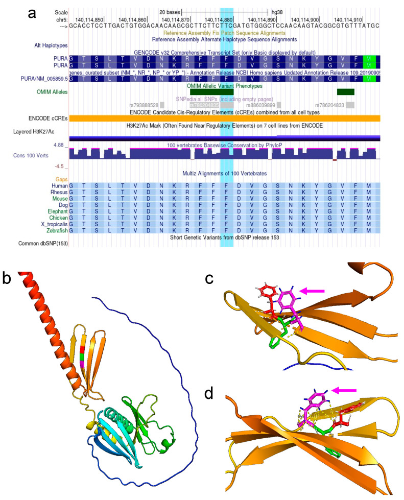
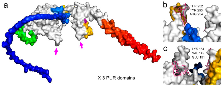
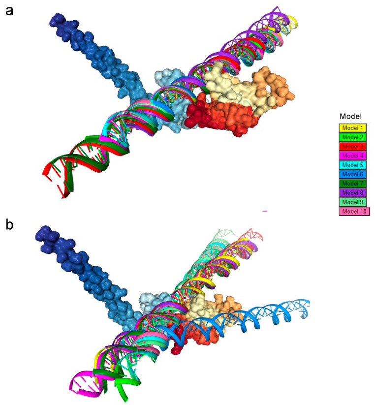
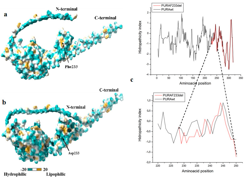
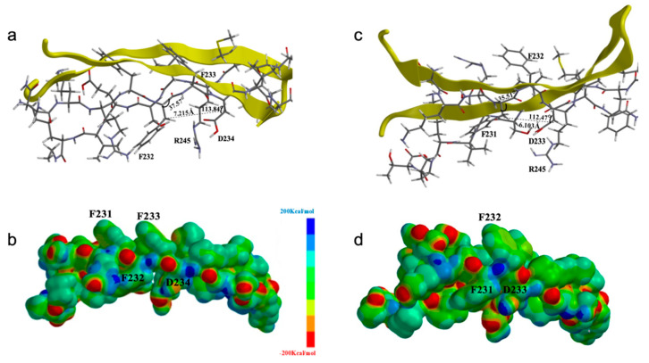
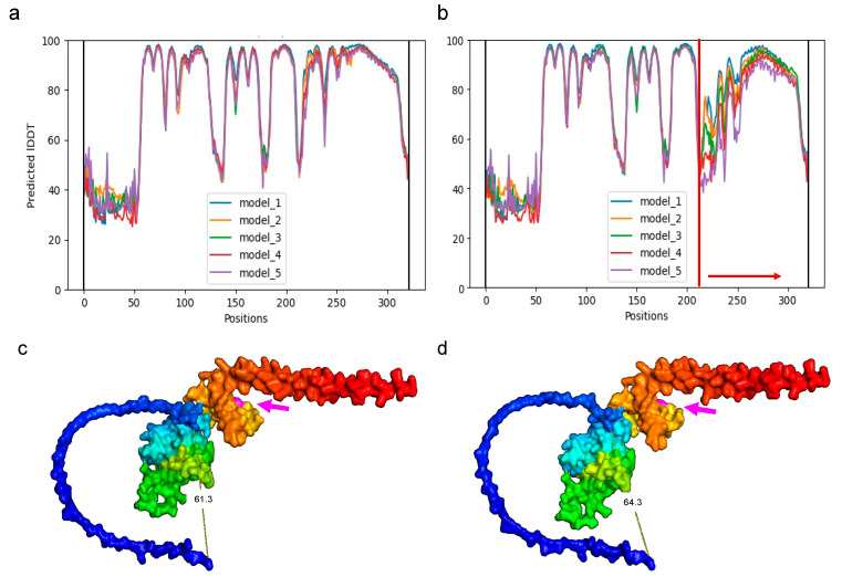
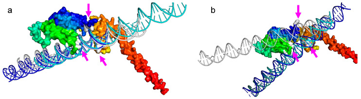
A whole-exome capture and next-generation sequencing was applied to an 11 y/o patient with a clinical history of congenital hypotonia, generalized motor and cognitive neurodevelopmental delay, and severe cognitive deficit, and without any identifiable Syndromic pattern, and to her parents, we disclosed a de novo heterozygous pathogenic mutation, c.697_699del p.Phe233del (rs786204835)(ACMG classification PS2, PM1, PM2, PP5), harbored in the PURA gene (MIM*600473) (5q31.3), associated with Autosomal Dominant Mental Retardation 31 (MIM # 616158). We used the significant improvement in the accuracy of protein structure prediction recently implemented in AlphaFold that incorporates novel neural network architectures and training procedures based on the evolutionary, physical, and geometric constraints of protein structures. The wild-type (WT) sequence and the mutated sequence, missing the Phe233, were reconstructed. The predicted local Distance Difference Test (lDDT) for the PURAwt and the PURA–Phe233del showed that the occurrence of the Phe233del affects between 220–320 amino acids. The distortion in the PURA structural conformation in the ~5 Å surrounding area after the p.Phe233del produces a conspicuous disruption of the repeat III, where the DNA and RNA helix unwinding capability occurs. PURA Protein–DNA docking corroborated these results in an in silico analysis that showed a loss of the contact of the PURA–Phe233del III repeat domain model with the DNA. Together, (i) the energetic and stereochemical, (ii) the hydropathic indexes and polarity surfaces, and (iii) the hybrid Quantum Mechanics–Molecular Mechanics (QM–MM) analyses of the PURA molecular models demarcate, at the atomic resolution, the specific surrounding region affected by these mutations and pave the way for future cell-based functional analysis. To the best of our knowledge, this is the first report of a de novo mutation underpinning a PURA syndrome in a Latin American patient and highlights the importance of predicting the molecular effects in protein structure using artificial intelligence algorithms and molecular and atomic resolution stereochemical analyses.
Overview
The claims and statistics of this paper haven’t been checked yet.
Run the check — free, no account needed. It reads the open-access text and checks the sources it cites.
Integrity signals
Registry
Cites
Statistics
Figures
Verification
Claims the paper makes
The claims and statistics of this paper haven’t been checked yet.
Run the check — free, no account needed. It reads the open-access text and checks the sources it cites.
Self-check
Percentages, sums and reported differences re-computed from the paper’s own numbers.
The self-check needs the paper’s full text.
Citations
Cites
Highly accurate protein structure prediction with AlphaFold
Jumper, J., Evans, R., Pritzel, A., Green, T., Figurnov, M., Ronneberger, O., Tunyasuvunakool, K., Bates, R., Žídek, A., Potapenko, A., et al. (2021) · Nature · doi:10.1038/s41586-021-03819-2 · Cited by 43,764
Richards, S., Aziz, N., Bale, S., Bick, D., Das, S., Gastier-Foster, J., Grody, W. W., Hegde, M., Lyon, E., Spector, E., et al. (2015) · Genetics in Medicine · doi:10.1038/gim.2015.30 · Cited by 32,594
Pervasive Sharing of Genetic Effects in Autoimmune Disease
Cotsapas, C., Voight, B. F., Rossin, E., Lage, K., Neale, B. M., Wallace, C., Abecasis, G. R., Barrett, J. C., Behrens, T., Cho, J., et al. (2011) · PLoS Genetics · doi:10.1371/journal.pgen.1002254 · Cited by 606
Research works printed but not linked: 51
- Tanaka, 2015, De novo mutations in PURA are associated with hypotonia and developmental delay, Mol. Case Stud., 1, a000356, 10.1101/mcs.a000356 · DOI
- Lalani, 2014, Mutations in PURA cause profound neonatal hypotonia, seizures, and encephalopathy in 5q31.3 microdeletion syndrome, Am. J. Hum. Genet., 95, 579, 10.1016/j.ajhg.2014.09.014 · DOI
- Hunt, 2014, Whole exome sequencing in family trios reveals de novo mutations in PURA as a cause of severe neurodevelopmental delay and learning disability, J. Med. Genet., 51, 806, 10.1136/jmedgenet-2014-102798 · DOI · Paper page
- Khalili, 2003, Pur Is Essential for Postnatal Brain Development and Developmentally Coupled Cellular Proliferation As Revealed by Genetic Inactivation in the Mouse, Mol. Cell. Biol., 23, 6857, 10.1128/MCB.23.19.6857-6875.2003 · DOI
- Wortman, 2010, Regulation of PURA gene transcription by three promoters generating distinctly spliced 5-prime leaders: A novel means of fine control over tissue specificity and viral signals, BMC Mol. Biol., 11, 1, 10.1186/1471-2199-11-81 · DOI
- White, 2009, Multiple roles for Purα in cellular and viral regulation, Cell Cycle, 8, 1, 10.4161/cc.8.3.7585 · DOI
- Hokkanen, 2012, Lack of Pur-alpha alters postnatal brain development and causes megalencephaly, Hum. Mol. Genet., 21, 473, 10.1093/hmg/ddr476 · DOI
- Reijnders, 2018, PURA syndrome: Clinical delineation and genotype-phenotype study in 32 individuals with review of published literature, J. Med. Genet., 55, 104, 10.1136/jmedgenet-2017-104946 · DOI
- Okamoto, 2017, Patient with a novel purine-rich element binding protein A mutation, Congenit. Anom., 57, 201, 10.1111/cga.12214 · DOI
- Shimojima, 2018, Infantile spasms related to a 5q31.2-q31.3 microdeletion including PURA, Hum. Genome Var., 5, 18007, 10.1038/hgv.2018.7 · DOI
- Bonaglia, 2015, Long-term follow-up of a patient with 5q31.3 microdeletion syndrome and the smallest de novo 5q31.2q31.3 deletion involving PURA, Mol. Cytogenet., 8, 89, 10.1186/s13039-015-0193-9 · DOI
- Qiao, 2019, Exome sequencing identified a de novo mutation of PURA gene in a patient with familial Xp22.31 microduplication, Eur. J. Med. Genet., 62, 103, 10.1016/j.ejmg.2018.06.010 · DOI
- Okorie, 2020, 1233 Mitochondrial Myopathy Making It Hard To Sleep! OSA management in Mitochondrial Myopathy with a variant in SNAPC4 and PURA genes, Sleep, 43, A470, 10.1093/sleep/zsaa056.1227 · DOI
- Spangenberg, 2021, Novel frameshift mutation in PURA gene causes severe encephalopathy of unclear cause, Mol. Genet. Genom. Med., 9, e1622, 10.1002/mgg3.1622 · DOI
- Mayorga, 2018, A frame-shift deletion in the PURA gene associates with a new clinical finding: Hypoglycorrhachia. Is GLUT1 a new PURA target?, Mol. Genet. Metab., 123, 331, 10.1016/j.ymgme.2017.12.436 · DOI
- 2020, A novel de novo mutation in the PURA gene associated with a new clinical finding: Large brainstem, J. Genet., 99, 7, 10.1007/s12041-019-1165-3 · DOI
- Senior, A.W., Evans, R., Jumper, J., Kirkpatrick, J., Sifre, L., and Green, T. (2020). AlphaFold. Nature, 577. · DOI · Paper page
- Al-Numan, H.H., Jan, R.M., Al-Saud, N.B.S., Rashidi, O.M., Alrayes, N.M., Alsufyani, H.A., Mujalli, A., Shaik, N.A., Mosli, M.H., and Elango, R. (2022). Exome Sequencing Identifies the Extremely Rare ITGAV and FN1 Variants in Early Onset Inflammatory Bowel Disease Patients. Front. Pediatr., 10. · DOI
- Buel, 2022, Can AlphaFold2 predict the impact of missense mutations on structure?, Nat. Struct. Mol. Biol., 29, 1, 10.1038/s41594-021-00714-2 · DOI · Paper page
- McKenna, 2010, The Genome Analysis Toolkit: A MapReduce framework for analyzing next-generation DNA sequencing data, Genome Res., 20, 1297, 10.1101/gr.107524.110 · DOI · Paper page
- Johar, 2015, Candidate gene discovery in autoimmunity by using extreme phenotypes, next generation sequencing and whole exome capture, Autoimmun. Rev., 14, 204, 10.1016/j.autrev.2014.10.021 · DOI
- Mastronardi, 2017, Genetics of non-syndromic childhood obesity and the use of high-throughput DNA sequencing technologies, J. Diabetes Complicat., 31, 1549, 10.1016/j.jdiacomp.2017.04.026 · DOI
- Landires, I., Núñez-Samudio, V., Fernandez, J., Sarria, C., Villareal, V., Córdoba, F., Apráez-Ippolito, G., Martínez, S., Vidal, O.M., and Vélez, J.I. (2020). Calpainopathy: Description of a Novel Mutation and Clinical Presentation with Early Severe Contractures. Genes, 11. · DOI
- Boguszewski, 2014, Whole Exome Sequencing of Extreme Morbid Obesity Patients: Translational Implications for Obesity and Related Disorders, Genes, 5, 709, 10.3390/genes5030709 · DOI
- Tayeh, 2022, Clinical pharmacogenomic testing and reporting: A technical standard of the American College of Medical Genetics and Genomics (ACMG), Genet. Med., 24, 759, 10.1016/j.gim.2021.12.009 · DOI
- Kearney, 2011, American College of Medical Genetics standards and guidelines for interpretation and reporting of postnatal constitutional copy number variants, Genet. Med., 13, 680, 10.1097/GIM.0b013e3182217a3a · DOI · Paper page
- Kalia, 2017, Recommendations for reporting of secondary findings in clinical exome and genome sequencing, 2016 update (ACMG SF v2.0): A policy statement of the American College of Medical Genetics and Genomics, Genet. Med., 19, 249, 10.1038/gim.2016.190 · DOI
- Yan, 2020, The HDOCK server for integrated protein–protein docking, Nat. Protoc., 15, 1829, 10.1038/s41596-020-0312-x · DOI
- Bergemann, 1992, The HeLa Pur factor binds single-stranded DNA at a specific element conserved in gene flanking regions and origins of DNA replication, Mol. Cell. Biol., 12, 1257
- Bergemann, 1992, Sequence of cDNA comprising the human pur gene and sequence-specific single-stranded-DNA-binding properties of the encoded protein, Mol. Cell. Biol., 12, 5673
- Wortman, 2005, Mechanism of DNA binding and localized strand separation by Purα and comparison with Pur family member, Purβ, Biochim. Biophys. Acta Mol. Cell Res., 1743, 64, 10.1016/j.bbamcr.2004.08.010 · DOI
- Ma, 1994, Conservation in human and mouse Purα of a motif common to several proteins involved in initiation of DNA replication, Gene, 149, 311, 10.1016/0378-1119(94)90167-8 · DOI
- Haas, 1993, A developmentally regulated DNA-binding protein from mouse brain stimulates myelin basic protein gene expression, Mol. Cell. Biol., 13, 3103
- Studer, 2014, Assessing the local structural quality of transmembrane protein models using statistical potentials (QMEANBrane), Bioinformatics, 30, i505, 10.1093/bioinformatics/btu457 · DOI
- Benkert, 2008, QMEAN: A comprehensive scoring function for model quality assessment, Proteins: Struct. Funct. Bioinform., 71, 261, 10.1002/prot.21715 · DOI
- Laskowski, 1993, PROCHECK: A program to check the stereochemical quality of protein structures, J. Appl. Crystallogr., 26, 283, 10.1107/S0021889892009944 · DOI · Paper page
- Gasteiger, E., Hoogland, C., Gattiker, A., Wilkins, M.R., Appel, R.D., and Bairoch, A. (2005). The Proteomics Protocols Handbook-Chapter 52: Protein Identification and Analysis Tools on the ExPASy Server. Proteom. Protoc. Handb., 571–607. · DOI
- Pettersen, 2004, UCSF Chimera?A visualization system for exploratory research and analysis, J. Comput. Chem., 25, 1605, 10.1002/jcc.20084 · DOI
- Guex, 1997, SWISS-MODEL and the Swiss-PdbViewer: An environment for comparative protein modeling, Electrophoresis, 18, 2714, 10.1002/elps.1150181505 · DOI
- Humphrey, 1996, VMD: Visual molecular dynamics, J. Mol. Graph., 14, 33, 10.1016/0263-7855(96)00018-5 · DOI · Paper page
- Phillips, 2005, Scalable molecular dynamics with NAMD, J. Comput. Chem., 26, 1781, 10.1002/jcc.20289 · DOI · Paper page
- 2020, Redox mechanism of Trypanosoma cruzi Resistance to Nitro Prodrugs Benznidazole and Nifurtimox, Int. J. Bioinform. Comput. Biol., 5, 1
- Bedoya, 2021, Structural Predictive Model of Presenilin-2 Protein and Analysis of Structural Effects of Familial Alzheimer’s Disease Mutations, Biochem. Res. Int., 2021, 9542038
- Bedoya, 2021, Protein Predictive Modeling and Simulation of Mutations of Presenilin-1 Familial Alzheimer’s Disease on the Orthosteric Site, Front. Mol. Biosci., 8, 387
- Cano, 2021, Diffusion Mechanism Modeling of Metformin in Human Organic Cationic Amino Acid Transporter one and Functional Impact of S189L, R206C, and G401S Mutation, Front. Pharmacol., 11, 587590, 10.3389/fphar.2020.587590 · DOI
- Ahmadi, 2018, Multiscale modeling of enzymes: QM-cluster, QM/MM, and QM/MM/MD: A tutorial review, Int. J. Quantum Chem., 118, e25558, 10.1002/qua.25558 · DOI
- Dewar, 1985, Development and use of quantum mechanical molecular models. 76. AM1: A new general purpose quantum mechanical molecular model, J. Am. Chem. Soc., 107, 3902, 10.1021/ja00299a024 · DOI
- Dewar, M.J.S., Zoebisch, E.G., Healy, E.F., and Stewart, J.J.P. (1985). AM1: A New General Purpose Quantum Mechanical Molecular Model1. J. Am. Chem. Soc., 107. · DOI
- Baker, 2007, Role of aromatic amino acids in protein-nucleic acid recognition, Biopolymers, 85, 456, 10.1002/bip.20682 · DOI
- Molitor, 2021, The Molecular Function of PURA and Its Implications in Neurological Diseases, Front. Genet., 12, 638217, 10.3389/fgene.2021.638217 · DOI
- Grzybowska, 2012, Human intronless genes: Functional groups, associated diseases, evolution, and mRNA processing in absence of splicing, Biochem. Biophys. Res. Commun., 424, 1, 10.1016/j.bbrc.2012.06.092 · DOI
Metadata
- Authors
- López-Rivera, J. J., Rodríguez-Salazar, L., Soto-Ospina, A., Estrada-Serrato, C., Serrano, D., Chaparro-Solano, H. M., Londoño, O., Rueda, P. A., Ardila, G., Villegas-Lanau, A., Godoy-Corredor, M., Cuartas, M., Vélez, J. I., Vidal, O. M., Isaza-Ruget, M. A., & Arcos-Burgos, M.
- Affiliations
- INPAC Research Group, Fundación Universitaria Sanitas, Bogotá 111321, Colombia
- Grupo de Bioinformática, Laboratorio de Clínica Colsanitas, Bogotá 110221, Colombia
- Universidad de Antioquia (CO)
- Grupo de Genética Médica, Clínica Universitaria Colombia y Clínica Pediátrica Colsanitas, Bogotá 111321, Colombia
- Genética Molecular (GenMol), Facultad de Ciencias Exactas y Naturales, Universidad de Antioquia, Medellín 050012, Colombia
- Laboratorio Clínico y de Patología, Clínica Colsanitas, Bogotá 110221, Colombia
- Grupo de Investigación Estudios en Psicología, Departamento de Psicología, Escuela de Humanidades, Universidad EAFIT, Medellín 050022, Colombia
- Universidad del Norte, Barranquilla 080001, Colombia
- Universidad del Norte (CO)
- Fundación Universitaria Sanitas (CO)
- Grupo de Investigación en Psiquiatría (GIPSI), Departamento de Psiquiatría, Instituto de Investigaciones Médicas, Facultad de Medicina, Universidad de Antioquia, Medellín 050012, Colombia
- ORCID
- Venue
- Brain Sciences
- Publisher
- MDPI AG
- Published
- 2022-06-30
- Volume
- 12
- Issue
- 7
- Pages
- 871
- ISSN
- 2076-3425 (ISSN-L)
- PMC
- PMC9313109
- OpenAlex
- W4283757303
- Cited by
- 17
- References
- 3
- Citations by year
- 2026: 2 · 2025: 1 · 2024: 9 · 2023: 4
- FWCI
- 2.10
- Type
- journal article
- Language
- en
- Open access
- gold
- doi.org
- Licence
- Locations
- Brain Sciences (publishedVersion, cc-by)
- Preprints.org (acceptedVersion, cc-by)
- PubMed Central (publishedVersion, cc-by)
- PubMed (publishedVersion)
- Institutional Repository University of Antioquia (University of Antioquia) (publishedVersion, cc-by)
- DOAJ (DOAJ: Directory of Open Access Journals) (submittedVersion, cc-by-sa)
- Funders
- Keralty
- Topics
- Life Sciences › Biochemistry, Genetics and Molecular Biology › Genetics › Genomics and Rare Diseases
- Life Sciences › Biochemistry, Genetics and Molecular Biology › Molecular Biology › Protein Structure and Dynamics
- Life Sciences › Biochemistry, Genetics and Molecular Biology › Genetics › Genetics and Neurodevelopmental Disorders
- Keywords
- PURA gene, de novo mutations, congenital hypotonia, neurodevelopmental delay, protein structure prediction, AlphaFold, protein-DNA docking
About these checks
Registry, retraction, Cites and statistics results are computed, not judged, with one exception: a printed reference that no index lists is read once by a model before it counts as a citation error. Claims are extracted by a model and kept only when their quote appears verbatim in the text; each cited source is then checked, with a model’s help, for whether it says what the claim relies on. Those verdicts can be wrong — read the claim’s quote and the cited source before relying on one.
Statistics and arithmetic are recomputed, not judged.
Confidence bands are shown only for fields calibrated on our annotated test set. None is calibrated yet, so no band is shown.
What counts as a claim ›How we measure confidence ›How we check this ›
Are you an author, or do you see a problem with this page? Request a review
