

The DOI matches its Crossref record; 3 of the 3 references it cites were found in a registry or an index.
Expression Analysis of Autophagy Related Markers LC3B, p62 and HMGB1 Indicate an Autophagy-Independent Negative Prognostic Impact of High p62 Expression in Pulmonary Squamous Cell Carcinomas
Abstract
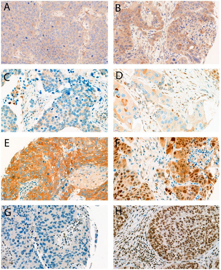
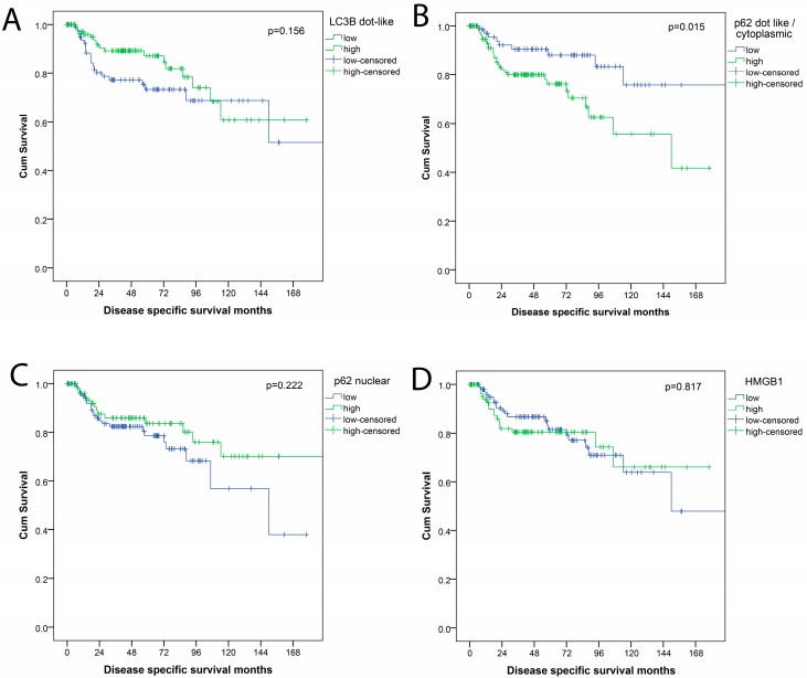
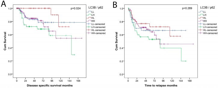
Autophagy is involved in maintaining cellular homeostasis under stress conditions. It also plays an important role in various diseases including cancer. Pulmonary squamous cell carcinomas (pSQCC) at present lack targetable molecular alterations, and demand alternative therapeutic options. We assessed the expression levels of autophagy related proteins LC3B, p62, and HMGB1 in 271 primary resected pSQCC by immunohistochemistry, in correlation with clinical and pathological parameters, as a rationale for a potential autophagy directed therapy. LC3B, p62, and HMGB1 staining showed various patterns. LC3Bhighp62low levels, suggested to indicate intact activated autophagy, were associated with prolonged disease specific survival (DSS) and LC3Bhighp62high levels, indicating activated but late stage impaired autophagy, with shorter DSS (p = 0.024). p62high expression regardless of LC3B, however, showed an even stronger association with shorter DSS (p = 0.015) and was also an independent negative prognostic factor in multivariate analysis (HR = 2.99; 95% CI 1.38–6.52; p = 0.006). HMGB1 expression correlated neither with the expression of LC3B and p62, nor with patients’ outcome. Different states of autophagy characterized by distinct p62 and LC3B expression patterns may be linked to patient’s prognosis in pSQCC. Our results, however, point also to an autophagy independent role of p62 with an even more pronounced prognostic impact compared to autophagy related p62.
Overview
The claims and statistics of this paper haven’t been checked yet.
Run the check — free, no account needed. It reads the open-access text and checks the sources it cites.
Integrity signals
Registry
Cites
Statistics
Figures
Verification
Claims the paper makes
The claims and statistics of this paper haven’t been checked yet.
Run the check — free, no account needed. It reads the open-access text and checks the sources it cites.
Claims other papers make about this one
Each is checked against this paper’s own text: does it say what the citing paper says it does?
The source backs part of the claim, not all of it — or backs it for a different population, condition or comparison.
In the citing paper
“Assessing LC3 puncta fails to show prognostic significance in non-small cell lung cancer [528, 529].”
From Guidelines for the use and interpretation of assays for monitoring autophagy (4th edition) 1 (2021) · cites this paper as [528]
Experimental — accuracy not yet measured
Self-check
Percentages, sums and reported differences re-computed from the paper’s own numbers.
The self-check needs the paper’s full text.
Citations
Cites
Guidelines for the use and interpretation of assays for monitoring autophagy (3rd edition).
Klionsky, D. J., Abdelmohsen, K., Abe, A., Abedin, M. J., Abeliovich, H., Acevedo Arozena, A., Adachi, H., Adams, C. M., Adams, P. D., Adeli, K., et al. (2016) · Autophagy · doi:10.1080/15548627.2015.1100356 · Cited by 5,894
Self-citationRizvi, N. A., Mazières, J., Planchard, D., Stinchcombe, T. E., Dy, G. K., Antonia, S. J., Horn, L., Lena, H., Minenza, E., Mennecier, B., et al. (2015) · The Lancet Oncology · doi:10.1016/s1470-2045(15)70054-9 · Cited by 1,415
Ladoire, S., Enot, D., Senovilla, L., Ghiringhelli, F., Poirier-Colame, V., Chaba, K., Semeraro, M., Chaix, M., Penault-Llorca, F., Arnould, L., et al. (2016) · Autophagy · doi:10.1080/15548627.2016.1154244 · Cited by 117
Research works printed but not linked: 47
- Langer, 2016, Incremental innovation and progress in advanced squamous cell lung cancer: Current status and future impact of treatment, J. Thorac. Oncol., 11, 2066, 10.1016/j.jtho.2016.08.138 · DOI
- Brambilla, 2012, Squamous cell carcinoma of the lung: Molecular subtypes and therapeutic opportunities, Clin. Cancer Res., 18, 2443, 10.1158/1078-0432.CCR-11-2370 · DOI
- Reck, 2016, Pembrolizumab versus chemotherapy for pd-l1–positive non–small-cell lung cancer, N. Engl. J. Med., 375, 1823, 10.1056/NEJMoa1606774 · DOI · Paper page
- Mizushima, 2010, Autophagy, FEBS Lett., 584, 1279, 10.1016/j.febslet.2010.02.053 · DOI
- Mizushima, 2018, A brief history of autophagy from cell biology to physiology and disease, Nat. Cell Biol., 20, 521, 10.1038/s41556-018-0092-5 · DOI · Paper page
- Jiang, 2014, Autophagy and human diseases, Cell Res., 24, 69, 10.1038/cr.2013.161 · DOI
- Levine, 2008, Autophagy in the pathogenesis of disease, Cell, 132, 27, 10.1016/j.cell.2007.12.018 · DOI · Paper page
- Meijer, 2009, Autophagy: Regulation and role in disease, Crit. Rev. Clin. Lab. Sci., 46, 210, 10.1080/10408360903044068 · DOI
- Chen, 2009, Role and regulation of autophagy in cancer, Biochim. Biophys. Acta, 1793, 1516, 10.1016/j.bbamcr.2008.12.013 · DOI
- Mathew, 2007, Role of autophagy in cancer, Nat. Rev. Cancer, 7, 961, 10.1038/nrc2254 · DOI · Paper page
- Martinet, 2013, Immunohistochemical analysis of macroautophagy: Recommendations and limitations, Autophagy, 9, 386, 10.4161/auto.22968 · DOI
- Jiang, 2015, Lc3- and p62-based biochemical methods for the analysis of autophagy progression in mammalian cells, Methods, 75, 13, 10.1016/j.ymeth.2014.11.021 · DOI
- Ladoire, 2015, Combined evaluation of lc3b puncta and hmgb1 expression predicts residual risk of relapse after adjuvant chemotherapy in breast cancer, Autophagy, 11, 1878, 10.1080/15548627.2015.1082022 · DOI
- Duffy, 2015, Autophagy modulation: A target for cancer treatment development, Cancer Chemother. Pharmacol., 75, 439, 10.1007/s00280-014-2637-z · DOI
- Sui, 2013, Autophagy and chemotherapy resistance: A promising therapeutic target for cancer treatment, Cell Death Dis., 4, e838, 10.1038/cddis.2013.350 · DOI
- Zhong, 2016, Autophagy, inflammation, and immunity: A troika governing cancer and its treatment, Cell, 166, 288, 10.1016/j.cell.2016.05.051 · DOI
- Onorati, A.V., Dyczynski, M., Ojha, R., and Amaravadi, R.K. (2018). Targeting autophagy in cancer. Cancer. · DOI
- Schlafli, 2016, Prognostic value of the autophagy markers lc3 and p62/sqstm1 in early-stage non-small cell lung cancer, Oncotarget, 7, 39544, 10.18632/oncotarget.9647 · DOI · Paper page
- Keller, 2017, Adverse prognostic value of pd-l1 expression in primary resected pulmonary squamous cell carcinomas and paired mediastinal lymph node metastases, Mod. Pathol., 31, 101, 10.1038/modpathol.2017.111 · DOI
- Brierley, J.D., Gospodarowicz, M.K., and Wittekind, C. (2017). The TNM Classification of Calignant Tumours, John Wiley & Sons. [8th ed.]. · DOI · Paper page
- Liu, 2014, Prognostic significance of p62/sqstm1 subcellular localization and lc3b in oral squamous cell carcinoma, Br. J. Cancer, 111, 944, 10.1038/bjc.2014.355 · DOI
- White, 2009, The double-edged sword of autophagy modulation in cancer, Clin. Cancer Res., 15, 5308, 10.1158/1078-0432.CCR-07-5023 · DOI
- Galluzzi, 2015, Autophagy in malignant transformation and cancer progression, EMBO J., 34, 856, 10.15252/embj.201490784 · DOI · Paper page
- Amaravadi, 2011, Principles and current strategies for targeting autophagy for cancer treatment, Clin. Cancer Res., 17, 654, 10.1158/1078-0432.CCR-10-2634 · DOI
- Chude, C.I., and Amaravadi, R.K. (2017). Targeting autophagy in cancer: Update on clinical trials and novel inhibitors. Int. J. Mol. Sci., 18. · DOI
- Adams, 2016, Prognostic relevance of autophagy markers lc3b and p62 in esophageal adenocarcinomas, Oncotarget, 7, 39241, 10.18632/oncotarget.9649 · DOI
- Niklaus, 2017, Expression analysis of lc3b and p62 indicates intact activated autophagy is associated with an unfavorable prognosis in colon cancer, Oncotarget, 8, 54604, 10.18632/oncotarget.17554 · DOI
- Bortnik, S., and Gorski, S.M. (2017). Clinical applications of autophagy proteins in cancer: From potential targets to biomarkers. Int. J. Mol. Sci., 18. · DOI
- Martinet, 2006, Detection of autophagy in tissue by standard immunohistochemistry: Possibilities and limitations, Autophagy, 2, 55, 10.4161/auto.2217 · DOI · Paper page
- Martinet, W., Roth, L., and De Meyer, G.R.Y. (2017). Standard immunohistochemical assays to assess autophagy in mammalian tissue. Cells, 30. · DOI
- Schlafli, 2015, Reliable lc3 and p62 autophagy marker detection in formalin fixed paraffin embedded human tissue by immunohistochemistry, Eur. J. Histochem., 59, 2481, 10.4081/ejh.2015.2481 · DOI
- He, 2017, The dual role and therapeutic potential of high-mobility group box 1 in cancer, Oncotarget, 8, 64534, 10.18632/oncotarget.17885 · DOI
- Wang, 2015, Beclin 1 and p62 expression in non-small cell lung cancer: Relation with malignant behaviors and clinical outcome, Int. J. Clin. Exp. Pathol., 8, 10644
- Daisuke, 2012, Accumulation of p62/sqstm1 is associated with poor prognosis in patients with lung adenocarcinoma, Cancer Sci., 103, 760, 10.1111/j.1349-7006.2012.02216.x · DOI
- Ruan, 2018, The prognostic value of p62 in solid tumor patients: A meta-analysis, Oncotarget, 9, 4258, 10.18632/oncotarget.23101 · DOI
- Katsuragi, 2015, P62/sqstm1 functions as a signaling hub and an autophagy adaptor, FEBS J., 282, 4672, 10.1111/febs.13540 · DOI
- Nezis, 2011, P62 at the interface of autophagy, oxidative stress signaling, and cancer, Antioxid. Redox Signal., 17, 786, 10.1089/ars.2011.4394 · DOI
- Jiang, 2015, P62 links autophagy and nrf2 signaling, Free Radic. Biol. Med., 88, 199, 10.1016/j.freeradbiomed.2015.06.014 · DOI
- Fan, 2018, Role of p62 in the regulation of cell death induction, Apoptosis, 23, 187, 10.1007/s10495-018-1445-z · DOI
- Pankiv, 2010, Nucleocytoplasmic shuttling of p62/sqstm1 and its role in recruitment of nuclear polyubiquitinated proteins to promyelocytic leukemia bodies, J. Biol. Chem., 285, 5941, 10.1074/jbc.M109.039925 · DOI
- Adams, O., Janser, F.A., Dislich, B., Berezowska, S., Humbert, M., Seiler, C.A., Kroell, D., Slotta-Huspenina, J., Feith, M., and Ott, K. (2018). A specific expression profile of lc3b and p62 is associated with nonresponse to neoadjuvant chemotherapy in esophageal adenocarcinomas. PLoS ONE, 13. · DOI
- Islam, M.A., Sooro, M.A., and Zhang, P. (2018). Autophagic regulation of p62 is critical for cancer therapy. Int. J. Mol. Sci., 19. · DOI
- Li, Y.Y., Feun, L.G., Thongkum, A., Tu, C.H., Chen, S.M., Wangpaichitr, M., Wu, C., Kuo, M.T., and Savaraj, N. (2017). Autophagic mechanism in anti-cancer immunity: Its pros and cons for cancer therapy. Int. J. Mol. Sci., 18. · DOI
- Hossain, 2012, A possible cross-talk between autophagy and apoptosis in generating an immune response in melanoma, Apoptosis, 17, 1066, 10.1007/s10495-012-0745-y · DOI
- Bloy, 2017, Immunogenic stress and death of cancer cells: Contribution of antigenicity vs adjuvanticity to immunosurveillance, Immunol. Rev., 280, 165, 10.1111/imr.12582 · DOI
- Ivan, D., and Fang, F. (2013). Tumors of the lungs and pleura. Cancer Grading Manual, Springer.
- Zlobec, 2013, Next-generation tissue microarray (ngTMA) increases the quality of biomarker studies: An example using CD3, CD8, and CD45RO in the tumor microenvironment of six different solid tumor types, J. Transl. Med., 11, 104, 10.1186/1479-5876-11-104 · DOI
Cited by
Sorted by citations
Guidelines for the use and interpretation of assays for monitoring autophagy (4th edition) 1
Klionsky, D. J., Abdel-Aziz, A. K., Abdelfatah, S., Abdellatif, M., Abdoli, A., Abel, S., Abeliovich, H., Abildgaard, M. H., Abudu, Y. P., Acevedo-Arozena, A., et al. (2021) · Autophagy · doi:10.1080/15548627.2020.1797280 · Cited by 2,897
Self-citationWang, P., Feng, Y.-B., Wang, L., Li, Y., Fan, C., Song, Q., & Yu, S. Y. (2019) · Brain, Behavior, and Immunity · doi:10.1016/j.bbi.2019.08.002 · Cited by 38
Metadata
- Authors
- Langer, R., Neppl, C., Keller, M. D., Schmid, R. A., Tschan, M. P., & Berezowska, S.
- Affiliations
- Institute of Pathology, University of Bern, Murtenstrasse 31, 3008 Bern, Switzerland
- Division of General Thoracic Surgery, Inselspital University Hospital Bern, 3010 Bern, Switzerland
- Venue
- Cancers
- Publisher
- MDPI AG
- Published
- 2018-08-21
- Volume
- 10
- Issue
- 9
- Pages
- 281
- ISSN
- 2072-6694
- PMC
- PMC6162479
- Cited by
- 17
- References
- 3
- Language
- en
- Funders
- Bernische Krebsliga (no number)
About these checks
Registry, retraction, Cites and statistics results are computed, not judged, with one exception: a printed reference that no index lists is read once by a model before it counts as a citation error. Claims are extracted by a model and kept only when their quote appears verbatim in the text; each cited source is then checked, with a model’s help, for whether it says what the claim relies on. Those verdicts can be wrong — read the claim’s quote and the cited source before relying on one.
Statistics and arithmetic are recomputed, not judged.
Confidence bands are shown only for fields calibrated on our annotated test set. None is calibrated yet, so no band is shown.
What counts as a claim ›How we measure confidence ›How we check this ›
Are you an author, or do you see a problem with this page? Request a review
