

The DOI matches its Crossref record; 60 of the 60 references it cites were found in a registry or an index.
Natural Killer Cell Activation Receptor NKp30 Oligomerization Depends on Its N-Glycosylation
Open accessAbstract
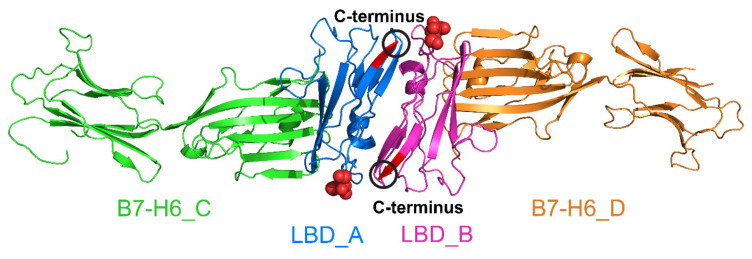
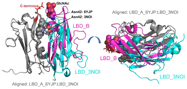
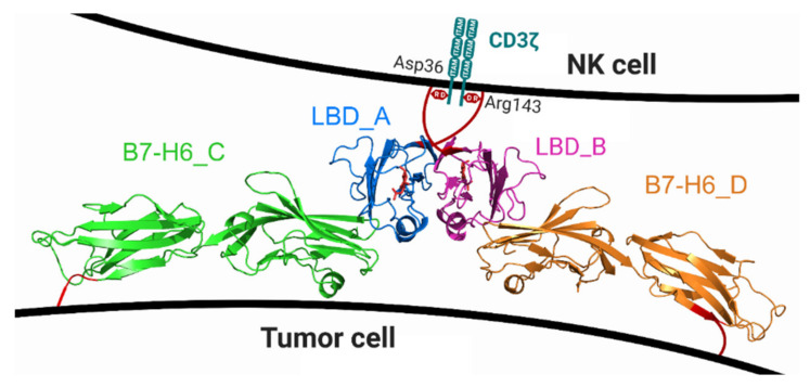
NKp30 is one of the main human natural killer (NK) cell activating receptors used in directed immunotherapy. The oligomerization of the NKp30 ligand binding domain depends on the length of the C-terminal stalk region, but our structural knowledge of NKp30 oligomerization and its role in signal transduction remains limited. Moreover, ligand binding of NKp30 is affected by the presence and type of N-glycosylation. In this study, we assessed whether NKp30 oligomerization depends on its N-glycosylation. Our results show that NKp30 forms oligomers when expressed in HEK293S GnTI− cell lines with simple N-glycans. However, NKp30 was detected only as monomers after enzymatic deglycosylation. Furthermore, we characterized the interaction between NKp30 and its best-studied cognate ligand, B7-H6, with respect to glycosylation and oligomerization, and we solved the crystal structure of this complex with glycosylated NKp30, revealing a new glycosylation-induced mode of NKp30 dimerization. Overall, this study provides new insights into the structural basis of NKp30 oligomerization and explains how the stalk region and glycosylation of NKp30 affect its ligand affinity. This furthers our understanding of the molecular mechanisms involved in NK cell activation, which is crucial for the successful design of novel NK cell-based targeted immunotherapeutics.
Overview
The claims and statistics of this paper haven’t been checked yet.
Run the check — free, no account needed. It reads the open-access text and checks the sources it cites.
Integrity signals
Registry
Cites
Statistics
Figures
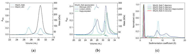
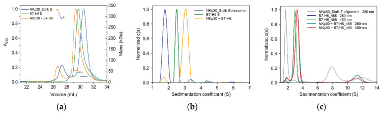
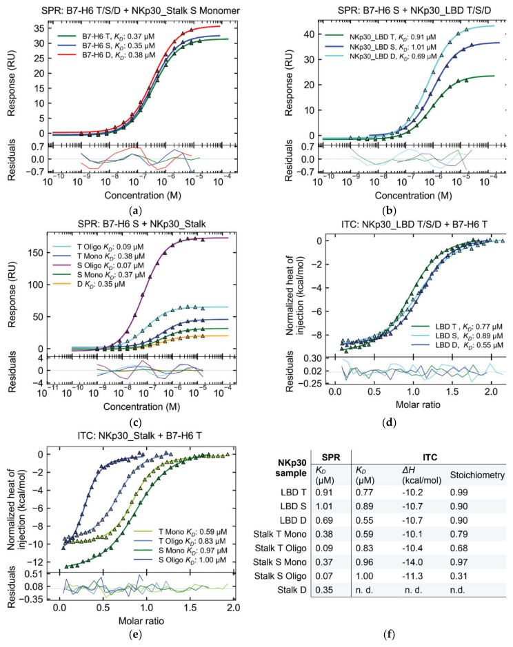
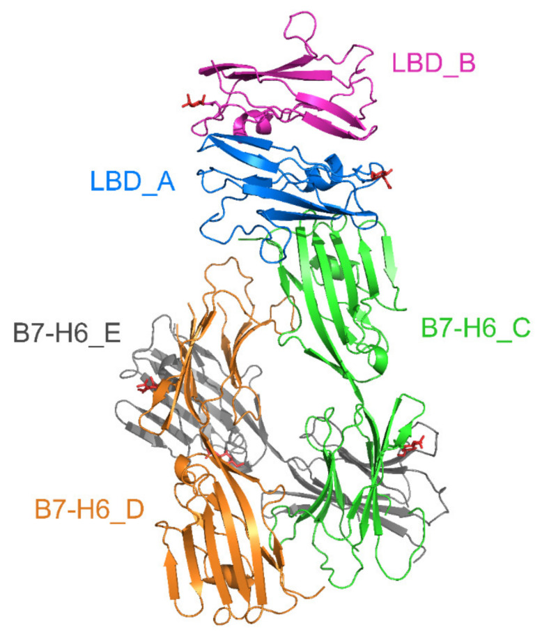
Verification
Claims the paper makes
The claims and statistics of this paper haven’t been checked yet.
Run the check — free, no account needed. It reads the open-access text and checks the sources it cites.
Claims other papers make about this one
Each is checked against this paper’s own text: does it say what the citing paper says it does?
The cited source says what the claim attributes to it.
In the citing paper
“Finally, N-linked glycans also play an important role in cell–cell interactions. This is the case of the CD337 receptor on Natural Killer cells indicating the recognized cell as cancerous [157].”
From GANAB and N-Glycans Substrates Are Relevant in Human Physiology, Polycystic Pathology and Multiple Sclerosis: A Review (2022) · cites this paper as [157]
Self-check
Percentages, sums and reported differences re-computed from the paper’s own numbers.
The self-check needs the paper’s full text.
Citations
Cites
UCSF Chimera—A visualization system for exploratory research and analysis
Pettersen, E. F., Goddard, T. D., Huang, C. C., Couch, G. S., Greenblatt, D. M., Meng, E. C., & Ferrin, T. E. (2004) · Journal of Computational Chemistry · doi:10.1002/jcc.20084 · Cited by 45,882
Features and development of Coot
Emsley, P., Lohkamp, B., Scott, W. G., & Cowtan, K. (2010) · Acta Crystallographica Section D Biological Crystallography · doi:10.1107/s0907444910007493 · Cited by 29,915
Phaser crystallographic software
McCoy, A. J., Grosse-Kunstleve, R. W., Adams, P. D., Winn, M. D., Storoni, L. C., & Read, R. J. (2007) · Journal of Applied Crystallography · doi:10.1107/s0021889807021206 · Cited by 20,612
Kabsch, W. (2010) · Acta Crystallographica Section D Biological Crystallography · doi:10.1107/s0907444909047337 · Cited by 16,463
MolProbity : all-atom structure validation for macromolecular crystallography
Chen, V. B., Arendall, W. B., Headd, J. J., Keedy, D. A., Immormino, R. M., Kapral, G. J., Murray, L. W., Richardson, J. S., & Richardson, D. C. (2010) · Acta Crystallographica Section D Biological Crystallography · doi:10.1107/s0907444909042073 · Cited by 14,115
Overview of the CCP 4 suite and current developments
Winn, M. D., Ballard, C. C., Cowtan, K. D., Dodson, E. J., Emsley, P., Evans, P. R., Keegan, R. M., Krissinel, E. B., Leslie, A. G. W., McCoy, A., et al. (2011) · Acta Crystallographica Section D Biological Crystallography · doi:10.1107/s0907444910045749 · Cited by 12,353
Inference of Macromolecular Assemblies from Crystalline State
Krissinel, E., & Henrick, K. (2007) · Journal of Molecular Biology · doi:10.1016/j.jmb.2007.05.022 · Cited by 10,007
REFMAC 5 for the refinement of macromolecular crystal structures
Murshudov, G. N., Skubák, P., Lebedev, A. A., Pannu, N. S., Steiner, R. A., Nicholls, R. A., Winn, M., Long, F., & Vagin, A. A. (2011) · Acta Crystallographica Section D Biological Crystallography · doi:10.1107/s0907444911001314 · Cited by 8,373
Schuck, P. (2000) · Biophysical Journal · doi:10.1016/s0006-3495(00)76713-0 · Cited by 3,868
Kiessling, R. W., Klein, E., & Wigzell, H. (1975) · European Journal of Immunology · doi:10.1002/eji.1830050208 · Cited by 1,827
Franke, D., Petoukhov, M. V., Konarev, P. V., Panjkovich, A., Tuukkanen, A. T., Mertens, H. D. T., Kikhney, A., Hajizadeh, N. R., Franklin, J. M., Jeffries, C. M., et al. (2017) · Journal of Applied Crystallography · doi:10.1107/s1600576717007786 · Cited by 1,271
Durocher, Y. (2002) · Nucleic Acids Research · doi:10.1093/nar/30.2.e9 · Cited by 1,041
Herberman, R. B., Nunn, M. E., Holden, H. T., & Lavrin, D. H. (1975) · International Journal of Cancer · doi:10.1002/ijc.2910160205 · Cited by 885
Pende, D., Parolini, S., Pessino, A. M., Sivori, S., Augugliaro, R., Morelli, L., Marcenaro, E., Accame, L., Malaspina, A., Biassoni, R., et al. (1999) · The Journal of Experimental Medicine · doi:10.1084/jem.190.10.1505 · Cited by 794
Brandt, C. S., Baratin, M., Yi, E. C., Kennedy, J. J., Gao, Z., Fox, B. A., Haldeman, B. A., Ostrander, C. D., Kaifu, T., Chabannon, C., et al. (2009) · The Journal of Experimental Medicine · doi:10.1084/jem.20090681 · Cited by 747
A time- and cost-efficient system for high-level protein production in mammalian cells
Aricescu, A. R., Lu, W., & Jones, E. Y. (2006) · Acta Crystallographica Section D Biological Crystallography · doi:10.1107/s0907444906029799 · Cited by 746
Reeves, P. J., Callewaert, N., Contreras, R., & Khorana, H. G. (2002) · Proceedings of the National Academy of Sciences · doi:10.1073/pnas.212519299 · Cited by 641
Calculations and Publication-Quality Illustrations for Analytical Ultracentrifugation Data
Brautigam, C. A. (2015) · Methods in enzymology on CD-ROM/Methods in enzymology · doi:10.1016/bs.mie.2015.05.001 · Cited by 590
Activating and inhibitory receptors of natural killer cells
Pegram, H. J., Andrews, D. M., Smyth, M. J., Darcy, P. K., & Kershaw, M. H. (2010) · Immunology and Cell Biology · doi:10.1038/icb.2010.78 · Cited by 583
Young, M. M., Tang, N., Hempel, J. C., Oshiro, C. M., Taylor, E. W., Kuntz, I. D., Gibson, B. W., & Dollinger, G. D. (2000) · Proceedings of the National Academy of Sciences · doi:10.1073/pnas.090099097 · Cited by 460
Inhibition of the NKp30 activating receptor by pp65 of human cytomegalovirus
Arnon, T. I., Achdout, H., Levi, O., Markel, G., Saleh, N., Katz, G., Gazit, R., Gonen‐Gross, T., Hanna, J. H., Nahari, E., et al. (2005) · Nature Immunology · doi:10.1038/ni1190 · Cited by 396
Zhao, H., Piszczek, G., & Schuck, P. (2014) · Methods · doi:10.1016/j.ymeth.2014.11.012 · Cited by 348
Natural cytotoxicity receptors and their ligands
Kruse, P. H., Matta, J., Ugolini, S., & Vivier, É. (2013) · Immunology and Cell Biology · doi:10.1038/icb.2013.98 · Cited by 281
New B7 Family Checkpoints in Human Cancers
Ni, L., & Dong, C. (2017) · Molecular Cancer Therapeutics · doi:10.1158/1535-7163.mct-16-0761 · Cited by 245
Schlecker, E., Fiegler, N., Arnold, A., Altevogt, P., Rose‐John, S., Moldenhauer, G., Sucker, A., Paschen, A., von Strandmann, E. P., Textor, S., et al. (2014) · Cancer Research · doi:10.1158/0008-5472.can-13-3017 · Cited by 216
Scheuermann, T. H., & Brautigam, C. A. (2014) · Methods · doi:10.1016/j.ymeth.2014.11.024 · Cited by 211
Backliwal, G., Hildinger, M., Kuettel, I., Delegrange, F., Hacker, D. L., & Wurm, F. M. (2008) · Biotechnology and Bioengineering · doi:10.1002/bit.21882 · Cited by 176
MoRDa, an automatic molecular replacement pipeline
Vagin, A. A., & Lebedev, A. A. (2015) · Acta Crystallographica Section A Foundations and Advances · doi:10.1107/s2053273315099672 · Cited by 161
Matta, J., Baratin, M., Chiche, L., Forel, J.-M., Cognet, C., Thomas, G., Farnarier, C., Pipéroglou, C., Papazian, L., Chaussabel, D., et al. (2013) · Blood · doi:10.1182/blood-2013-01-481705 · Cited by 151
Wang, W., Guo, H., Geng, J., et al. (2014) · Journal of Biological Chemistry · doi:10.1074/jbc.m114.603464 · Cited by 140
Li, Y., Wang, Q., & Mariuzza, R. A. (2011) · The Journal of Experimental Medicine · doi:10.1084/jem.20102548 · Cited by 133
B7-H6/NKp30 interaction: a mechanism of alerting NK cells against tumors
Kaifu, T., Escalière, B., Gastinel, L. N., Vivier, É., & Baratin, M. (2011) · Cellular and Molecular Life Sciences · doi:10.1007/s00018-011-0802-7 · Cited by 128
Chisholm, S. E., & Reyburn, H. T. (2006) · Journal of Virology · doi:10.1128/jvi.80.5.2225-2233.2006 · Cited by 92
Human Natural Killer Receptors, Co‐Receptors, and Their Ligands
Biassoni, R. (2009) · Current Protocols in Immunology · doi:10.1002/0471142735.im1410s84 · Cited by 84
Joyce, M. G., Tran, P., Zhuravleva, M. A., et al. (2011) · Proceedings of the National Academy of Sciences · doi:10.1073/pnas.1100622108 · Cited by 76
Automated refinement of macromolecular structures at low resolution using prior information
Kovalevskiy, O. V., Nicholls, R. A., & Murshudov, G. N. (2016) · Acta Crystallographica Section D Structural Biology · doi:10.1107/s2059798316014534 · Cited by 72
Deglycosylation of proteins for crystallization using recombinant fusion protein glycosidases
Grueninger‐Leitch, F., D'Arcy, A., D’Arcy, B., et al. (1996) · Protein Science · doi:10.1002/pro.5560051224 · Cited by 70
B7H6-Specific Bispecific T Cell Engagers Lead to Tumor Elimination and Host Antitumor Immunity
Wu, M.-R., Zhang, T., Gacerez, A. T., Coupet, T. A., DeMars, L. R., & Sentman, C. L. (2015) · The Journal of Immunology · doi:10.4049/jimmunol.1402517 · Cited by 68
NKp46 Clusters at the Immune Synapse and Regulates NK Cell Polarization
Hadad, U., Thauland, T. J., Martinez, O. M., Butte, M. J., Porgador, A., & Krams, S. M. (2015) · Frontiers in Immunology · doi:10.3389/fimmu.2015.00495 · Cited by 64
BAG-6, a jack of all trades in health and disease
Binici, J., & Koch, J. (2013) · Cellular and Molecular Life Sciences · doi:10.1007/s00018-013-1522-y · Cited by 63
Hershkovitz, O., Jarahian, M., Zilka, A., Bar-Ilan, A., Landau, G., Jivov, S., Tekoah, Y., Lichtenstein, R. G., Gallagher, J. T., Hoffmann, S. C., et al. (2007) · Glycobiology · doi:10.1093/glycob/cwm125 · Cited by 62
Chaturvedi, S. K., Ma, J., Brown, P. H., Zhao, H., & Schuck, P. (2018) · Nature Communications · doi:10.1038/s41467-018-06902-x · Cited by 52
Kellner, C., Maurer, T., Hallack, D., Repp, R., van de Winkel, J. G. J., Parren, P. W. H. I., Valerius, T., Humpe, A., Gramatzki, M., & Peipp, M. (2012) · The Journal of Immunology · doi:10.4049/jimmunol.1201321 · Cited by 50
Binici, J., Hartmann, J., Herrmann, J. E., et al. (2013) · Journal of Biological Chemistry · doi:10.1074/jbc.m113.483602 · Cited by 47
Hartmann, J., Tran, T.-V., Kaudeer, J., et al. (2012) · Journal of Biological Chemistry · doi:10.1074/jbc.m111.304238 · Cited by 42
The B7 Family Member B7-H6: a New Bane of Tumor
Chen, Y., Mo, J., Jia, X., & He, Y. (2017) · Pathology & Oncology Research · doi:10.1007/s12253-017-0357-5 · Cited by 39
Peipp, M., Derer, S., Lohse, S., Staudinger, M., Klausz, K., Valerius, T., Gramatzki, M., & Kellner, C. (2015) · Oncotarget · doi:10.18632/oncotarget.5135 · Cited by 38
Kellner, C., Guenther, A., Humpe, A., Repp, R., Klausz, K., Derer, S., Valerius, T., Ritgen, M., Brüggemann, M., van de Winkel, J. G. J., et al. (2015) · OncoImmunology · doi:10.1080/2162402x.2015.1058459 · Cited by 35
Pompach, P., Man, P., Kavan, D., et al. (2009) · Journal of Mass Spectrometry · doi:10.1002/jms.1609 · Cited by 34
Harnessing Soluble NK Cell Killer Receptors for the Generation of Novel Cancer Immune Therapy
Arnon, T. I., Markel, G., Bar-Ilan, A., Hanna, J. H., Fima, E., Benchetrit, F., Galili, R., Cerwenka, A., Benharroch, D., Sion‐Vardy, N., et al. (2008) · PLoS ONE · doi:10.1371/journal.pone.0002150 · Cited by 32
Mapping protein structural changes by quantitative cross-linking
Kukačka, Z., Rosůlek, M., Strohalm, M., Kavan, D., & Novák, P. (2015) · Methods · doi:10.1016/j.ymeth.2015.05.027 · Cited by 29
Self-citationGacerez, A. T., Hua, C. K., Ackerman, M. E., & Sentman, C. L. (2018) · Cancer Immunology Immunotherapy · doi:10.1007/s00262-018-2124-1 · Cited by 29
Immunological role and underlying mechanisms of B7‐H6 in tumorigenesis
Hu, Y., Zeng, T., Zheng, X. W., Hu, Q.-H., Li, Y., Tan, X., Yue, H., Wang, W., Tan, H., & Zou, J. (2020) · Clinica Chimica Acta · doi:10.1016/j.cca.2019.12.030 · Cited by 28
Bláha, J., Pachl, P., Novák, P., et al. (2015) · Protein Expression and Purification · doi:10.1016/j.pep.2015.01.006 · Cited by 25
Self-citationJaron‐Mendelson, M., Yossef, R., Appel, M. Y., et al. (2012) · The Journal of Immunology · doi:10.4049/jimmunol.1102496 · Cited by 19
Xu, X.-P., Narni-Mancinelli, É., Cantoni, C., Li, Y., Guia, S., Gauthier, L., Chen, Q., Moretta, A., Vely, F., Eisenstein, E., et al. (2016) · Journal of Molecular Biology · doi:10.1016/j.jmb.2016.09.011 · Cited by 16
Memmer, S., Weil, S., Beyer, S., et al. (2016) · Journal of Biological Chemistry · doi:10.1074/jbc.m116.742981 · Cited by 14
Xu, X.-P., Li, Y., Gauthier, L., et al. (2015) · Acta Crystallographica Section F Structural Biology Communications · doi:10.1107/s2053230x15006755 · Cited by 12
Herrmann, J. E., Berberich, H., Hartmann, J., et al. (2013) · Journal of Biological Chemistry · doi:10.1074/jbc.m113.514786 · Cited by 10
Arnon, T. I., Markel, G., Bar-Ilan, A., Hanna, J. H., Fima, E., Benchetrit, F., Galili, R., Cerwenka, A., Benharroch, D., Sion‐Vardy, N., et al. (2008) · PLoS ONE · doi:10.1371/annotation/eaf9794a-9325-4977-952d-51285c3f6c6a · Cited by 3
Cited by
Sorted by citations
De Masi, R., & Orlando, S. (2022) · International Journal of Molecular Sciences · doi:10.3390/ijms23137373 · Cited by 20
“This is the case of the CD337 receptor on Natural Killer cells indicating the recognized cell as cancerous [157].”
Background
Metadata
- Authors
- Skořepa, O., Pazicky, S., Kalousková, B., Bláha, J., Abreu, C., Ječmen, T., Rosůlek, M., Fish, A., Sedivy, A., Harlos, K., Dohnálek, J., Skálová, T., & Vaněk, O.
- Affiliations
- Charles University (CZ)
- Department of Biochemistry, Faculty of Science, Charles University, Hlavova 2030, 12840 Prague, Czech Republic
- Czech Academy of Sciences (CZ)
- The Netherlands Cancer Institute (NL)
- Vienna BioCenter Core Facilities (AT)
- Centre for Human Genetics (GB)
- Corresponding author
- Vaněk, Ondřej
- ORCID
- Venue
- Cancers
- Publisher
- MDPI AG
- Published
- 2020-07-21
- Volume
- 12
- Issue
- 7
- Pages
- 1998
- ISSN
- 2072-6694 (ISSN-L)
- PMC
- PMC7409301
- OpenAlex
- W3044139241
- Cited by
- 21
- References
- 60
- Citations by year
- 2026: 3 · 2025: 5 · 2024: 1 · 2023: 3 · 2022: 5 · 2021: 4
- FWCI
- 1.05
- Type
- journal article
- Language
- en
- Open access
- gold
- doi.org
- Licence
- Locations
- Cancers (publishedVersion, cc-by)
- PubMed Central (publishedVersion, cc-by)
- PubMed (publishedVersion)
- DOAJ (DOAJ: Directory of Open Access Journals) (submittedVersion, cc-by-sa)
- Funders
- Grantová Agentura České Republiky (18-10687S)
- Ministerstvo Školství, Mládeže a Tělovýchovy (LTC17065, CZ.02.1.01/0.0/0.0/16_013/0001776, LM2015043)
- European Regional Development Fund (CZ.1.05/1.1.00/02.0109)
- Grantová Agentura, Univerzita Karlova (927916)
- Univerzita Karlova v Praze (SVV 260427/2020)
- Wellcome Trust (203141/Z/16/Z)
- European Cooperation in Science and Technology (CA15126)
- Topics
- Life Sciences › Immunology and Microbiology › Immunology › Immune Cell Function and Interaction
- Life Sciences › Biochemistry, Genetics and Molecular Biology › Molecular Biology › Glycosylation and Glycoproteins Research
- Life Sciences › Immunology and Microbiology › Immunology › Monoclonal and Polyclonal Antibodies Research
- Keywords
- NKp30, receptor oligomerization, N-glycosylation, B7-H6, ligand binding, crystal structure, deglycosylation, NK cell activation, NK cell therapy, stalk region
About these checks
Registry, retraction, Cites and statistics results are computed, not judged, with one exception: a printed reference that no index lists is read once by a model before it counts as a citation error. Claims are extracted by a model and kept only when their quote appears verbatim in the text; each cited source is then checked, with a model’s help, for whether it says what the claim relies on. Those verdicts can be wrong — read the claim’s quote and the cited source before relying on one.
Statistics and arithmetic are recomputed, not judged.
Confidence bands are shown only for fields calibrated on our annotated test set. None is calibrated yet, so no band is shown.
What counts as a claim ›How we measure confidence ›How we check this ›
Are you an author, or do you see a problem with this page? Request a review
