

The DOI matches its Crossref record; 3 of the 3 references it cites were found in a registry or an index.
Targeted Oral Delivery of Paclitaxel Using Colostrum-Derived Exosomes
Abstract
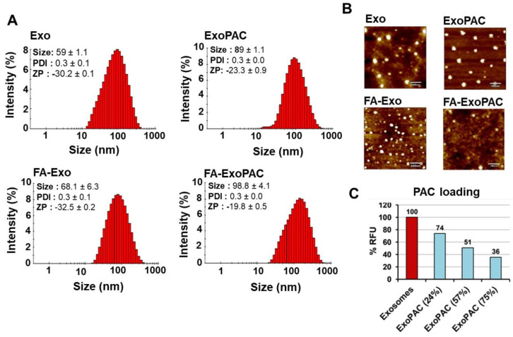
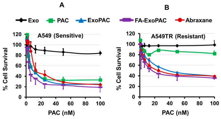
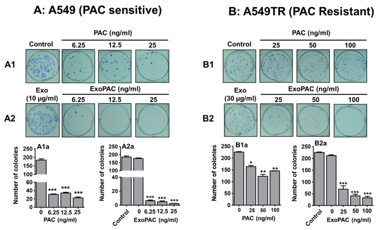
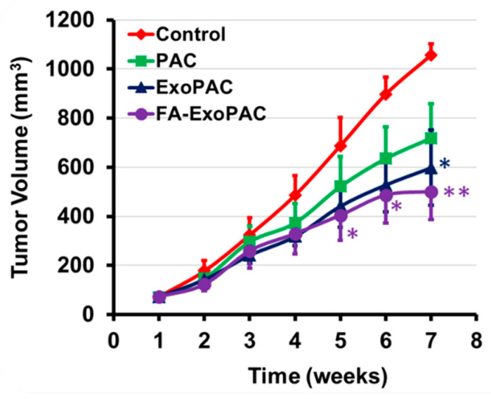
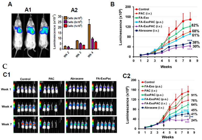
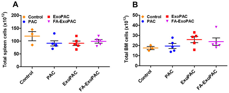
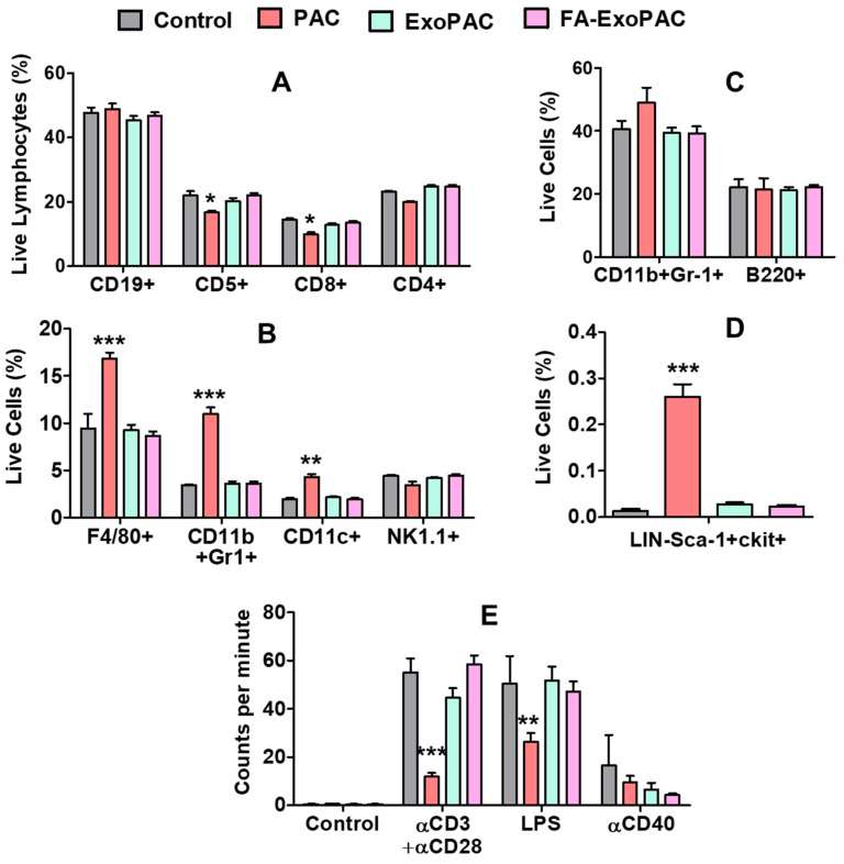
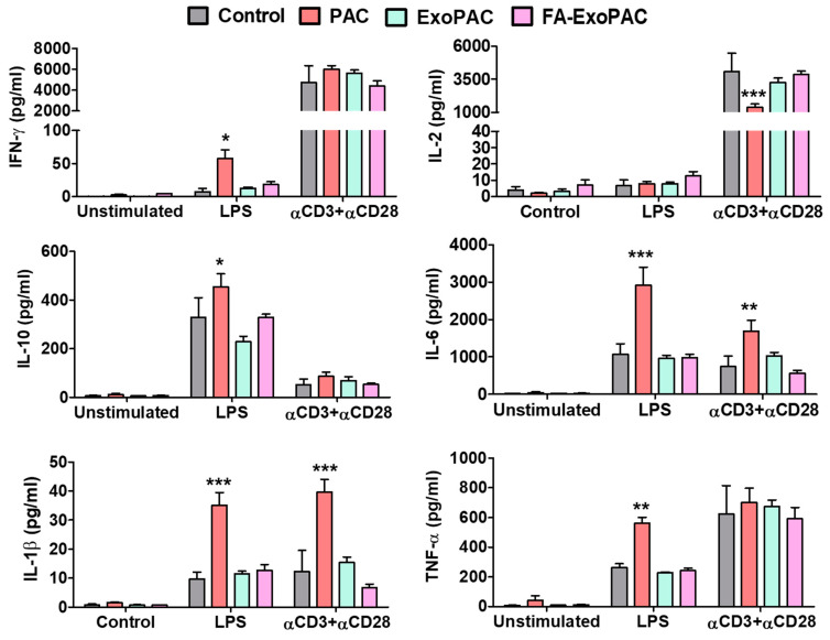
Lung cancer is the leading cause of cancer-related deaths worldwide. Non-small-cell lung cancer (NSCLC) is the most common type accounting for 84% of all lung cancers. Paclitaxel (PAC) is a widely used drug in the treatment of a broad spectrum of human cancers, including lung. While efficacious, PAC generally is not well tolerated and its limitations include low aqueous solubility, and significant toxicity. To overcome the dose-related toxicity of solvent-based PAC, we utilized bovine colostrum-derived exosomes as a delivery vehicle for PAC for the treatment of lung cancer. Colostrum provided higher yield of exosomes and could be loaded with higher amount of PAC compared to mature milk. Exosomal formulation of PAC (ExoPAC) showed higher antiproliferative activity and inhibition of colony formation against A549 cells compared with PAC alone, and also showed antiproliferative activity against a drug-resistant variant of A549. To further enhance its efficacy, exosomes were attached with a tumor-targeting ligand, folic acid (FA). FA-ExoPAC given orally showed significant inhibition (>50%) of subcutaneous tumor xenograft while similar doses of PAC showed insignificant inhibition. In the orthotopic lung cancer model, oral dosing of FA-ExoPAC achieved greater efficacy (55% growth inhibition) than traditional i.v. PAC (24–32% growth inhibition) and similar efficacy as i.v. Abraxane (59% growth inhibition). The FA-ExoPAC given i.v. exceeded the therapeutic efficacy of Abraxane (76% growth inhibition). Finally, wild-type animals treated with p.o. ExoPAC did not show gross, systemic or immunotoxicity. Solvent-based PAC caused immunotoxicity which was either reduced or completely mitigated by its exosomal formulations. These studies show that a tumor-targeted oral formulation of PAC (FA-ExoPAC) significantly improved the overall efficacy and safety profile while providing a user-friendly, cost-effective alternative to bolus i.v. PAC and i.v. Abraxane.
Overview
The claims and statistics of this paper haven’t been checked yet.
Run the check — free, no account needed. It reads the open-access text and checks the sources it cites.
Integrity signals
Registry
Cites
Statistics
Figures
Verification
Claims the paper makes
The claims and statistics of this paper haven’t been checked yet.
Run the check — free, no account needed. It reads the open-access text and checks the sources it cites.
Claims other papers make about this one
Each is checked against this paper’s own text: does it say what the citing paper says it does?
The cited source says what the claim attributes to it.
In the citing paper
“while intravenous administration of FA-ExoPAC surpassed the efficacy of Abraxane (albumin-bound paclitaxel), which is FDA-approved for similar use.”
From Emerging role of exosomes in cancer therapy: progress and challenges (2025) · cites this paper as [74]
In the citing paper
“| Lung Cancer | FA-ExoPAC | Improved drug delivery efficiency and significantly reduced toxicity | [74] |”
From Emerging role of exosomes in cancer therapy: progress and challenges (2025) · cites this paper as [74]
In the citing paper
“In animal models, oral FA-ExoPAC achieved a tumor inhibition rate of 55%”
From Emerging role of exosomes in cancer therapy: progress and challenges (2025) · cites this paper as [74]
In the citing paper
“Compared to traditional intravenous paclitaxel, exosome-loaded paclitaxel not only improves drug delivery efficiency but also significantly reduces toxicity [74].”
From Emerging role of exosomes in cancer therapy: progress and challenges (2025) · cites this paper as [74]
Self-check
Percentages, sums and reported differences re-computed from the paper’s own numbers.
The self-check needs the paper’s full text.
Citations
Cites
Théry, C., Witwer, K. W., Aikawa, E., Alcaraz, M. J., Anderson, J. D., Andriantsitohaina, R., Antoniou, A., Arab, T., Archer, F., Atkin‐Smith, G. K., et al. (2018) · Journal of Extracellular Vesicles · doi:10.1080/20013078.2018.1535750 · Cited by 11,099
The biology , function , and biomedical applications of exosomes
Kalluri, R., & LeBleu, V. S. (2020) · Science · doi:10.1126/science.aau6977 · Cited by 10,770
Exosomes facilitate therapeutic targeting of oncogenic KRAS in pancreatic cancer
Kamerkar, S., LeBleu, V. S., Sugimoto, H., Yang, S., Ruivo, C. F., Melo, S. A., Lee, J. J., & Kalluri, R. (2017) · Nature · doi:10.1038/nature22341 · Cited by 2,639
Research works printed but not linked: 40
- Siegel, 2021, Cancer Statistics, 2021, CA Cancer J. Clin., 71, 7
- Siegel, 2014, Cancer statistics, 2014, CA Cancer J. Clin., 64, 9
- Horwitz, 1994, Taxol (paclitaxel): Mechanisms of action, Ann. Oncol., 5, S3
- Constantinides, 2000, Formulation development and antitumor activity of a filter-sterilizable emulsion of paclitaxel, Pharm. Res., 17, 175, 10.1023/A:1007565230130 · DOI
- Sarpotdar, 1996, Formulation and antitumor activity evaluation of nanocrystalline suspensions of poorly soluble anticancer drugs, Pharm. Res., 13, 272, 10.1023/A:1016051316815 · DOI
- Palumbo, 2015, Nanoparticle albumin-bound paclitaxel (nab-paclitaxel) as second-line chemotherapy in HER2-negative, taxane-pretreated metastatic breast cancer patients: Prospective evaluation of activity, safety, and quality of life, Drug Des. Dev. Ther., 9, 2189, 10.2147/DDDT.S79563 · DOI
- Chou, 2020, Improvement of Paclitaxel-Associated Adverse Reactions (ADRs) via the Use of Nano-Based Drug Delivery Systems: A Systematic Review and Network Meta-Analysis, Int. J. Nanomed., 15, 1731, 10.2147/IJN.S231407 · DOI
- Kooijmans, 2012, Exosome mimetics: A novel class of drug delivery systems, Int. J. Nanomed., 7, 1525
- Lakhal, 2011, Exosome nanotechnology: An emerging paradigm shift in drug delivery: Exploitation of exosome nanovesicles for systemic in vivo delivery of RNAi heralds new horizons for drug delivery across biological barriers, Bioessays, 33, 737, 10.1002/bies.201100076 · DOI · Paper page
- Aisner, 2007, Overview of the changing paradigm in cancer treatment: Oral chemotherapy, Am. J. Health Syst. Pharm., 64, S4, 10.2146/ajhp070035 · DOI
- Joo, 2008, Oral paclitaxel chemotherapy for brain tumors: Ideal combination treatment of paclitaxel and P-glycoprotein inhibitor, Oncol. Rep., 19, 17
- Browder, 2000, Antiangiogenic scheduling of chemotherapy improves efficacy against experimental drug-resistant cancer, Cancer Res., 60, 1878
- Bobrie, 2011, Exosome secretion: Molecular mechanisms and roles in immune responses, Traffic, 12, 1659, 10.1111/j.1600-0854.2011.01225.x · DOI
- Kandimalla, 2021, Milk exosomes: A biogenic nanocarrier for small molecules and macromolecules to combat cancer, Am. J. Reprod. Immunol., 85, e13349, 10.1111/aji.13349 · DOI
- Vader, 2016, Extracellular vesicles for drug delivery, Adv. Drug Deliv. Rev., 106, 148, 10.1016/j.addr.2016.02.006 · DOI · Paper page
- Agrawal, 2017, Milk-derived exosomes for oral delivery of paclitaxel, Nanomed. Nanotechnol. Biol. Med., 13, 1627, 10.1016/j.nano.2017.03.001 · DOI
- Aqil, 2017, Exosomal delivery of berry anthocyanidins for the management of ovarian cancer, Food Funct., 8, 4100, 10.1039/C7FO00882A · DOI
- Munagala, 2017, Exosomal formulation of anthocyanidins against multiple cancer types, Cancer Lett., 393, 94, 10.1016/j.canlet.2017.02.004 · DOI
- Aqil, 2016, Exosomal formulation enhances therapeutic response of celastrol against lung cancer, Exp. Mol. Pathol., 101, 12, 10.1016/j.yexmp.2016.05.013 · DOI
- Aqil, 2019, Milk exosomes—Natural nanoparticles for siRNA delivery, Cancer Lett., 449, 186, 10.1016/j.canlet.2019.02.011 · DOI
- Munagala, 2021, Exosome-mediated delivery of RNA and DNA for gene therapy, Cancer Lett., 505, 58, 10.1016/j.canlet.2021.02.011 · DOI
- Yang, 2013, Insights into the binding of paclitaxel to human serum albumin: Multispectroscopic studies, Lumin. J. Biol. Chem. Lumin., 28, 427, 10.1002/bio.2522 · DOI
- Kyakulaga, 2020, Synergistic combinations of paclitaxel and withaferin A against human non-small cell lung cancer cells, Oncotarget, 11, 1399, 10.18632/oncotarget.27519 · DOI
- Fan, 2011, Stable isotope resolved metabolomics of lung cancer in a SCID mouse model, Metab. Off. J. Metab. Soc., 7, 257
- Zhong, 2009, Effect of yeast-derived beta-glucan in conjunction with bevacizumab for the treatment of human lung adenocarcinoma in subcutaneous and orthotopic xenograft models, J. Immunother., 32, 703, 10.1097/CJI.0b013e3181ad3fcf · DOI
- Umanzor, G., Rugo, H.S., Barrios, F.J., Vasallo, R.H., Chivalan, M.A., Bejarano, S., Ramirez, J.R., Fein, L., Kowalyszyn, R.D., and Cutler, D.L. (2020, January 8–11). Abstract PD1-08: Oral Paclitaxel and Encequidar (oPac + E) versus IV paclitaxel (IVPac) in the Treatment of Metastatic Breast Cancer (mBC) Patients (Study KX-ORAX-001): Progression Free Survival (PFS) and Overall Survival (OS) Updates. Proceedings of the San Antonio Breast Cancer Symposium, San Antonio, TX, USA. · DOI
- Rugo, H.S., Umanzor, G., Barrios, F.J., Vasallo, R.H., Chivalan, M.A., Bejarano, S., Ramirez, J.R., Fein, L., Kowalyszyn, R.D., and Cutler, D.L. (2020, January 8–11). Lower Rates of Neuropathy with Oral Paclitaxel and Encequidar (oPac + E) Compared to IV Paclitaxel (IVPac) in Treatment of Metastatic Breast Cancer (mBC): Study KX-ORAX-001. Proceedings of the San Antonio Breast Cancer Symposium, San Antonio, TX, USA.
- Mendt, M., Kamerkar, S., Sugimoto, H., McAndrews, K.M., Wu, C.C., Gagea, M., Yang, S., Blanko, E.V.R., Peng, Q., and Ma, X. (2018). Generation and testing of clinical-grade exosomes for pancreatic cancer. JCI Insight., 3. · DOI
- Ledermann, 2015, Targeting the folate receptor: Diagnostic and therapeutic approaches to personalize cancer treatments, Ann. Oncol., 26, 2034, 10.1093/annonc/mdv250 · DOI
- Shi, 2015, A current review of folate receptor alpha as a potential tumor target in non-small-cell lung cancer, Drug Des. Dev. Ther., 9, 4989
- Cheung, 2016, Targeting folate receptor alpha for cancer treatment, Oncotarget, 7, 52553, 10.18632/oncotarget.9651 · DOI · Paper page
- Low, 2009, Folate-targeted therapeutic and imaging agents for cancer, Curr. Opin. Chem. Biol., 13, 256, 10.1016/j.cbpa.2009.03.022 · DOI
- Sega, 2008, Tumor detection using folate receptor-targeted imaging agents, Cancer Metastasis Rev., 27, 655, 10.1007/s10555-008-9155-6 · DOI
- Morelli, 2004, Endocytosis, intracellular sorting, and processing of exosomes by dendritic cells, Blood, 104, 3257, 10.1182/blood-2004-03-0824 · DOI
- Aqil, 2017, Exosomes for the Enhanced Tissue Bioavailability and Efficacy of Curcumin, AAPS J., 19, 1691, 10.1208/s12248-017-0154-9 · DOI
- Parolini, 2009, Microenvironmental pH is a key factor for exosome traffic in tumor cells, J. Biol. Chem., 284, 34211, 10.1074/jbc.M109.041152 · DOI
- Feng, 2010, Cellular internalization of exosomes occurs through phagocytosis, Traffic, 11, 675, 10.1111/j.1600-0854.2010.01041.x · DOI
- Killion, 1998, Orthotopic models are necessary to predict therapy of transplantable tumors in mice, Cancer Metastasis Rev., 17, 279, 10.1023/A:1006140513233 · DOI
- Kerbel, 2003, Human tumor xenografts as predictive preclinical models for anticancer drug activity in humans: Better than commonly perceived-but they can be improved, Cancer Biol. Ther., 2, S134, 10.4161/cbt.213 · DOI
- Fauzee, 2011, Taxanes: Promising Anti-Cancer Drugs, Asian Pac. J. Cancer Prev., 12, 837
Cited by
Sorted by citations
Emerging role of exosomes in cancer therapy: progress and challenges
Li, J., Wang, J., & Chen, Z. (2025) · Molecular Cancer · doi:10.1186/s12943-024-02215-4 · Cited by 297
Exosome-Based Drug Delivery: Translation from Bench to Clinic
Koh, H. B., Kim, H. J., Kang, S.-W., & Yoo, T.-H. (2023) · Pharmaceutics · doi:10.3390/pharmaceutics15082042 · Cited by 259
Metadata
- Authors
- Kandimalla, R., Aqil, F., Alhakeem, S., Jeyabalan, J., Tyagi, N., Agrawal, A., Yan, J., Spencer, W., Bondada, S., & Gupta, R.
- Venue
- Cancers
- Publisher
- MDPI AG
- Published
- 2021-07-23
- Volume
- 13
- Issue
- 15
- Pages
- 3700
- ISSN
- 2072-6694
- PMC
- PMC8345039
- Cited by
- 99
- References
- 3
- Language
- en
- Update notices
- correction (2021-07-23)
- Funders
- National Institutes of Health (R44-CA-221487)
About these checks
Registry, retraction, Cites and statistics results are computed, not judged, with one exception: a printed reference that no index lists is read once by a model before it counts as a citation error. Claims are extracted by a model and kept only when their quote appears verbatim in the text; each cited source is then checked, with a model’s help, for whether it says what the claim relies on. Those verdicts can be wrong — read the claim’s quote and the cited source before relying on one.
Statistics and arithmetic are recomputed, not judged.
Confidence bands are shown only for fields calibrated on our annotated test set. None is calibrated yet, so no band is shown.
What counts as a claim ›How we measure confidence ›How we check this ›
Are you an author, or do you see a problem with this page? Request a review
