

The DOI matches its Crossref record; 266 of the 267 references it cites were found in a registry or an index.
The Urokinase Receptor (uPAR) as a “Trojan Horse” in Targeted Cancer Therapy: Challenges and Opportunities
Open accessAbstract
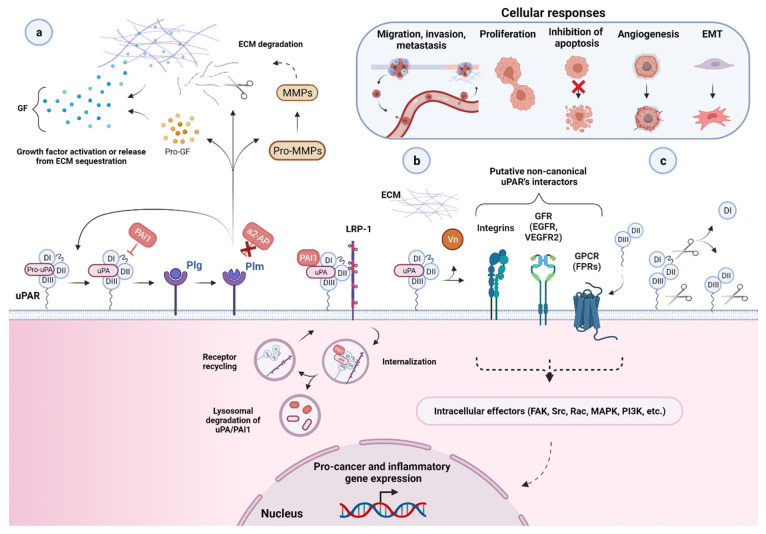
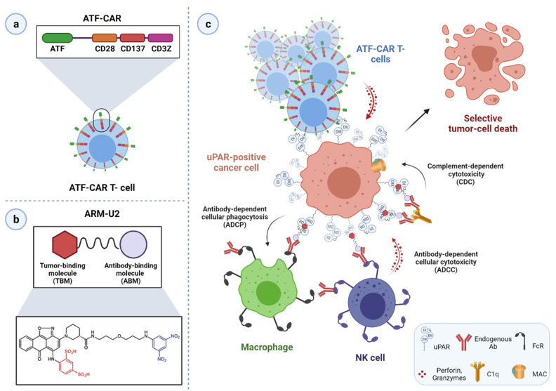
One of the largest challenges to the implementation of precision oncology is identifying and validating selective tumor-driving targets to enhance the therapeutic efficacy while limiting off-target toxicity. In this context, the urokinase-type plasminogen activator receptor (uPAR) has progressively emerged as a promising therapeutic target in the management of aggressive malignancies. By focalizing the plasminogen activation cascade and subsequent extracellular proteolysis on the cell surface of migrating cells, uPAR endows malignant cells with a high proteolytic and migratory potential to dissolve the restraining extracellular matrix (ECM) barriers and metastasize to distant sites. uPAR is also assumed to choreograph multiple other neoplastic stages via a complex molecular interplay with distinct cancer-associated signaling pathways. Accordingly, high uPAR expression is observed in virtually all human cancers and is frequently associated with poor patient prognosis and survival. The promising therapeutic potential unveiled by the pleiotropic nature of this receptor has prompted the development of distinct targeted intervention strategies. The present review will focus on recently emerged cytotoxic approaches emphasizing the novel technologies and related limits hindering their application in the clinical setting. Finally, future research directions and emerging opportunities in the field of uPAR targeting are also discussed.
Overview
The claims and statistics of this paper haven’t been checked yet.
Run the check — free, no account needed. It reads the open-access text and checks the sources it cites.
Integrity signals
Registry
Cites
Statistics
Figures
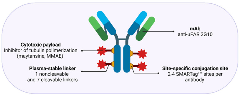
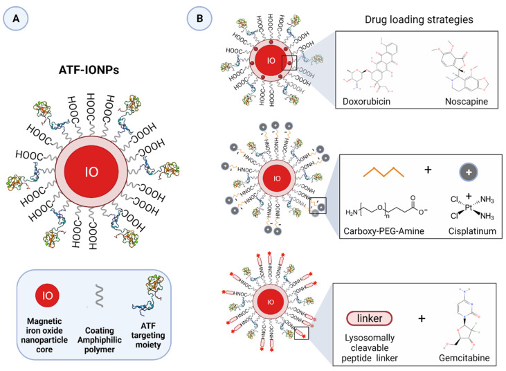
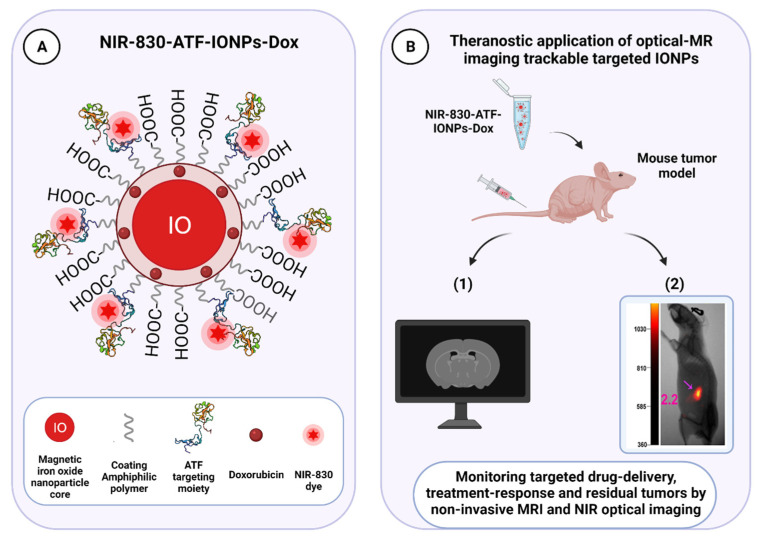
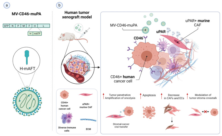
Verification
Claims the paper makes
The claims and statistics of this paper haven’t been checked yet.
Run the check — free, no account needed. It reads the open-access text and checks the sources it cites.
Claims other papers make about this one
Each is checked against this paper’s own text: does it say what the citing paper says it does?
The source backs part of the claim, not all of it — or backs it for a different population, condition or comparison.
In the citing paper
“Radiopharmaceutical case studies vividly illustrate instances where this approach resulted in tumor regression and prolonged survival, underscoring the promise of such tailored strategies [97].”
From Radiopharmaceuticals: navigating the frontier of precision medicine and therapeutic innovation (2024) · cites this paper as [97]
Experimental — accuracy not yet measured
Self-check
Percentages, sums and reported differences re-computed from the paper’s own numbers.
The self-check needs the paper’s full text.
Citations
Cites
A guide to cancer immunotherapy: from T cell basic science to clinical practice
Waldman, A. D., Fritz, J. M., & Lenardo, M. J. (2020) · Nature Reviews Immunology · doi:10.1038/s41577-020-0306-5 · Cited by 4,154
Targeting macrophages: therapeutic approaches in cancer
Cassetta, L., & Pollard, J. W. (2018) · Nature Reviews Drug Discovery · doi:10.1038/nrd.2018.169 · Cited by 2,086
uPAR: a versatile signalling orchestrator
Blasi, F., & Carmeliet, P. (2002) · Nature Reviews Molecular Cell Biology · doi:10.1038/nrm977 · Cited by 1,224 · cited 5×
“Although impairment of uPAR function or inhibition of its expression was associated with impaired invasive, metastatic, and tumorigenic potential of many tumor models in vitro and in vivo [4,11,12,14,16,17,20,52,57,79,84,85], one study in transgenic mice with uPAR gene ablation showed no pronounced causal effects on d…”
BackgroundInfluentialUnlocking the potential of antibody–drug conjugates for cancer therapy
Drago, J. Z., Modi, S., & Chandarlapaty, S. (2021) · Nature Reviews Clinical Oncology · doi:10.1038/s41571-021-00470-8 · Cited by 1,209
Senolytic CAR T cells reverse senescence-associated pathologies
Amor, C., Feucht, J., Leibold, J., Ho, Y.-J., Zhu, C., Alonso-Curbelo, D., Mansilla-Soto, J., Boyer, J. A., Li, X., Giavridis, T., et al. (2020) · Nature · doi:10.1038/s41586-020-2403-9 · Cited by 1,098
Targeting the tumour stroma to improve cancer therapy
Valkenburg, K. C., de Groot, A. E., & Pienta, K. J. (2018) · Nature Reviews Clinical Oncology · doi:10.1038/s41571-018-0007-1 · Cited by 1,084 · cited 2×
“Indeed, while having an indirect anti-cancer effect by attenuating the tumor-promoting effect of the stroma, cytotoxic targeting of this compartment may also increase drug-delivery efficiency by breaking down the dense stromal barrier, which severely hampers tumor perfusion by therapeutic agents, ultimately leading to…”
BackgroundRadiopharmaceutical therapy in cancer: clinical advances and challenges
Sgouros, G., Bodei, L., McDevitt, M. R., & Nedrow, J. R. (2020) · Nature Reviews Drug Discovery · doi:10.1038/s41573-020-0073-9 · Cited by 1,032
Regulation of cell signalling by uPAR
Smith, H. W., & Marshall, C. (2009) · Nature Reviews Molecular Cell Biology · doi:10.1038/nrm2821 · Cited by 983 · cited 9×
“Although impairment of uPAR function or inhibition of its expression was associated with impaired invasive, metastatic, and tumorigenic potential of many tumor models in vitro and in vivo [4,11,12,14,16,17,20,52,57,79,84,85], one study in transgenic mice with uPAR gene ablation showed no pronounced causal effects on d…”
BackgroundInfluentialProgress in tumor-associated macrophage (TAM)-targeted therapeutics
Ngambenjawong, C., Gustafson, H. H., & Pun, S. H. (2017) · Advanced Drug Delivery Reviews · doi:10.1016/j.addr.2017.04.010 · Cited by 858
Tumor-associated stromal cells as key contributors to the tumor microenvironment
Bussard, K. M., Mutkus, L., Stumpf, K., Gomez-Manzano, C., & Marini, F. C. (2016) · Breast Cancer Research · doi:10.1186/s13058-016-0740-2 · Cited by 746
Research works printed but not linked: 1
- Sampson, J.H. (2017). Chapter 10-Immunotoxin Therapy for Brain Tumors. Translational Immunotherapy of Brain Tumors, Academic Press.
Cited by
Sorted by citations
Radiopharmaceuticals: navigating the frontier of precision medicine and therapeutic innovation
Dhoundiyal, S., Srivastava, S., Kumar, S., Singh, G., Ashique, S., Pal, R., Mishra, N., & Taghizadeh-Hesary, F. (2024) · European Journal of Medical Research · doi:10.1186/s40001-023-01627-0 · Cited by 144
“Radiopharmaceuti-cal case studies vividly illustrate instances where this approach resulted in tumor regression and prolonged survival, underscoring the promise of such tailored strategies [97].”
BackgroundUrokinase-type plasminogen activator receptor (uPAR) as a therapeutic target in cancer
Zhai, B., Tian, H., Sun, J., Zou, J., Zhang, X., Cheng, J., Shi, Y., Fan, Y. B., & Guo, D. (2022) · Journal of Translational Medicine · doi:10.1186/s12967-022-03329-3 · Cited by 123
Hamada, M., Varkoly, K., Riyadh, O., Beladi, R. N., Munuswamy‐Ramanujam, G., Rawls, A., Wilson‐Rawls, J., Chen, H., McFadden, G., & Lucas, A. R. (2024) · Biomedicines · doi:10.3390/biomedicines12061167 · Cited by 61
“Cleavage of the D1–D2 linker domains by other proteases irreversibly blocks uPAR’s interaction with other proteins [27,28].”
BackgroundModulation of Cellular Function by the Urokinase Receptor Signalling: A Mechanistic View
Alfano, D., Franco, P., & Stoppelli, M. P. (2022) · Frontiers in Cell and Developmental Biology · doi:10.3389/fcell.2022.818616 · Cited by 59
“However, despite the advances in the molecular design and generation of uPAR-derived peptides or novel interactors, no uPAR-targeting therapeutics have currently progressed to clinical trials (Metrangolo et al., 2021).”
BackgroundBogdanović, B., Fagret, D., Ghezzi, C., & Montemagno, C. (2024) · Pharmaceuticals · doi:10.3390/ph17111556 · Cited by 49
“uPAR is a cell surface receptor crucial for cell migration, tissue remodeling, and inflammation. uPAR is frequently overexpressed in cancers such as breast, prostate, and lung cancer, where it is associated with increased tumor invasion and poor prognosis [174].”
The roles of proteases in prostate cancer
Koistinen, H. K., Kovanen, R., Hollenberg, M. D., Dufour, A., Radisky, E. S., Stenman, U., Batra, J., Clements, J. A., Hooper, J. D., Diamandis, E. P., et al. (2023) · IUBMB Life · doi:10.1002/iub.2700 · Cited by 46
suPAR: An Inflammatory Mediator for Kidneys
Sudhini, Y. R., Wei, C., & Reiser, J. (2022) · Kidney Diseases · doi:10.1159/000524965 · Cited by 42
Verhulst, E., Garnier, D., Meester, I. D., & Bauvois, B. (2022) · Cancers · doi:10.3390/cancers14030624 · Cited by 28
“The list of ectoproteases as potential targets for cancer therapy is further expanding, as has already been seen with other ADAMs (including ADAM8, ADAM10, ADAM28), MT1-MMP/MMP14, the complex urokinase plasminogen (uPA)/uPAR, and neutral endopeptidase N/CD10 overexpressed in solid and haematological tumours [19,101,29…”
BackgroundReliable Hallmarks and Biomarkers of Senescent Lymphocytes
Сергеевна, М. Ю., Tereshchenko, V., Bogdanova, D. A., & Александрович, Р. С. (2023) · International Journal of Molecular Sciences · doi:10.3390/ijms242115653 · Cited by 28
Fibroblast Activation Protein Targeting Probe with Gly–Pro Sequence for PET of Glioblastoma
Lai, C., Cao, R., Li, R., He, C., Wang, X., Shi, H., Qu, C., Qian, K., Song, S., Chen, W., et al. (2023) · Molecular Pharmaceutics · doi:10.1021/acs.molpharmaceut.3c00248 · Cited by 26
Iftikhar, A., Mahmood, N., & Rabbani, S. A. (2025) · Cancers · doi:10.3390/cancers17203309 · Cited by 23
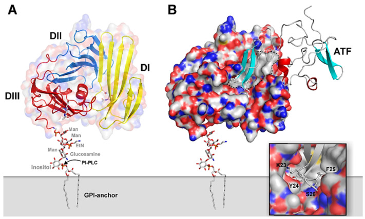
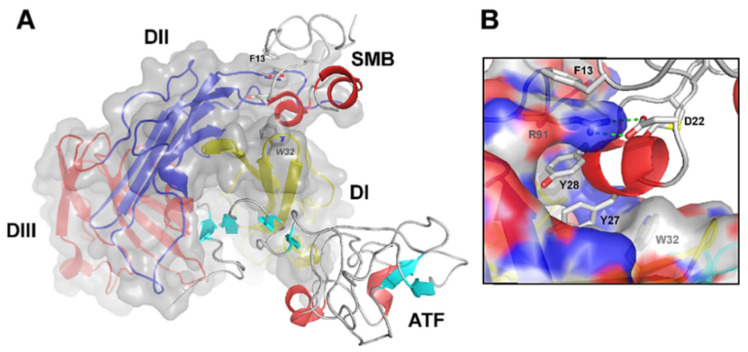
“This binding is stabilized by several key residues within the DI domain, including Tyr57, Tyr92, and Arg91 and Trp32 which form critical hydrophobic contacts with uPA [53,55–58] (Figure 1).”
Application of the transgenic pig model in biomedical research: A review
Wei, J., Zhang, W., Li, J., Jin, Y., & Qiu, Z. (2022) · Frontiers in Cell and Developmental Biology · doi:10.3389/fcell.2022.1031812 · Cited by 23
uPAR, beyond regulating physiological functions, has orchestrated roles in cancer (Review)
Wang, L., Lin, X., & Sun, P. (2022) · International Journal of Oncology · doi:10.3892/ijo.2022.5441 · Cited by 19
Metrangolo, V., Blomquist, M. H., Dutta, A., Gårdsvoll, H., Krigslund, O., Nørregaard, K. S., Jürgensen, H. J., Ploug, M., Flick, M. J., Behrendt, N., et al. (2025) · Science Advances · doi:10.1126/sciadv.adq0513 · Cited by 17
“2), thus allowing both studies in xenografted and syngeneic tumors and an initial assessment of ADC on-target toxicity ( 11 , 45 ).”
InfluentialSelf-citationShmakova, A., Сергеевна, К. П., Rysenkova, K. D., Popov, V. S., Gorbunova, A. S., Karpukhina, A. A., Karagyaur, M. N., Rubina, K. A., Va, T., & Semina, E. V. (2022) · Cancers · doi:10.3390/cancers14040994 · Cited by 18
Cui, X., Pei, X., Wang, H., Feng, P., Qin, H., Liu, S., Yan, Q., & Liu, J. (2022) · Cells · doi:10.3390/cells11193141 · Cited by 16
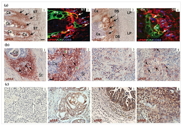
“The urokinase plasminogen activator receptor (uPAR), a glycoprotein linked to glycosylphosphatidylinositol (GPI) anchored on the cell surface, is highly expressed in the invasive areas of the tumor-stromal microenvironment in cancers [20,21].”
BackgroundA convergent uPAR-positive tumor ecosystem creates broad vulnerability to CAR T cell therapy
Zhang, Z., Ho, Y.-J., Fang, X., Kim, M., Li, M., Luan, W., Hinterleitner, C., Haubner, S., Kogel, F., Pratt, E. C., et al. (2026) · Cell · doi:10.1016/j.cell.2026.03.002 · Cited by 15
Peng, L., Li, Y., Yao, S., Gaedcke, J., Baart, V. M., Sier, C. F. M., Neeße, A., Ellenrieder, V., Bohnenberger, H., Fuchs, F., et al. (2023) · Cancers · doi:10.3390/cancers15051587 · Cited by 15
The inflammatory response of human pancreatic cancer samples compared to normal controls
Brayer, K. J., Hanson, J. A., Cingam, S. R., Martinez, C. G., Ness, S. A., & Rabinowitz, I. (2023) · PLoS ONE · doi:10.1371/journal.pone.0284232 · Cited by 14
Conilh, L., Metrangolo, V., Crescioli, S., Reichert, J. M., & Dumontet, C. (2026) · Cell · doi:10.1016/j.cell.2026.04.016 · Cited by 14
Self-citationMateusiak, Ł., Floru, S., Groof, T. W. M. D., Wouters, J., Declerck, N. B., Debie, P., Janssen, S., Zeven, K., Puttemans, J., Vincke, C., et al. (2024) · Advanced Science · doi:10.1002/advs.202400700 · Cited by 12
Dual modified ferritin nanocages for tumor-targeted and microenvironment-responsive drug delivery
Li, H., Qu, Y., Guo, Z., Chen, D., Jiang, L., Xu, P., Huang, M., & Yuan, C. (2025) · International Journal of Biological Macromolecules · doi:10.1016/j.ijbiomac.2025.140694 · Cited by 10
Current strategies for senescence treatment: Focused on theranostic performance of nanomaterials
Cong, Y., Li, X., & Hong, H. (2025) · Journal of Controlled Release · doi:10.1016/j.jconrel.2025.113710 · Cited by 9
Skin Tape Strip Proteomics in Mycosis Fungoides Identifies Tumor-Associated Biomarkers
Techner, J., Hooper, M. J., Evans, S. T., LeWitt, T. M., Paller, A. S., Guitart, J., Lu, K. Q., & Zhou, X. A. (2022) · Journal of Investigative Dermatology · doi:10.1016/j.jid.2022.07.025 · Cited by 8
Targeted imaging of uPAR expression in vivo with cyclic AE105 variants
Leth, J. M., Newcombe, E. A., Grønnemose, A. L., Jørgensen, J. T., Qvist, K., Clausen, A. S., Knudsen, L. B. S., Kjær, A., Kragelund, B. B., Jørgensen, T. J. D., et al. (2023) · Scientific Reports · doi:10.1038/s41598-023-43934-w · Cited by 9
Self-citationRecent Advances in Targeting the Urokinase Plasminogen Activator with Nanotherapeutics
Kumar, A. A., Vine, K. L., & Ranson, M. (2023) · Molecular Pharmaceutics · doi:10.1021/acs.molpharmaceut.3c00055 · Cited by 9
Zhang, H., Xie, Z., Xie, S., Wu, J., Khan, M., Gao, P., & Li, J. (2025) · Bioorganic Chemistry · doi:10.1016/j.bioorg.2025.108773 · Cited by 6
Development of Cyclic Peptide‐Based Radiotracers for uPAR
Wang, X., Zhang, X., Zhang, S., Zhang, L., Shen, J., Zhang, Y., Zhang, H., Zhang, M., Wang, R., & Hu, K. (2025) · ChemMedChem · doi:10.1002/cmdc.202500061 · Cited by 4
García-Pérez, O., Melgar-Vilaplana, L., Sifaoui, I., Garcia-Bello, M. A., Córdoba-Lanús, E., & Fernández‐de‐Misa, R. (2023) · Melanoma Research · doi:10.1097/cmr.0000000000000904 · Cited by 3
Serrat, J., Torres-Valle, M., Veríssimo, C. D. M., Siles‐Lucas, M., & González‐Miguel, J. (2025) · Veterinary Research · doi:10.1186/s13567-025-01449-4 · Cited by 3
Genome Engineering as a Therapeutic Approach in Cancer Therapy: A Comprehensive Review
Gemayel, J., Chebly, A., Kourié, H. R., Hanna, C., Mheidly, K., Mhanna, M., Karam, F., Ghoussaini, D., Najjar, P. E., & Khalil, C. (2024) · Advanced Genetics · doi:10.1002/ggn2.202300201 · Cited by 2
Bruun, S. B., Madsen, J. B., & Brasen, C. L. (2024) · Practical Laboratory Medicine · doi:10.1016/j.plabm.2024.e00371 · Cited by 3
“…urokinase plasminogen activator receptor (uPAR) [1]. uPAR is expressed in various cells, such as neutrophils, monocytes, macrophages, endothelial cells, fibroblasts, smooth muscle cells, and some tumor cells [2]. uPAR expression increases during inflammation, immune responses, and injury [3].”
BackgroundAlbumin-based drug carrier targeting urokinase receptor for cancer therapy
Li, H., Wang, Z., Yu, S., Chen, S., Zhou, Y., Qu, Y., Xu, P., Jiang, L., Yuan, C., & Huang, M. (2023) · International Journal of Pharmaceutics · doi:10.1016/j.ijpharm.2023.122636 · Cited by 3
Endothelial-specific Enhancer as a Cis Element of PLAUR Regulation by TNF-alpha, IL-1beta, and VEGF
Penkov, D., Beloglazova, I. B., & Parfyonova, Y. V. (2024) · Current Pharmaceutical Design · doi:10.2174/0113816128296376240424072322 · Cited by 2
uPAR-targeted senolytic delivery to enhance cancer therapy
Nguyen, T. O. O., Nguyễn, B. L., Patil, B. R., Le, N. D., Kim, B., Jin, S. G., Choi, H., Ku, S., Xuan, H. L., Kim, J., et al. (2025) · Journal of Pharmaceutical Investigation · doi:10.1007/s40005-025-00757-3 · Cited by 2
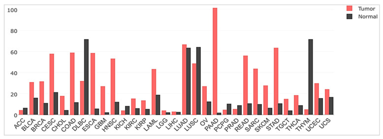
“…carcinoma exhibit pronounced differences in mRNA levels of PLAUR (gene encoding uPAR) between tumor and normal tissues, while adenoid cystic carcinoma and diffuse large B-cell lymphoma show relatively similar or lower uPAR gene expression compared to healthy tissues (Metrangolo et al. 2021).”
Clinical Efficacy of Xueshuantong plus Urokinase in the Treatment of Sudden Deafness
Wang, Y., Wang, J., & Chen, X. (2022) · Evidence-based Complementary and Alternative Medicine · doi:10.1155/2022/7775556 · Cited by 2
RetractedHan, Y., Tu, L., Zhang, Y., Liu, Q., Dong, Q., & Sun, Z. (2023) · ChemistrySelect · doi:10.1002/slct.202204504 · Cited by 1
Schulte, A. J., Lewellen, M., Durose, W. W., Nolan, E. E., Taghizadeh, L. A., Todhunter, D., Bush, C., Lang, H. P., Brown, M. E., DePauw, T. A., et al. (2025) · Journal of Pharmacology and Experimental Therapeutics · doi:10.1016/j.jpet.2025.103674 · Cited by 1
“46 Recently, Amor et al 47 showed that uPAR-targeted chimeric antigen receptor-redirected T cells eliminated senescent cells, contributing to the reversal of chemical-or diet-induced liver fibrosis and life extension of mice with lung adenocarcinomas.”
Research on precision treatment of pancreatic cancer targeted by antibody-drug conjugates
Liu, X., Liu, W., Wu, C., Yuan, Y., Chang, A., & Hao, J. (2025) · BioScience Trends · doi:10.5582/bst.2025.01271 · Cited by 1
suPARnostic: an advanced predictive tool for detecting recurrence in renal cell carcinoma
Azawi, N., Mosholt, K. S. S., Fryd, N. D., Lund, L. C., Brignone, J. I., Hvid, N., Wulf-Johansson, H., Pedersen, O. B., Sækmose, S. G., & Dabestani, S. (2023) · BMC Urology · doi:10.1186/s12894-023-01337-z
“Upregulation of uPAR proteolysis is seen in various inflammatory and pathological tissue-remod-eling processes, including cancer metastasis, fibrinolysis, responses to infectious challenges, and wound healing [19].”
BackgroundHaemostatic Proteins as Markers of Disease Progression and Prognosis in Breast CancerPreprint
Izuegbuna, O. (2024) · Qeios · doi:10.32388/ufgk1o
Luo, T., Zhang, K., Wu, C., Wu, X., Yang, H., Chen, Z., & Jiang, F. (2025) · Frontiers in Pharmacology · doi:10.3389/fphar.2025.1681718
“…facilitating the conversion of plasminogen into plasmin and thereby promoting proteolytic activity. uPAR is overexpressed in both the parenchyma and invasive margins of pancreatic cancer tissues, while its expression in normal pancreatic tissue is minimal to undetectable (Metrangolo et al., 2021).”
Anane, R. F., Kumari, A., Venugopal, H., Trépout, S., Whisstock, J. C., Engelholm, L. H., Law, R. H. P., & Ploug, M. (2026) · Protein Science · doi:10.1002/pro.70473
“…(Lund et al., 2011) prompted a change in the research focus on uPAR-targeting strategies — from being mostly function-inhibition-centric to relying more on the cytotoxic eradication of dividing cells with high uPAR expression (Kriegbaum et al., 2011; Metrangolo et al., 2021; Yuan et al., 2021).”
Self-citationuPAR-directed immunotherapies in solid tumors: current advances and future perspectives
Napolitano, F., & Montuori, N. (2026) · Frontiers in Pharmacology · doi:10.3389/fphar.2026.1961182
“…the restricted expression of uPAR in normal tissues, except for subsets of myeloid cells, reduces the likelihood of severe systemic toxicity and supports the development of selective therapeutic strategies with a potentially favorable safety profile (Metrangolo et al., 2021; Eefsen et al., 2025).”
Influential
Metadata
- Authors
- Metrangolo, V., Ploug, M., & Engelholm, L. H.
- Affiliations
- University of Copenhagen (DK)
- Corresponding author
- Engelholm, Lars H.
- Venue
- Cancers
- Publisher
- MDPI AG
- Published
- 2021-10-27
- Volume
- 13
- Issue
- 21
- Pages
- 5376
- ISSN
- 2072-6694 (ISSN-L)
- PMC
- PMC8582577
- OpenAlex
- W3209572831
- Cited by
- 44
- References
- 267
- Citations by year
- 2026: 4 · 2025: 11 · 2024: 8 · 2023: 11 · 2022: 10
- FWCI
- 0.87
- Type
- journal article
- Language
- en
- Open access
- gold
- doi.org
- Licence
- Locations
- Cancers (publishedVersion, cc-by)
- Europe PMC (PubMed Central) (publishedVersion, other-oa)
- PubMed Central (publishedVersion, cc-by)
- PubMed (publishedVersion)
- Research at the University of Copenhagen (University of Copenhagen) (publishedVersion, cc-by)
- DOAJ (DOAJ: Directory of Open Access Journals) (submittedVersion, cc-by-sa)
- Funders
- European Union’s Horizon 2020 research and innovation programme under the Marie Skłodowska Curie (801481)
- Danish Cancer Society (R231-A13832)
- Topics
- Life Sciences › Biochemistry, Genetics and Molecular Biology › Cancer Research › Protease and Inhibitor Mechanisms
- Life Sciences › Biochemistry, Genetics and Molecular Biology › Biochemistry › Peptidase Inhibition and Analysis
- Health Sciences › Medicine › Hematology › Blood Coagulation and Thrombosis Mechanisms
- Keywords
- urokinase-type plasminogen activator receptor, uPAR, targeted cancer therapy, precision oncology, plasminogen activation, extracellular matrix degradation, metastasis, cell migration, off-target toxicity, prognostic factors
About these checks
Registry, retraction, Cites and statistics results are computed, not judged, with one exception: a printed reference that no index lists is read once by a model before it counts as a citation error. Claims are extracted by a model and kept only when their quote appears verbatim in the text; each cited source is then checked, with a model’s help, for whether it says what the claim relies on. Those verdicts can be wrong — read the claim’s quote and the cited source before relying on one.
Statistics and arithmetic are recomputed, not judged.
Confidence bands are shown only for fields calibrated on our annotated test set. None is calibrated yet, so no band is shown.
What counts as a claim ›How we measure confidence ›How we check this ›
Are you an author, or do you see a problem with this page? Request a review
