

The DOI matches its Crossref record; 266 of the 267 references it cites were found in a registry or an index.
The Urokinase Receptor (uPAR) as a “Trojan Horse” in Targeted Cancer Therapy: Challenges and Opportunities
Open accessAbstract
One of the largest challenges to the implementation of precision oncology is identifying and validating selective tumor-driving targets to enhance the therapeutic efficacy while limiting off-target toxicity. In this context, the urokinase-type plasminogen activator receptor (uPAR) has progressively emerged as a promising therapeutic target in the management of aggressive malignancies. By focalizing the plasminogen activation cascade and subsequent extracellular proteolysis on the cell surface of migrating cells, uPAR endows malignant cells with a high proteolytic and migratory potential to dissolve the restraining extracellular matrix (ECM) barriers and metastasize to distant sites. uPAR is also assumed to choreograph multiple other neoplastic stages via a complex molecular interplay with distinct cancer-associated signaling pathways. Accordingly, high uPAR expression is observed in virtually all human cancers and is frequently associated with poor patient prognosis and survival. The promising therapeutic potential unveiled by the pleiotropic nature of this receptor has prompted the development of distinct targeted intervention strategies. The present review will focus on recently emerged cytotoxic approaches emphasizing the novel technologies and related limits hindering their application in the clinical setting. Finally, future research directions and emerging opportunities in the field of uPAR targeting are also discussed.
Overview
The claims and statistics of this paper haven’t been checked yet.
Run the check — free, no account needed. It reads the open-access text and checks the sources it cites.
Integrity signals
Registry
Cites
Statistics
Figures
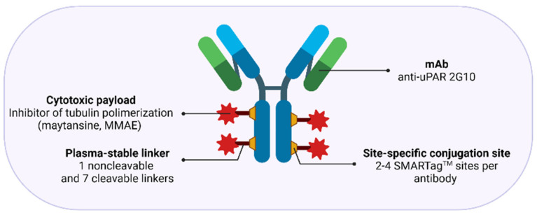
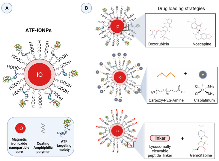
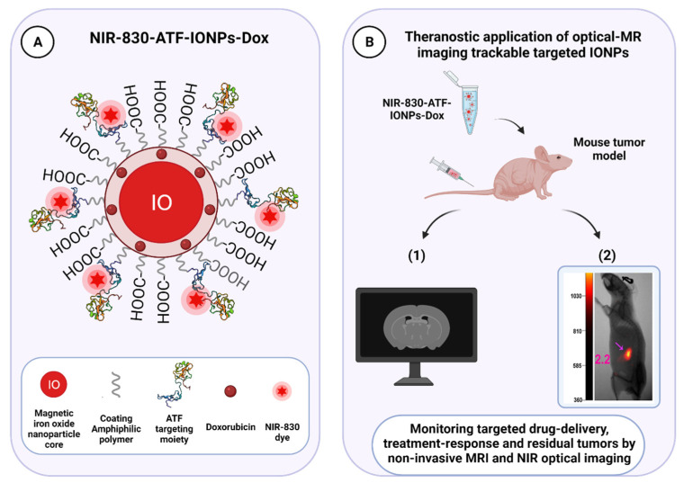
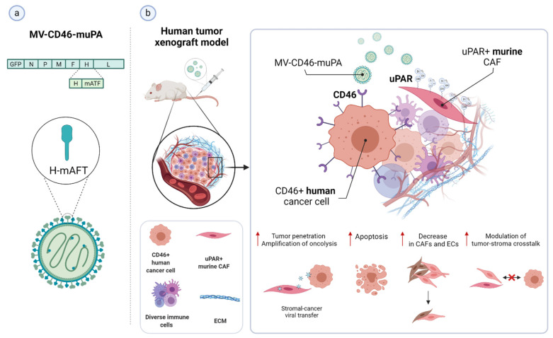
Verification
Claims the paper makes
The claims and statistics of this paper haven’t been checked yet.
Run the check — free, no account needed. It reads the open-access text and checks the sources it cites.
Claims other papers make about this one
Each is checked against this paper’s own text: does it say what the citing paper says it does?
The source backs part of the claim, not all of it — or backs it for a different population, condition or comparison.
In the citing paper
“Radiopharmaceutical case studies vividly illustrate instances where this approach resulted in tumor regression and prolonged survival, underscoring the promise of such tailored strategies [97].”
From Radiopharmaceuticals: navigating the frontier of precision medicine and therapeutic innovation (2024) · cites this paper as [97]
Experimental — accuracy not yet measured
Self-check
Percentages, sums and reported differences re-computed from the paper’s own numbers.
The self-check needs the paper’s full text.
Citations
Cites
A guide to cancer immunotherapy: from T cell basic science to clinical practice
Waldman, A. D., Fritz, J. M., & Lenardo, M. J. (2020) · Nature Reviews Immunology · doi:10.1038/s41577-020-0306-5 · Cited by 4,154
Targeting macrophages: therapeutic approaches in cancer
Cassetta, L., & Pollard, J. W. (2018) · Nature Reviews Drug Discovery · doi:10.1038/nrd.2018.169 · Cited by 2,086
uPAR: a versatile signalling orchestrator
Blasi, F., & Carmeliet, P. (2002) · Nature Reviews Molecular Cell Biology · doi:10.1038/nrm977 · Cited by 1,224 · cited 5×
“Although impairment of uPAR function or inhibition of its expression was associated with impaired invasive, metastatic, and tumorigenic potential of many tumor models in vitro and in vivo [4,11,12,14,16,17,20,52,57,79,84,85], one study in transgenic mice with uPAR gene ablation showed no pronounced causal effects on d…”
BackgroundInfluentialUnlocking the potential of antibody–drug conjugates for cancer therapy
Drago, J. Z., Modi, S., & Chandarlapaty, S. (2021) · Nature Reviews Clinical Oncology · doi:10.1038/s41571-021-00470-8 · Cited by 1,209
Senolytic CAR T cells reverse senescence-associated pathologies
Amor, C., Feucht, J., Leibold, J., Ho, Y.-J., Zhu, C., Alonso-Curbelo, D., Mansilla-Soto, J., Boyer, J. A., Li, X., Giavridis, T., et al. (2020) · Nature · doi:10.1038/s41586-020-2403-9 · Cited by 1,098
Targeting the tumour stroma to improve cancer therapy
Valkenburg, K. C., de Groot, A. E., & Pienta, K. J. (2018) · Nature Reviews Clinical Oncology · doi:10.1038/s41571-018-0007-1 · Cited by 1,084 · cited 2×
“Indeed, while having an indirect anti-cancer effect by attenuating the tumor-promoting effect of the stroma, cytotoxic targeting of this compartment may also increase drug-delivery efficiency by breaking down the dense stromal barrier, which severely hampers tumor perfusion by therapeutic agents, ultimately leading to…”
BackgroundRadiopharmaceutical therapy in cancer: clinical advances and challenges
Sgouros, G., Bodei, L., McDevitt, M. R., & Nedrow, J. R. (2020) · Nature Reviews Drug Discovery · doi:10.1038/s41573-020-0073-9 · Cited by 1,032
Regulation of cell signalling by uPAR
Smith, H. W., & Marshall, C. (2009) · Nature Reviews Molecular Cell Biology · doi:10.1038/nrm2821 · Cited by 983 · cited 9×
“Although impairment of uPAR function or inhibition of its expression was associated with impaired invasive, metastatic, and tumorigenic potential of many tumor models in vitro and in vivo [4,11,12,14,16,17,20,52,57,79,84,85], one study in transgenic mice with uPAR gene ablation showed no pronounced causal effects on d…”
BackgroundInfluentialProgress in tumor-associated macrophage (TAM)-targeted therapeutics
Ngambenjawong, C., Gustafson, H. H., & Pun, S. H. (2017) · Advanced Drug Delivery Reviews · doi:10.1016/j.addr.2017.04.010 · Cited by 858
Tumor-associated stromal cells as key contributors to the tumor microenvironment
Bussard, K. M., Mutkus, L., Stumpf, K., Gomez-Manzano, C., & Marini, F. C. (2016) · Breast Cancer Research · doi:10.1186/s13058-016-0740-2 · Cited by 746
One molecule of diphtheria toxin fragment a introduced into a cell can kill the cell
Yamaizumi, M., Mekada, E., Uchida, T., & Okada, Y. (1978) · Cell · doi:10.1016/0092-8674(78)90099-5 · Cited by 745
A cellular binding site for the Mr 55,000 form of the human plasminogen activator, urokinase.
Vassalli, J. D., Baccino, D., & Belin, D. (1985) · The Journal of Cell Biology · doi:10.1083/jcb.100.1.86 · Cited by 711
Strategic Approaches to Optimizing Peptide ADME Properties
Di, L. (2015) · The AAPS Journal · doi:10.1208/s12248-014-9687-3 · Cited by 669
Roldan, A. L., Cubellis, M. V., Masucci, M. T., Behrendt, N., Lund, L. R., Danø, K., Appella, E., & Blasi, F. (1990) · The EMBO Journal · doi:10.1002/j.1460-2075.1990.tb08132.x · Cited by 602
Identification of the urokinase receptor as an adhesion receptor for vitronectin.
Wei, Y., Waltz, D. A., Rao, N. L., Drummond, R. J., Rosenberg, S., & Chapman, H. A. (1994) · Journal of Biological Chemistry · doi:10.1016/s0021-9258(18)31646-6 · Cited by 579
Ploug, M., Rønne, E., Behrendt, N., Jensen, A. L., Blasi, F., & Danø, K. (1991) · Journal of Biological Chemistry · doi:10.1016/s0021-9258(18)52382-6 · Cited by 573 · cited 2×
“t the outer leaflet of the lip d bilayer of the c ll me brane by a C-terminal glycosyl-phosphatidylinositol (GPI) anchor [35,47].”
BackgroundSelf-citationNanoparticles: Emerging carriers for drug delivery
Mudshinge, S. R., Deore, A. B., Patil, S. R., & Bhalgat, C. M. (2011) · Saudi Pharmaceutical Journal · doi:10.1016/j.jsps.2011.04.001 · Cited by 557
Killing Mechanisms of Chimeric Antigen Receptor (CAR) T Cells
Benmebarek, M.-R., Karches, C. H., Cadilha, B. L., Lesch, S., Endres, S., & Kobold, S. (2019) · International Journal of Molecular Sciences · doi:10.3390/ijms20061283 · Cited by 557
Ghiso, J. A. A., Kovalski, K., & Ossowski, L. (1999) · The Journal of Cell Biology · doi:10.1083/jcb.147.1.89 · Cited by 546 · cited 1×
“This association may have a bearing on the ability of uPAR to drive the transition between single-cell tumor dormancy and proliferation, which allows for the long-term survival of residual tumor cells during dormancy and reactivation of their proliferation years after primary treatment upon favorable conditions [13,85…”
BackgroundpH-responsive polymer nanoparticles for drug delivery
Deirram, N., Zhang, C., Kermaniyan, S. S., Johnston, A. P. R., & Such, G. K. (2019) · Macromolecular Rapid Communications · doi:10.1002/marc.201800917 · Cited by 531
Orth, M., Metzger, P., Gerum, S., Mayerle, J. V., Schneider, G., Belka, C., Schnurr, M., & Lauber, K. (2019) · Radiation Oncology · doi:10.1186/s13014-019-1345-6 · Cited by 523
Ellis, V., Behrendt, N., & Danø, K. (1991) · Journal of Biological Chemistry · doi:10.1016/s0021-9258(18)98963-5 · Cited by 504
Mahmood, N., Mihalcioiu, C., & Rabbani, S. A. (2018) · Frontiers in Oncology · doi:10.3389/fonc.2018.00024 · Cited by 478 · cited 8×
“Although impairment of uPAR function or inhibition of its expression was associated with impaired invasive, metastatic, and tumorigenic potential of many tumor models in vitro and in vivo [4,11,12,14,16,17,20,52,57,79,84,85], one study in transgenic mice with uPAR gene ablation showed no pronounced causal effects on d…”
BackgroundInfluentialAppella, E., Robinson, E. A., Ullrich, S. J., Stoppelli, M. P., Corti, A., Cassani, G., & Blasi, F. (1987) · Journal of Biological Chemistry · doi:10.1016/s0021-9258(18)61211-6 · Cited by 475
Targeted magnetic iron oxide nanoparticles for tumor imaging and therapy
Shin, D. (2008) · International Journal of Nanomedicine · doi:10.2147/ijn.s2824 · Cited by 467
Stoppelli, M. P., Corti, A., Soffientini, A., Cassani, G., Blasi, F., & Assoian, R. K. (1985) · Proceedings of the National Academy of Sciences · doi:10.1073/pnas.82.15.4939 · Cited by 443
Plasminogen deficiency causes severe thrombosis but is compatible with development and reproduction.Preprint
Bugge, T. H., Flick, M. J., Daugherty, C. C., & Degen, J. L. (1995) · Genes & Development · doi:10.1101/gad.9.7.794 · Cited by 437 · cited 1×
“Surprisingly, no early-onset overt phenotypic abnormalities were observed in uPAR null mice (Plaur −/−) [74–76], in contrast to those genetically deficient in plasminogen (Plg −/−) [77], implicating the existence of a functional redundancy by which other proteases, besides the physiological bona fide activators, uPA a…”
BackgroundOncolytic viruses as engineering platforms for combination immunotherapy
Twumasi‐Boateng, K., Pettigrew, J. L., Kwok, Y. Y. E., Bell, J. C., & Nelson, B. H. (2018) · Nature reviews. Cancer · doi:10.1038/s41568-018-0009-4 · Cited by 430
CAR-T cells: the long and winding road to solid tumors
D’Aloia, M. M., Zizzari, I. G., Sacchetti, B., Pierelli, L., & Alimandi, M. (2018) · Cell Death and Disease · doi:10.1038/s41419-018-0278-6 · Cited by 412
Ossowski, L., & Aguirre‐Ghiso, J. A. (2000) · Current Opinion in Cell Biology · doi:10.1016/s0955-0674(00)00140-x · Cited by 380
Cubellis, M. V., Wun, T.-C., & Blasi, F. (1990) · The EMBO Journal · doi:10.1002/j.1460-2075.1990.tb08213.x · Cited by 377 · cited 1×
“Its internalization mechanism, which remains still controversial for most of Type I RIPs, has been demonstrated to involve the low-density lipoprotein receptors (LDLR) family and, in particular, LRP1 [201,202], which is also known to mediate the physiological internalization of receptor-bound urokinase/PAI-1 complexes…”
BackgroundResnati, M., Pallavicini, I., Wang, J. M., Oppenheim, J. J., Serhan, C. N., Romano, M., & Blasi, F. (2002) · Proceedings of the National Academy of Sciences · doi:10.1073/pnas.022652999 · Cited by 359
Mehta, A. M., Sonabend, A. M., & Bruce, J. N. (2017) · Neurotherapeutics · doi:10.1007/s13311-017-0520-4 · Cited by 359
Plasminogen Activation Initiated by Single-chain Urokinase-type Plasminogen Activator
Ellis, V., SCULLY, M. F., & KAKKAR, V. V. (1989) · Journal of Biological Chemistry · doi:10.1016/s0021-9258(18)94159-1 · Cited by 336
Lee, G. Y., Qian, W. P., Wang, L., Wang, Y. A., Staley, C. A., Satpathy, M., Nie, S., Mao, H. Z., & Yang, L. P. H. (2013) · ACS Nano · doi:10.1021/nn3043463 · Cited by 323
Stephens, R. W., Pöllänen, J., Tapiovaara, H., Leung, K. C., Sim, P. S., Salonen, E., Rønne, E., Behrendt, N., Danø, K., & Vaheri, A. I. (1989) · The Journal of Cell Biology · doi:10.1083/jcb.108.5.1987 · Cited by 320
The urokinase plasminogen activator system: a target for anti-cancer therapy
Ulisse, S., Baldini, E., Sorrenti, S., & D'Armiento, M. (2009) · Current Cancer Drug Targets · doi:10.2174/156800909787314002 · Cited by 312 · cited 6×
“Although impairment of uPAR function or inhibition of its expression was associated with impaired invasive, metastatic, and tumorigenic potential of many tumor models in vitro and in vivo [4,11,12,14,16,17,20,52,57,79,84,85], one study in transgenic mice with uPAR gene ablation showed no pronounced causal effects on d…”
BackgroundInfluentialRecycling of the urokinase receptor upon internalization of the uPA:serpin complexes
Nykjær, A., Conese, M., Christensen, E., Olson, D. P., Cremona, O., Gliemann, J., & Blasi, F. (1997) · The EMBO Journal · doi:10.1093/emboj/16.10.2610 · Cited by 309
Structure of Human Urokinase Plasminogen Activator in Complex with Its Receptor
Huai, Q., Mazar, A. P., Kuo, A., Parry, G. C., Shaw, D. E., Callahan, J. A., Li, Y., Yuan, C., Bian, C., Chen, L., et al. (2006) · Science · doi:10.1126/science.1121143 · Cited by 309 · cited 3×
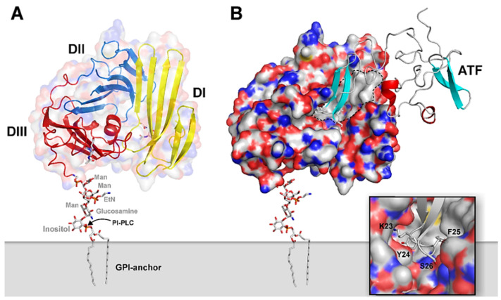
“Figure 1A illustrates a schematic model of the multidomain assembly of GPI- anchored uPAR, based on the crystal structure of the bimolecular complex of uPAR with the receptor binding fragment of uPA (ATF) (Figure 1B) [36].”
BackgroundMethodInfluentialResnati, M., Guttinger, M., Valcamonica, S., Sidénius, N., Blasi, F., & Fazioli, F. (1996) · The EMBO Journal · doi:10.1002/j.1460-2075.1996.tb00502.x · Cited by 298
Cancer invasion and tissue remodeling‐cooperation of protease systems and cell types
Danø, K., Rømer, J., Nielsen, B. S., Bjørn, S. F., Pyke, C., Rygaard, J., & Lund, L. R. (1999) · Apmis · doi:10.1111/j.1699-0463.1999.tb01534.x · Cited by 291
Vascular leak syndrome: a side effect of immunotherapy
Baluna, R. G., & Vitetta, E. S. (1997) · Immunopharmacology · doi:10.1016/s0162-3109(97)00041-6 · Cited by 282
Bugge, T. H., Flick, M. J., Danton, M. J. S., Daugherty, C. C., Rømer, J., Danø, K., Carmeliet, P., Collen, D., & Degen, J. L. (1996) · Proceedings of the National Academy of Sciences · doi:10.1073/pnas.93.12.5899 · Cited by 275
The urokinase receptor: Focused cell surface proteolysis, cell adhesion and signaling
Blasi, F., & Sidénius, N. (2009) · FEBS Letters · doi:10.1016/j.febslet.2009.12.039 · Cited by 258 · cited 4×
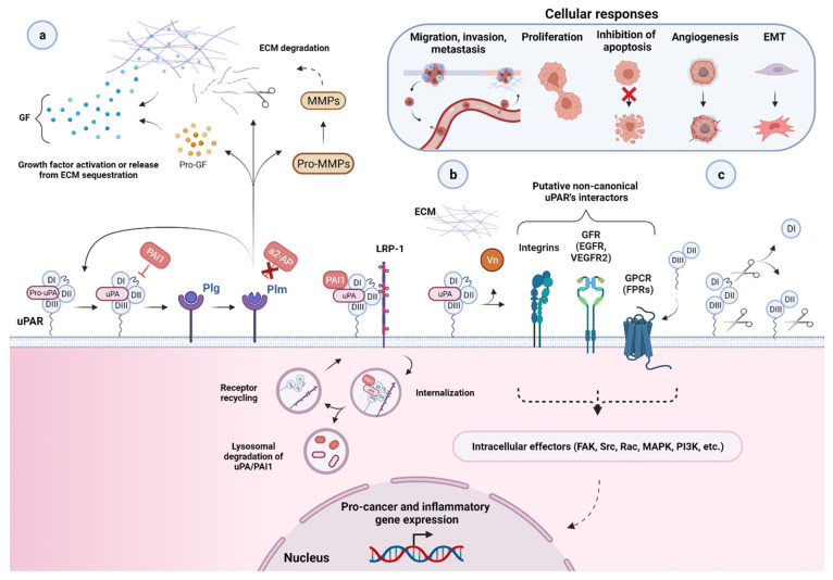
“Besides uPAR’s well-established role in pericellular proteolysis, so-called non-proteolytic functions have emerged over the years, implying uPAR’s involvement in multiple intracellularsignaling pathways affecting several aspects of the neoplastic evolution such as cell proliferation, differentiation, survival, adhesio…”
BackgroundInfluentialuPAR induces epithelial–mesenchymal transition in hypoxic breast cancer cells
Lester, R. D., Jo, M., Montel, V., Takimoto, S., & Gonias, S. L. (2007) · The Journal of Cell Biology · doi:10.1083/jcb.200701092 · Cited by 256
Antibody–Drug Conjugates—A Tutorial Review
Baah, S., Laws, M., & Rahman, K. M. (2021) · Molecules · doi:10.3390/molecules26102943 · Cited by 254
Ploug, M., & Ellis, V. (1994) · FEBS Letters · doi:10.1016/0014-5793(94)00674-1 · Cited by 248
Self-citationNanocarriers and Their Loading Strategies
Wang, N., Cheng, X., Li, N., Wang, H., & Chen, H. (2018) · Advanced Healthcare Materials · doi:10.1002/adhm.201801002 · Cited by 248
Llinas, P., Du, M. L., Gårdsvoll, H., Danø, K., Ploug, M., Gilquin, B., Stura, E. A., & Ménèz, A. (2005) · The EMBO Journal · doi:10.1038/sj.emboj.7600635 · Cited by 239 · cited 1×
“, Vn, as detailed in the next paragraph [38,41,51].”
BackgroundSelf-citationFazioli, F., Resnati, M., Sidénius, N., Higashimoto, Y., Appella, E., & Blasi, F. (1997) · The EMBO Journal · doi:10.1093/emboj/16.24.7279 · Cited by 237
The Urokinase Receptor. Protein Structure and Role in Plasminogen Activation and Cancer Invasion
Danø, K., Behrendt, N., Brunner, B., Ellis, V., Ploug, M., & PYKE (1995) · Japanese Journal of Thrombosis and Hemostasis · doi:10.2491/jjsth.6.212 · Cited by 234
Self-citationuPAR-induced cell adhesion and migration: vitronectin provides the key
Madsen, C. D., Ferraris, G. M. S., Andolfo, A., Cunningham, O., & Sidénius, N. (2007) · The Journal of Cell Biology · doi:10.1083/jcb.200612058 · Cited by 232
Generation and characterization of urokinase receptor-deficient mice.
DEWERCHIN, M., Nuffelen, A. V., Wallays, G., Bouché, A., Moons, L., Carmeliet, P., Mulligan, R. C., & Collen, D. (1996) · Journal of Clinical Investigation · doi:10.1172/jci118489 · Cited by 230
Bugge, T. H., Suh, T. T., Flick, M. J., Daugherty, C. C., John, R., Solberg, H., Ellis, V., Danø, K., & Degen, J. L. (1995) · Journal of Biological Chemistry · doi:10.1074/jbc.270.28.16886 · Cited by 218
Reversible cellular adhesion to vitronectin linked to urokinase receptor occupancy.
Waltz, D. A., & Chapman, H. A. (1994) · Journal of Biological Chemistry · doi:10.1016/s0021-9258(17)36688-7 · Cited by 216
Receptor-Targeted Nanoparticles for In vivo Imaging of Breast Cancer
Yang, L. P. H., Peng, X.-H., Wang, Y. A., Wang, X., Cao, Z., Ni, C., Karna, P., Zhang, X., Wood, W. C., Gao, X., et al. (2009) · Clinical Cancer Research · doi:10.1158/1078-0432.ccr-08-3289 · Cited by 216
The urokinase receptor. Protein structure and role in plasminogen activation and cancer invasion
Danø, K., Behrendt, N., Brünner, N., Ellis, V., Ploug, M., & Pyke, C. (1994) · Fibrinolysis and Proteolysis · doi:10.1016/0268-9499(94)90717-x · Cited by 214
Self-citationEnhanced expression of urokinase plasminogen activator and its receptor in pancreatic carcinoma
Cantero, D., Frieß, H., Deflorin, J., Zimmermann, A., Bründler, M.-A., Riesle, E., Korc, M., & Büchler, M. (1997) · British Journal of Cancer · doi:10.1038/bjc.1997.63 · Cited by 208
Bulle, A. S., & Lim, K. (2020) · Signal Transduction and Targeted Therapy · doi:10.1038/s41392-020-00341-1 · Cited by 208
Conese, M., Nykjær, A., Petersen, C. M., Cremona, O., Pardi, R., Andreasen, P. A., Gliemann, J., Christensen, E., & Blasi, F. (1995) · The Journal of Cell Biology · doi:10.1083/jcb.131.6.1609 · Cited by 191
Kjøller, L., & Hall, A. (2001) · The Journal of Cell Biology · doi:10.1083/jcb.152.6.1145 · Cited by 188
Receptor for urokinase is present in tumor-associated macrophages in ductal breast carcinoma.
Pyke, C., Græm, N., Ralfkiaer, E. M., Rønne, E., Høyer‐Hansen, G., Brünner, N., & Danø, K. (1993) · PubMed · Cited by 186
Ploug, M., Østergaard, S., Gårdsvoll, H., Kovalski, K., Holst‐Hansen, C., Holm, A., Ossowski, L., & Danø, K. (2001) · Biochemistry · doi:10.1021/bi010662g · Cited by 178
Self-citationMurphy, J. R. (2011) · Toxins · doi:10.3390/toxins3030294 · Cited by 177
Jo, M., Lester, R. D., Montel, V., Eastman, B. M., Takimoto, S., & Gonias, S. L. (2009) · Journal of Biological Chemistry · doi:10.1074/jbc.m109.023960 · Cited by 176
Immunotoxins and Anticancer Drug Conjugate Assemblies: The Role of the Linkage between Components
Dosio, F., Brusa, P., & Cattel, L. (2011) · Toxins · doi:10.3390/toxins3070848 · Cited by 174
Arora, N., & Leppla, S. H. (1993) · Journal of Biological Chemistry · doi:10.1016/s0021-9258(18)53698-x · Cited by 170
Targeting of Tumor Cells by Cell Surface Urokinase Plasminogen Activator-dependent Anthrax Toxin
Liu, S., Bugge, T. H., & Leppla, S. H. (2001) · Journal of Biological Chemistry · doi:10.1074/jbc.m011085200 · Cited by 168 · cited 7×
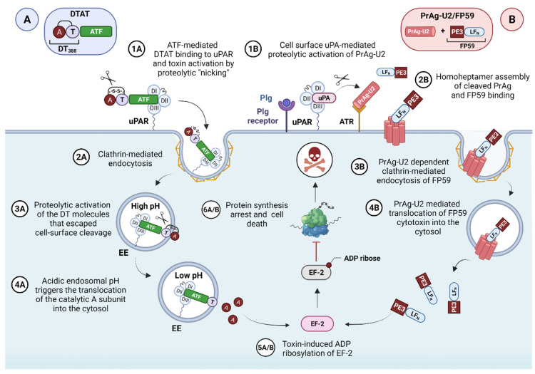
“The absolute requirement for the functional assembly of an active uPA/uPAR cell surface template for uPA-mediated plasminogen and subsequent PrAg-U2 activation in vivo was impeccably demonstrated by the complete lack of treatment response in tumor-bearing plg, uPA, and uPAR single-deficient mice challenged with the en…”
BackgroundMethodInfluentialShi, T., Song, X., Wang, Y., Liu, F., & Wei, J. (2020) · Frontiers in Immunology · doi:10.3389/fimmu.2020.00683 · Cited by 168
Ploug, M. (2003) · Current Pharmaceutical Design · doi:10.2174/1381612033454630 · Cited by 160 · cited 6×
“The biochemical aspects of this cascade have been ext nsively r viewed in [43] and schematically illustrated in Figure 3, which provides a graphical overview of PAR functional involveme t in cancer biology.”
BackgroundInfluentialSelf-citationBorley, N. R., Mortensen, N. J. M., Jewell, D. P., & Warren, B. F. (2000) · The Journal of Pathology · doi:10.1002/(sici)1096-9896(200002)190:2 · Cited by 158
Toxin-Based Therapeutic Approaches
Shapira, A., & Benhar, I. (2010) · Toxins · doi:10.3390/toxins2112519 · Cited by 155
Reduction in Surface Urokinase Receptor Forces Malignant Cells into a Protracted State of Dormancy
Yu, W., Kim, J., & Ossowski, L. (1997) · The Journal of Cell Biology · doi:10.1083/jcb.137.3.767 · Cited by 155 · cited 1×
“This association may have a bearing on the ability of uPAR to drive the transition between single-cell tumor dormancy and proliferation, which allows for the long-term survival of residual tumor cells during dormancy and reactivation of their proliferation years after primary treatment upon favorable conditions [13,85…”
BackgroundExpression and localization of urokinase-type plasminogen activator receptor in human gliomas.
Yamamoto, M., Sawaya, R. A., Mohanam, S., Rao, V. H., Bruner, J. M., Nicolson, G. L., & Rao, J. S. (1994) · PubMed · Cited by 149
Rømer, J., Lund, L. R., Eriksen, J., Pyke, C., Kristensen, P., & Danø, K. (1994) · Journal of Investigative Dermatology · doi:10.1111/1523-1747.ep12373187 · Cited by 143
Radionuclide Therapy for Neuroendocrine Tumors
Cives, M., & Strosberg, J. (2017) · Current Oncology Reports · doi:10.1007/s11912-017-0567-8 · Cited by 142
Targeting the Tumor Microenvironment: Focus on Angiogenesis
Fan, F., Schimming, A., Jaeger, D., & Podar, K. (2011) · Journal of Oncology · doi:10.1155/2012/281261 · Cited by 139
Therapeutics targeting the fibrinolytic system
Lin, H., Lu-ning, X., Yu, S., Hong, W., Huang, M., & Xu, P. (2020) · Experimental & Molecular Medicine · doi:10.1038/s12276-020-0397-x · Cited by 132 · cited 1×
“uPAR-targeted theranostic NPs have also been developed to selectively deliver phototherapeutic agents to tumor sites, with a few valuable examples reported in the literature [248] and reviewed [19].”
BackgroundSaporin-S6: A Useful Tool in Cancer Therapy
Polito, L., Bortolotti, M., Mercatelli, D., Battelli, M. G., & Bolognesi, A. (2013) · Toxins · doi:10.3390/toxins5101698 · Cited by 131
A critical role for plasminogen in inflammation
Baker, S. K., & Strickland, S. (2020) · The Journal of Experimental Medicine · doi:10.1084/jem.20191865 · Cited by 130
Role of Urokinase Receptor in Tumor Progression and Development
Noh, H., Hong, S., & Huang, S. (2013) · Theranostics · doi:10.7150/thno.4218 · Cited by 129 · cited 1×
“Distinct regulatory mechanisms control uPAR expression in tumor and associated stromal cells at multiple levels, respectively, transcriptional, post-transcriptional, and posttranslational, with the former being considered the principal one [12,14,52,84,85,103,149].”
BackgroundCarmeliet, P., Moons, L., Dewerchin, M. A., Rosenberg, S., Herbert, J., Lupu, F., & Collen, D. (1998) · The Journal of Cell Biology · doi:10.1083/jcb.140.1.233 · Cited by 126 · cited 1×
“Surprisingly, no early-onset overt phenotypic abnormalities were observed in uPAR null mice (Plaur −/−) [74–76], in contrast to those genetically deficient in plasminogen (Plg −/−) [77], implicating the existence of a functional redundancy by which other proteases, besides the physiological bona fide activators, uPA a…”
BackgroundPotent antitumor activity of a urokinase-activated engineered anthrax toxin
Liu, S., Aaronson, H., Mitola, D. J., Leppla, S. H., & Bugge, T. H. (2003) · Proceedings of the National Academy of Sciences · doi:10.1073/pnas.0236849100 · Cited by 124 · cited 30×
“The absolute requirement for the functional assembly of an active uPA/uPAR cell surface template for uPA-mediated plasminogen and subsequent PrAg-U2 activation in vivo was impeccably demonstrated by the complete lack of treatment response in tumor-bearing plg, uPA, and uPAR single-deficient mice challenged with the en…”
BackgroundMethodInfluentialProtein toxins: intracellular trafficking for targeted therapy
Johannes, L., & Decaudin, D. (2005) · Gene Therapy · doi:10.1038/sj.gt.3302557 · Cited by 124
Prevalence of mucosotropic human papillomaviruses in squamous-cell carcinomas of the head and neck
Snijders, P. J. F., Scholes, A. G. M., Hart, C. A., Jones, A. S., Vaughan, E. D., Woolgar, J. A., Meijer, C. J. L. M., Walboomers, J. M. M., & Field, J. K. (1996) · International Journal of Cancer · doi:10.1002/(sici)1097-0215(19960516)66:4 · Cited by 122
Crystal structures of two human vitronectin, urokinase and urokinase receptor complexes
Huai, Q., Zhou, A., Lin, L., Mazar, A. P., Parry, G. C., Callahan, J. A., Shaw, D. E., Furie, B., Furie, B. C., & Huang, M. (2008) · Nature Structural & Molecular Biology · doi:10.1038/nsmb.1404 · Cited by 120 · cited 3×
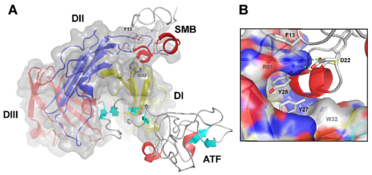
“Vn binds uPAR t a composite epitope xposed on the DI/ II interface, via its small, N-terminal somatoedin B (SM ) domain and with a relatively weaker binding affinity (KD of 2 μM) (Figure 2) [33,37,40,43].”
BackgroundMethodChaurasia, P., Aguirre‐Ghiso, J. A., Liang, O. D., Gårdsvoll, H., Ploug, M., & Ossowski, L. (2006) · Journal of Biological Chemistry · doi:10.1074/jbc.m512311200 · Cited by 120
Self-citationCannon, A. C., Thompson, C. M., Hall, B. R., Jain, M., Sushil Kumar, K., & Batra, S. K. (2018) · Genes & Cancer · doi:10.18632/genesandcancer.171 · Cited by 120
Bharadwaj, A. G., Holloway, R. W., Miller, V. A., & Waisman, D. M. (2021) · Cancers · doi:10.3390/cancers13081838 · Cited by 120
The urokinase receptor: a ligand or a receptor? Story of a sociable molecule
Ragno, P. (2006) · Cellular and Molecular Life Sciences · doi:10.1007/s00018-005-5428-1 · Cited by 118 · cited 1×
“Since their discovery about 30 years ago, a wide variety of uPAR non-canonical interactors have been proposed in the literature, leading to the definition of the so-called uPAR interactome, comprising over 42 alleged protein partners so far [16,52,56,73,79,94].”
BackgroundYuan, H., Che, S., Hui, B., Yang, Y.-Y., Wang, X., Zhang, X., Qiang, Y., & Ma, H. (2019) · Biomedicine & Pharmacotherapy · doi:10.1016/j.biopha.2019.108614 · Cited by 118
Yang, L. P. H., Cao, Z., Sajja, H. K., Mao, H. Z., Wang, L., Geng, H., Xu, H., Jiang, T., Wood, W. C., Nie, S., et al. (2008) · Journal of Biomedical Nanotechnology · doi:10.1166/jbn.2008.007 · Cited by 116
Clinical Efficacy and Toxicity of Anti-EGFR Therapy in Common Cancers
Harandi, A., Zaidi, A., Stocker, A. M., & Laber, D. A. (2009) · Journal of Oncology · doi:10.1155/2009/567486 · Cited by 116
The interaction between urokinase receptor and vitronectin in cell adhesion and signalling
Madsen, C. D., & Sidénius, N. (2008) · European Journal of Cell Biology · doi:10.1016/j.ejcb.2008.02.003 · Cited by 115
First-in-human uPAR PET: Imaging of Cancer Aggressiveness
Persson, M., Skovgaard, D. C., Brandt-Larsen, M., Christensen, C. L., Madsen, J., Nielsen, C. H., Thurison, T., Klausen, T. L., Holm, S., Loft, A., et al. (2015) · Theranostics · doi:10.7150/thno.12956 · Cited by 115 · cited 1×
“The design and implementation of theranostic approaches combining uPAR-targeted therapeutic and non-invasive imaging modalities, such as positron emission tomography (PET), magnetic resonance (MRI), or near-infrared (NIR) fluorescence imaging, are currently underway and hold clinical potential for significantly improv…”
BackgroundSelf-citationStructural Basis of Interaction between Urokinase-type Plasminogen Activator and its Receptor
Bařinka, C., Parry, G. C., Callahan, J. A., Shaw, D. E., Kuo, A., Bdeir, K. H., Cines, D. B., Mazar, A. P., & Lubkowski, J. T. (2006) · Journal of Molecular Biology · doi:10.1016/j.jmb.2006.08.063 · Cited by 114 · cited 1×
“, Vn, as detailed in the next paragraph [38,41,51].”
BackgroundFrankel, A. E., Powell, B. L., Hall, P. D., Case, L. D., & Kreitman, R. J. (2002) · PubMed · Cited by 113
Degryse, B., Resnati, M., Czekay, R., Loskutoff, D. J., & Blasi, F. (2005) · Journal of Biological Chemistry · doi:10.1074/jbc.m413954200 · Cited by 112
Solberg, H., Ploug, M., Høyer‐Hansen, G., Nielsen, B. S., & Lund, L. R. (2001) · Journal of Histochemistry & Cytochemistry · doi:10.1177/002215540104900211 · Cited by 111
Self-citationThe urokinase plasminogen activator receptor as a gene therapy target for cancer
Pillay, V., Dass, C. R., & Choong, P. (2006) · Trends in biotechnology · doi:10.1016/j.tibtech.2006.10.011 · Cited by 109 · cited 1×
“Although impairment of uPAR function or inhibition of its expression was associated with impaired invasive, metastatic, and tumorigenic potential of many tumor models in vitro and in vivo [4,11,12,14,16,17,20,52,57,79,84,85], one study in transgenic mice with uPAR gene ablation showed no pronounced causal effects on d…”
Backgroundde Paulis, A., Montuori, N., Prevete, N., Fiorentino, I., Rossi, F. W., Visconte, V., Rossi, G., Marone, G., & Ragno, P. (2004) · The Journal of Immunology · doi:10.4049/jimmunol.173.9.5739 · Cited by 109
Use of molecular markers for predicting therapy response in cancer patients
Duffy, M. J., O’Donovan, N., & Crown, J. P. (2010) · Cancer Treatment Reviews · doi:10.1016/j.ctrv.2010.07.004 · Cited by 107
Onda, M., Nagata, S., FitzGerald, D. J., Beers, R., Fisher, R. J., Vincent, J. J., Lee, B., Nakamura, M., Hwang, J., Kreitman, R. J., et al. (2006) · The Journal of Immunology · doi:10.4049/jimmunol.177.12.8822 · Cited by 107
Hu, Y., Chi, C., Wang, S., Wang, L., Liang, P., Liu, F., Shang, W., Wang, W., Zhang, F., Li, S., et al. (2017) · Advanced Materials · doi:10.1002/adma.201700448 · Cited by 105
Theranostic Nanoparticles for Tracking and Monitoring Disease State
Zavaleta, C. L., Ho, D., & Chung, E. J. (2017) · SLAS TECHNOLOGY · doi:10.1177/2472630317738699 · Cited by 105
Soluble and cleaved forms of the urokinase-receptor: degradation products or active molecules?
Montuori, N., Visconte, V., Rossi, G., & Ragno, P. (2004) · Thrombosis and Haemostasis · doi:10.1160/th04-09-0580 · Cited by 104
Edwards, D. R., Høyer‐Hansen, G., Blasi, F., & Sloane, B. F. (2008) · Springer eBooks · doi:10.1007/978-0-387-69057-5 · Cited by 104
He, Y., Liu, X., Chen, Z., Zhu, J., Xiong, Y., Li, K., Dong, J., & Li, X. (2007) · Clinical Cancer Research · doi:10.1158/1078-0432.ccr-06-2088 · Cited by 99
Mapping of the Vitronectin-binding Site on the Urokinase Receptor
Gårdsvoll, H., & Ploug, M. (2007) · Journal of Biological Chemistry · doi:10.1074/jbc.m610184200 · Cited by 98 · cited 2×
“Vn binds uPAR t a composite epitope xposed on the DI/ II interface, via its small, N-terminal somatoedin B (SM ) domain and with a relatively weaker binding affinity (KD of 2 μM) (Figure 2) [33,37,40,43].”
BackgroundSelf-citationClinical Applications of the Urokinase Receptor (uPAR) for Cancer Patients
Boonstra, M. C., Verspaget, H. W., Ganesh, S., Kubben, F. J. G. M., Vahrmeijer, A. L., van de Velde, C. J. �., Kuppen, P. J. K., Quax, P. H. A., & Sier, C. F. M. (2011) · Current Pharmaceutical Design · doi:10.2174/138161211796718233 · Cited by 98 · cited 1×
“Besides uPAR’s well-established role in pericellular proteolysis, so-called non-proteolytic functions have emerged over the years, implying uPAR’s involvement in multiple intracellularsignaling pathways affecting several aspects of the neoplastic evolution such as cell proliferation, differentiation, survival, adhesio…”
BackgroundAntibody-drug conjugates in clinical trials for lymphoid malignancies and multiple myeloma
Yu, B., & Liu, D. (2019) · Journal of Hematology & Oncology · doi:10.1186/s13045-019-0786-6 · Cited by 97
Urokinase‐type plasminogen activator and its receptor synergize to promote pathogenic proteolysis
Zhou, H., Nichols, A., Meda, P., & Vassalli, J. (2000) · The EMBO Journal · doi:10.1093/emboj/19.17.4817 · Cited by 94 · cited 2×
“The normal skin phenotype exhibited by either the single transgenic mice, bitransgenic mice overexpressing uPAR and a receptor-binding catalytically inactive form of uPA, or bitransgenic mice crossed into a Plg −/− background undoubtedly underscores the strict requirement for the assembly of a functional cell surface…”
BackgroundVallera, D. A. (2002) · JNCI Journal of the National Cancer Institute · doi:10.1093/jnci/94.8.597 · Cited by 94
The Urokinase Receptor Interactome
Eden, G. C., Archinti, M., Furlan, F., Murphy, R. P., & Degryse, B. (2011) · Current Pharmaceutical Design · doi:10.2174/138161211796718215 · Cited by 92 · cited 3×
“For an exhaustive overview of potential uPARinteractors, signaling networks, and functional implications in cancer, we invite the reader to refer to the following detailed reviews [4,16,73].”
BackgroundMazar, A. P. (2001) · Anti-Cancer Drugs · doi:10.1097/00001813-200106000-00001 · Cited by 91 · cited 4×
“Distinct regulatory mechanisms control uPAR expression in tumor and associated stromal cells at multiple levels, respectively, transcriptional, post-transcriptional, and posttranslational, with the former being considered the principal one [12,14,52,84,85,103,149].”
BackgroundInfluentialTranscriptional and posttranscriptional regulation of the plasminogen activator system
Medcalf, R. L., Muñoz‐Cánoves, P., & Nagamine, Y. (2005) · Thrombosis and Haemostasis · doi:10.1160/th04-12-0814 · Cited by 90
The urokinase receptor. A cell surface, regulated chemokine
Blasi, F. (1999) · Apmis · doi:10.1111/j.1699-0463.1999.tb01531.x · Cited by 90
Targeting uPAR with Antagonistic Recombinant Human Antibodies in Aggressive Breast Cancer
LeBeau, A. M., Duriseti, S., Murphy, S. T., Pepin, F., Hann, B. C., Gray, J. W., VanBrocklin, H. F., & Craik, C. S. (2013) · Cancer Research · doi:10.1158/0008-5472.can-12-3526 · Cited by 90
Jo, M., Eastman, B. M., Webb, D. L., Stoletov, K. V., Klemke, R. L., & Gonias, S. L. (2010) · Cancer Research · doi:10.1158/0008-5472.can-10-1936 · Cited by 89 · cited 1×
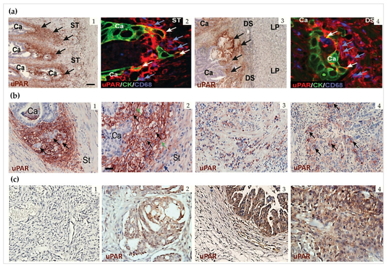
“In some tumors, uPAR expression has also been detected in cancer stem cells [93,125] and bone marrow cancer cells [131,150,151].”
BackgroundAbdalla, M. O. A., Karna, P., Sajja, H. K., Mao, H. Z., Yates, C. C., Turner, T., & Aneja, R. (2010) · Journal of Controlled Release · doi:10.1016/j.jconrel.2010.10.030 · Cited by 88
Huang, C., Xie, D., Cui, J., Li, Q., Gao, Y., & Xie, K. (2014) · Clinical Cancer Research · doi:10.1158/1078-0432.ccr-13-2311 · Cited by 87
Multiple activities of a multifaceted receptor: roles of cleaved and soluble uPAR
Montuori, N. (2009) · Frontiers in bioscience · doi:10.2741/3392 · Cited by 86 · cited 1×
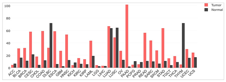
“The urokinase plasminogen activator receptor (uPAR) represents one of the emerging attractive tumor targets for such targeted therapy due to its sparse expression in healthy homeostatic tissues compared to the robust expression in most solid tumors [4,5].”
BackgroundFerraris, G. M. S., Schulte, C., Buttiglione, V., Lorenzi, V. D., Piontini, A., Galluzzi, M., Podestà, A., Madsen, C. D., & Sidénius, N. (2014) · The EMBO Journal · doi:10.15252/embj.201387611 · Cited by 86
Urokinase Receptor and Integrin Interactions
Kugler, M. C., Wei, Y., & Chapman, H. A. (2003) · Current Pharmaceutical Design · doi:10.2174/1381612033454658 · Cited by 86
Mazar, A. P., Ahn, R. W., & O’Halloran, T. V. (2011) · Current Pharmaceutical Design · doi:10.2174/138161211796718152 · Cited by 85 · cited 2×
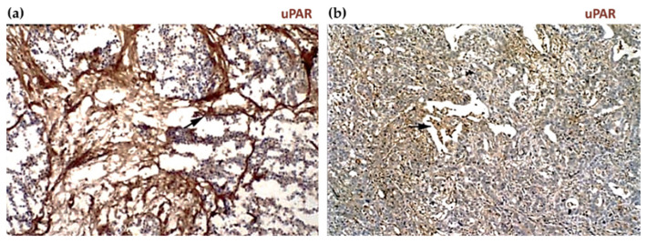
“The remarkable stromal expression of uPAR also allows targeting this compartment, which offers multiple opportunities to enhance the therapeutic efficacy compared to exclusively targeting the tumor cells, especially in tumors expressing uPAR in both cell types, such as pancreatic cancer, or those lacking a tumor-speci…”
BackgroundIllemann, M., Bird, N. C., Majeed, A. W., Lærum, O. D., Lund, L. R., Danø, K., & Nielsen, B. S. (2008) · International Journal of Cancer · doi:10.1002/ijc.24166 · Cited by 84
Characterization of the Functional Epitope on the Urokinase Receptor
Gårdsvoll, H., Gilquin, B., Du, M. L., Ménèz, A., Jørgensen, T. J. D., & Ploug, M. (2006) · Journal of Biological Chemistry · doi:10.1074/jbc.m513583200 · Cited by 84
Self-citationGao, N., Bozeman, E. N., Qian, W., Wang, L., Chen, H., Lipowska, M., Staley, C. A., Wang, Y. A., Mao, H. Z., & Yang, L. P. H. (2017) · Theranostics · doi:10.7150/thno.18125 · Cited by 83
Nozaki, S., Endo, Y., Kawashiri, S., Nakagawa, K., Yamamoto, E., Yonemura, Y., & Sasaki, T. (1998) · Oral Oncology · doi:10.1016/s1368-8375(97)00028-6 · Cited by 82
Lin, L., Gårdsvoll, H., Huai, Q., Huang, M., & Ploug, M. (2010) · Journal of Biological Chemistry · doi:10.1074/jbc.m109.093492 · Cited by 81 · cited 3×
“2 n ), which exhibits a pronounced species-specificity between humans and mice [49,50].”
BackgroundMethodSelf-citationRullo, A. F., Fitzgerald, K., Muthusamy, V., Liu, M., Yuan, C., Huang, M., Kim, M., Cho, A. E., & Spiegel, D. A. (2016) · Angewandte Chemie International Edition · doi:10.1002/anie.201510866 · Cited by 81
Nagata, S., & Pastan, I. H. (2009) · Advanced Drug Delivery Reviews · doi:10.1016/j.addr.2009.07.014 · Cited by 80
uPAR-targeted Optical Imaging Contrasts as Theranostic Agents for Tumor Margin Detection
Yang, L. P. H., Sajja, H. K., Cao, Z., Qian, W., Bender, L., Marcus, A. I., Lipowska, M., Wood, W. C., & Wang, Y. A. (2014) · Theranostics · doi:10.7150/thno.7409 · Cited by 79
Lund, I. K., Illemann, M., Thurison, T., Christensen, I. J., & Høyer‐Hansen, G. (2011) · Current Drug Targets · doi:10.2174/138945011797635902 · Cited by 75 · cited 4×
“Although impairment of uPAR function or inhibition of its expression was associated with impaired invasive, metastatic, and tumorigenic potential of many tumor models in vitro and in vivo [4,11,12,14,16,17,20,52,57,79,84,85], one study in transgenic mice with uPAR gene ablation showed no pronounced causal effects on d…”
BackgroundInfluentialGårdsvoll, H., Danø, K., & Ploug, M. (1999) · Journal of Biological Chemistry · doi:10.1074/jbc.274.53.37995 · Cited by 74
Self-citationHeiss, M. M., Simon, E. H., Beyer, B. C. M., Gruetzner, K. U., Tarabichi, A., Babic, R., Schildberg, F. W., & Allgayer, H. (2002) · Journal of Clinical Oncology · doi:10.1200/jco.2002.08.003 · Cited by 74
Magdolen, V., Rettenberger, P. M., Koppitz, M., Goretzkt, L., Kessler, H., Weidle, U. H., König, B., Graeff, H., Schmitt, M., & Wilhelm, O. G. (1996) · European Journal of Biochemistry · doi:10.1111/j.1432-1033.1996.0743p.x · Cited by 73 · cited 2×
“t the outer leaflet of the lip d bilayer of the c ll me brane by a C-terminal glycosyl-phosphatidylinositol (GPI) anchor [35,47].”
BackgroundThe plasminogen activation system in inflammation
Rosso, M. D. (2008) · Frontiers in bioscience · doi:10.2741/3032 · Cited by 73
Structure and ligand interactions of the urokinase receptor (uPAR)
Kjærgaard, M. (2008) · Frontiers in bioscience · doi:10.2741/3092 · Cited by 73
Usher, P. A., Thomsen, O., Iversen, P., Johnsen, M., Brünner, N., Høyer‐Hansen, G., Andreasen, P. A., Danø, K., & Nielsen, B. S. (2004) · International Journal of Cancer · doi:10.1002/ijc.20665 · Cited by 72
Ploug, M., Rahbek-Nielsen, H., Nielsen, P. F., Roepstorff, P., & Danø, K. (1998) · Journal of Biological Chemistry · doi:10.1074/jbc.273.22.13933 · Cited by 69 · cited 1×
“Human uPAR is a 55–60 kDa highly glycosylated protein (283 aa residues) [45,46], tethered to the outer leaflet of the lipid bilayer of the cell membrane by a C-terminal glycosyl-phosphatidylinositol (GPI) anchor [35,47].”
BackgroundSelf-citationStrategies to Reduce the Immunogenicity of Recombinant Immunotoxins
Mazor, R., King, E. M., & Pastan, I. H. (2018) · American Journal Of Pathology · doi:10.1016/j.ajpath.2018.04.016 · Cited by 69
Jacobsen, B., & Ploug, M. (2008) · Current Medicinal Chemistry · doi:10.2174/092986708785909012 · Cited by 68
Self-citationIntermolecular complementation achieves high-specificity tumor targeting by anthrax toxin
Liu, S., Redeye, V., Kuremsky, J. G., Kuhnen, M., Molinolo, A., Bugge, T. H., & Leppla, S. H. (2005) · Nature Biotechnology · doi:10.1038/nbt1091 · Cited by 68
Jing, Y., Tong, C., Zhang, J., Nakamura, T., Iankov, I. D., Russell, S. J., & Merchan, J. R. (2009) · Cancer Research · doi:10.1158/0008-5472.can-08-2628 · Cited by 68
Kriegbaum, M. C., Persson, M., Haldager, L., Alpízar‐Alpízar, W., Jacobsen, B., Gårdsvoll, H., Kjær, A., & Ploug, M. (2011) · Current Drug Targets · doi:10.2174/138945011797635812 · Cited by 67 · cited 5×
“Although impairment of uPAR function or inhibition of its expression was associated with impaired invasive, metastatic, and tumorigenic potential of many tumor models in vitro and in vivo [4,11,12,14,16,17,20,52,57,79,84,85], one study in transgenic mice with uPAR gene ablation showed no pronounced causal effects on d…”
BackgroundInfluentialSelf-citationMulthaupt, H. A. B., Mazar, A. P., Cines, D., Warhol, M. J., & McCrae, K. R. (1994) · PubMed · Cited by 67
Lindberg, P., Larsson, Å., & Nielsen, B. S. (2006) · International Journal of Cancer · doi:10.1002/ijc.21568 · Cited by 66
Taruno, K., Kurita, T., Kuwahata, A., Yanagihara, K., Enokido, K., Katayose, Y., Nakamura, S., Takei, H., Sekino, M., & Kusakabe, M. (2019) · Journal of Surgical Oncology · doi:10.1002/jso.25747 · Cited by 66
Tjwa, M., Sidénius, N., Moura, R., Jansen, S., Theunissen, K., Andolfo, A., Mol, M. D., Dewerchin, M. A., Moons, L., Blasi, F., et al. (2009) · Journal of Clinical Investigation · doi:10.1172/jci36010 · Cited by 65
Todhunter, D. A., Hall, W. A., Rustamzadeh, E., Shu, Y., Doumbia, S., & Vallera, D. A. (2004) · Protein Engineering Design and Selection · doi:10.1093/protein/gzh023 · Cited by 65
Ahmed, M. S. U., Salam, A. B., Yates, C. C., Willian, K. R., Jaynes, J. M., Turner, T., & Abdalla, M. O. A. (2017) · International Journal of Nanomedicine · doi:10.2147/ijn.s139011 · Cited by 64
Intact and cleaved uPAR forms: diagnostic and prognostic value in cancer
Rasch, M. G. (2008) · Frontiers in bioscience · doi:10.2741/3186 · Cited by 63 · cited 1×
“These uPAR forms may therefore allegedly play a role in the process of tumor progression, possibly as autocrine or paracrine signals for tumor cell motility and angiogenesis [59,65,67].”
Backgroundα2‐Macroglobulin Receptor Mediates Binding and Cytotoxicity of Plant Ribosome‐Inactivating Proteins
Cavallaro, U., Nykjær, A., Nielsen, M. S., & Soria, M. R. (1995) · European Journal of Biochemistry · doi:10.1111/j.1432-1033.1995.tb20795.x · Cited by 63
Gårdsvoll, H., Werner, F., Søndergaard, L., Danø, K., & Ploug, M. (2004) · Protein Expression and Purification · doi:10.1016/j.pep.2003.12.002 · Cited by 62 · cited 1×
“Human uPAR is a 55–60 kDa highly glycosylated protein (283 aa residues) [45,46], tethered to the outer leaflet of the lipid bilayer of the cell membrane by a C-terminal glycosyl-phosphatidylinositol (GPI) anchor [35,47].”
BackgroundSelf-citationDuriseti, S., Goetz, D. H., Hostetter, D. R., LeBeau, A. M., Wei, Y., & Craik, C. S. (2010) · Journal of Biological Chemistry · doi:10.1074/jbc.m109.077677 · Cited by 61
Clathrin and LRP-1-Independent Constitutive Endocytosis and Recycling of uPAR
Cortese, K., Sahores, M., Madsen, C. D., Tacchetti, C., & Blasi, F. (2008) · PLoS ONE · doi:10.1371/journal.pone.0003730 · Cited by 60 · cited 1×
“as responsible for uPAR constitutive endocytosis and recycling [8].”
BackgroundTargeting the Urokinase Plasminogen Activator Receptor with Synthetic Self-Assembly Nanoparticles
Wang, M., Löwik, D. W. P. M., Miller, A. D., & Thanou, M. (2008) · Bioconjugate Chemistry · doi:10.1021/bc8001908 · Cited by 59
Inhibitors of the Protease Domain of Urokinase-Type Plasminogen Activator
Rockway, T. W., Nienaber, V. L., & Giranda, V. L. (2002) · Current Pharmaceutical Design · doi:10.2174/1381612023392676 · Cited by 59
Madunić, J. (2018) · Thrombosis and Haemostasis · doi:10.1055/s-0038-1675399 · Cited by 59
Illemann, M., Lærum, O. D., Hasselby, J. P., Thurison, T., Høyer‐Hansen, G., Nielsen, H. J., the Danish Study Group on Early Detection of Colorectal Cancer, & Christensen, I. J. (2014) · Cancer Medicine · doi:10.1002/cam4.242 · Cited by 58
Targeting Receptors on Cancer Cells with Protein Toxins
Antignani, A., Ho, E. C. H., Bilotta, M. T., Qiu, R., Sarnvosky, R., & FitzGerald, D. J. (2020) · Biomolecules · doi:10.3390/biom10091331 · Cited by 58
Urokinase type plasminogen activator receptor (uPAR) as a new therapeutic target in cancer
Montuori, N., Pesapane, A., Rossi, F. W., Giudice, V., de Paulis, A., Selleri, C., & Ragno, P. (2016) · PubMed Central · doi:10.14273/unisa-2679 · Cited by 56 · cited 1×
“This association may have a bearing on the ability of uPAR to drive the transition between single-cell tumor dormancy and proliferation, which allows for the long-term survival of residual tumor cells during dormancy and reactivation of their proliferation years after primary treatment upon favorable conditions [13,85…”
BackgroundUrokinase receptor and resistance to targeted anticancer agents
Gonias, S. L., & Hu, J. (2015) · Frontiers in Pharmacology · doi:10.3389/fphar.2015.00154 · Cited by 55
Tumour endoproteases: the cutting edge of cancer drug delivery?
Atkinson, J. M., Siller, C. S., & Gill, J. H. (2008) · British Journal of Pharmacology · doi:10.1038/sj.bjp.0707657 · Cited by 55
Conformational Regulation of Urokinase Receptor Function
Gårdsvoll, H., Jacobsen, B., Kriegbaum, M. C., Behrendt, N., Engelholm, L. H., Østergaard, S., & Ploug, M. (2011) · Journal of Biological Chemistry · doi:10.1074/jbc.m111.220087 · Cited by 55
Self-citationSafe and Effective Sarcoma Therapy through Bispecific Targeting of EGFR and uPAR
Borgatti, A., Koopmeiners, J. S., Sarver, A. L., Winter, A. L., Stuebner, K., Todhunter, D., Rizzardi, A. E., Henriksen, J. C., Schmechel, S. C., Forster, C. L., et al. (2017) · Molecular Cancer Therapeutics · doi:10.1158/1535-7163.mct-16-0637 · Cited by 54
Hildenbrand, R., Niedergethmann, M., Marx, A., Belharazem, D., Allgayer, H., Schleger, C., & Ströbel, P. (2009) · American Journal Of Pathology · doi:10.2353/ajpath.2009.080785 · Cited by 53 · cited 1×
“Besides breast cancer, uPAR amplification has also been frequently reported in pancreatic cancer and analogously recognized as a significant adverse prognostic parameter identifying a subgroup of particularly aggressive tumors [10].”
BackgroundDesign and engineering of deimmunized biotherapeutics
Griswold, K. E., & Bailey‐Kellogg, C. (2016) · Current Opinion in Structural Biology · doi:10.1016/j.sbi.2016.06.003 · Cited by 53
Cavallaro, U., Vecchio, A. D., Lappi, D. A., & Soria, M. R. (1993) · Journal of Biological Chemistry · doi:10.1016/s0021-9258(19)49445-3 · Cited by 52
Tang, C., Hill, M. L., Brumwell, A. N., Chapman, H. A., & Wei, Y. (2008) · Journal of Cell Science · doi:10.1242/jcs.029769 · Cited by 51
Pierga, J., Bonneton, C., Magdelénat, H., Vincent‐Salomon, A., Nos, C., Boudou, E., Pouillart, P., Thiery, J. P., & de Crémoux, P. (2004) · International Journal of Cancer · doi:10.1002/ijc.20698 · Cited by 50
Mertens, H. D. T., Kjærgaard, M., Mysling, S., Gårdsvoll, H., Jørgensen, T. J. D., Svergun, D. I., & Ploug, M. (2012) · Journal of Biological Chemistry · doi:10.1074/jbc.m112.398404 · Cited by 50 · cited 4×
“There are also malignancies where the prominent uPAR expression is reported on cancer cells such as in esophageal [121,122] and cutaneous squamous cell carcinomas [40], as well as in gliomas such as anaplastic astrocytoma and glioblastomas [123,124].”
BackgroundInfluentialSelf-citationAllgayer, H., & Aguirre‐Ghiso, J. A. (2008) · Apmis · doi:10.1111/j.1600-0463.2008.00997.x · Cited by 50
Abi‐Habib, R. J., Singh, R., Liu, S., Bugge, T. H., Leppla, S. H., & Frankel, A. E. (2006) · Molecular Cancer Therapeutics · doi:10.1158/1535-7163.mct-06-0315 · Cited by 50
The Urokinase Receptor: A Multifunctional Receptor in Cancer Cell Biology. Therapeutic Implications
Santi, A. L., Napolitano, F., Montuori, N., & Ragno, P. (2021) · International Journal of Molecular Sciences · doi:10.3390/ijms22084111 · Cited by 50
Alpízar‐Alpízar, W., Christensen, I. J., Santoni‐Rugiu, E., Skarstein, A., Øvrebø, K., Illemann, M., & Lærum, O. D. (2011) · International Journal of Cancer · doi:10.1002/ijc.26417 · Cited by 49
Knör, S., Sato, S., Huber, T., Morgenstern, A., Bruchertseifer, F., Schmitt, M., Kessler, H., Senekowitsch–Schmidtke, R., Magdolen, V., & Seidl, C. (2007) · European Journal of Nuclear Medicine and Molecular Imaging · doi:10.1007/s00259-007-0582-3 · Cited by 48
Bifulco, K., Longanesi-Cattani, I., Gala, M. K., Carluccio, G. D., Masucci, M. T., Pavone, V., Lista, L., Arra, C., Stoppelli, M. P., & Carriero, M. V. (2010) · Journal of Thrombosis and Haemostasis · doi:10.1111/j.1538-7836.2010.04075.x · Cited by 47 · cited 1×
“These uPAR forms may therefore allegedly play a role in the process of tumor progression, possibly as autocrine or paracrine signals for tumor cell motility and angiogenesis [59,65,67].”
BackgroundEfficacy of antiangiogenic targeted toxins against glioblastoma multiforme
Hall, W. A., & Vallera, D. A. (2006) · Neurosurgical FOCUS · doi:10.3171/foc.2006.20.4.15 · Cited by 46
Crystal Structure of the Urokinase Receptor in a Ligand-Free Form
Xu, X., Gårdsvoll, H., Yuan, C., Lin, L., Ploug, M., & Huang, M. (2012) · Journal of Molecular Biology · doi:10.1016/j.jmb.2011.12.058 · Cited by 46
Self-citationSimultaneous Tumor and Stroma Targeting by Oncolytic Viruses
Everts, A., Bergeman, M. H., McFadden, G., & Kemp, V. (2020) · Biomedicines · doi:10.3390/biomedicines8110474 · Cited by 45 · cited 2×
“In this context, the direct targeting of specific stromal components is one of the strategies being explored, with uPAR representing one of the emerging targets due to its abundant and ubiquitous expression in the tumor stroma of most cancer types [2].”
BackgroundIntracranial therapy of glioblastoma with the fusion protein DTAT in immunodeficient mice
Rustamzadeh, E., Hall, W. A., Todhunter, D. A., Vallera, V. D., Low, W. C., Liu, H., Panoskaltsis‐Mortari, A., & Vallera, D. A. (2006) · International Journal of Cancer · doi:10.1002/ijc.22278 · Cited by 44
Melis, M. L., Bijster, M., de Visser, M., Konijnenberg, M., de Swart, J., Rolleman, E. J., Boerman, O. C., Krenning, E. P., & de Jong, M. (2009) · European Journal of Nuclear Medicine and Molecular Imaging · doi:10.1007/s00259-009-1196-8 · Cited by 43
Persson, M., & Kjær, A. (2013) · Clinical Physiology and Functional Imaging · doi:10.1111/cpf.12037 · Cited by 43
O’Halloran, T. V., Ahn, R. W., Hankins, P. L., Swindell, E. P., & Mazar, A. P. (2013) · Theranostics · doi:10.7150/thno.4953 · Cited by 43
Persson, M., Juhl, K., Rasmussen, P., Brandt-Larsen, M., Madsen, J., Ploug, M., & Kjær, A. (2014) · Molecular Pharmaceutics · doi:10.1021/mp500177c · Cited by 43
Self-citationOntak-like human IL-2 fusion toxin
Wang, Z., Zheng, Q., Zhang, H., Bronson, R. T., Madsen, J. C., Sachs, D. H., Huang, C. A., & Wang, Z. (2017) · Journal of Immunological Methods · doi:10.1016/j.jim.2017.05.008 · Cited by 43
Fabbrini, M. S., Carpani, D., Rivero, I. B., & Soria, M. R. (1997) · The FASEB Journal · doi:10.1096/fasebj.11.13.9367352 · Cited by 42
Clinical targeting recombinant immunotoxins for cancer therapy
Li, M., Liu, Z., Liu, X., Hui, Q., Lu, S., Qu, L.-L., Li, Y., Zhou, Y. J., Ren, H., & Hu, P. (2017) · OncoTargets and Therapy · doi:10.2147/ott.s134584 · Cited by 42
Shiomi, H., Eguchi, Y., Tani, T., Kodama, M., & Hattori, T. (2000) · American Journal Of Pathology · doi:10.1016/s0002-9440(10)64761-x · Cited by 39
Leth, J. M., & Ploug, M. (2021) · Frontiers in Cell and Developmental Biology · doi:10.3389/fcell.2021.732015 · Cited by 39 · cited 1×
“A thorough overview of the versatile applications of AE105 for non-invasive imaging of cancer is provided by the following review [26].”
BackgroundSelf-citationHu, J., Jo, M., Eastman, B. M., Gilder, A. S., Bui, J. D., & Gonias, S. L. (2014) · American Journal Of Pathology · doi:10.1016/j.ajpath.2014.08.003 · Cited by 38
Radioimmunoconjugates for treating cancer: recent advances and current opportunities
Bourgeois, M., Bailly, C., Frindel, M., Guérard, F., Chérel, M., Faivre-Chauvet, A., Kraeber‐Bodéré, F., & Bodet‐Milin, C. (2017) · Expert Opinion on Biological Therapy · doi:10.1080/14712598.2017.1322577 · Cited by 38
Evolution and Medical Significance of LU Domain−Containing Proteins
Leth, J. M., Leth-Espensen, K. Z., Kristensen, K. K., Kumari, A., Winther, A.-M. L., Young, S. G., & Ploug, M. (2019) · International Journal of Molecular Sciences · doi:10.3390/ijms20112760 · Cited by 38 · cited 1×
“These domains belong to the Ly-6/uPAR/α-neurotoxin (LU) protein domain family, which display a distinct conserved disulfide bridge pattern that creates the archetypical three-finger Ly6/uPAR (LU) domain [44,48].”
BackgroundSelf-citationAntitumor efficacy of a urokinase activation–dependent anthrax toxin
Rønø, B., Rømer, J., Liu, S., Bugge, T. H., Leppla, S. H., & Kristjansen, P. E. G. (2006) · Molecular Cancer Therapeutics · doi:10.1158/1535-7163.mct-05-0163 · Cited by 37
Jardı́, M., Inglés‐Esteve, J., Burgal, M., Azqueta, C., Velasco, F., López-Pedrera, C., Miles, L. A., & Félez, J. (1996) · Thrombosis and Haemostasis · doi:10.1055/s-0038-1650701 · Cited by 36
Mechanism of action of Pseudomonas exotoxin. Identification of a rate-limiting step.
Zdanovsky, A. G., Chiron, M., Pastan, I. H., & FitzGerald, D. J. (1993) · Journal of Biological Chemistry · doi:10.1016/s0021-9258(20)80612-7 · Cited by 36
Pichia pastoris as a host for secretion of toxic saporin chimeras
Lombardi, A., Bursomanno, S., Lopardo, T., Traini, R., Colombatti, M., Ippoliti, R., Flavell, D. J., Flavell, S. U., Ceriotti, A., & Fabbrini, M. S. (2009) · The FASEB Journal · doi:10.1096/fj.08-118042 · Cited by 36
Rømer, J., Pyke, C., Lund, L. R., Danø, K., & Ralfkiaer, E. M. (2001) · Journal of Investigative Dermatology · doi:10.1046/j.1523-1747.2001.01241.x · Cited by 36
Research works printed but not linked: 1
- Sampson, J.H. (2017). Chapter 10-Immunotoxin Therapy for Brain Tumors. Translational Immunotherapy of Brain Tumors, Academic Press.
Cited by
Sorted by citations
Radiopharmaceuticals: navigating the frontier of precision medicine and therapeutic innovation
Dhoundiyal, S., Srivastava, S., Kumar, S., Singh, G., Ashique, S., Pal, R., Mishra, N., & Taghizadeh-Hesary, F. (2024) · European Journal of Medical Research · doi:10.1186/s40001-023-01627-0 · Cited by 144
“Radiopharmaceuti-cal case studies vividly illustrate instances where this approach resulted in tumor regression and prolonged survival, underscoring the promise of such tailored strategies [97].”
BackgroundUrokinase-type plasminogen activator receptor (uPAR) as a therapeutic target in cancer
Zhai, B., Tian, H., Sun, J., Zou, J., Zhang, X., Cheng, J., Shi, Y., Fan, Y. B., & Guo, D. (2022) · Journal of Translational Medicine · doi:10.1186/s12967-022-03329-3 · Cited by 123
Hamada, M., Varkoly, K., Riyadh, O., Beladi, R. N., Munuswamy‐Ramanujam, G., Rawls, A., Wilson‐Rawls, J., Chen, H., McFadden, G., & Lucas, A. R. (2024) · Biomedicines · doi:10.3390/biomedicines12061167 · Cited by 61
“Cleavage of the D1–D2 linker domains by other proteases irreversibly blocks uPAR’s interaction with other proteins [27,28].”
BackgroundModulation of Cellular Function by the Urokinase Receptor Signalling: A Mechanistic View
Alfano, D., Franco, P., & Stoppelli, M. P. (2022) · Frontiers in Cell and Developmental Biology · doi:10.3389/fcell.2022.818616 · Cited by 59
“However, despite the advances in the molecular design and generation of uPAR-derived peptides or novel interactors, no uPAR-targeting therapeutics have currently progressed to clinical trials (Metrangolo et al., 2021).”
BackgroundBogdanović, B., Fagret, D., Ghezzi, C., & Montemagno, C. (2024) · Pharmaceuticals · doi:10.3390/ph17111556 · Cited by 49
“uPAR is a cell surface receptor crucial for cell migration, tissue remodeling, and inflammation. uPAR is frequently overexpressed in cancers such as breast, prostate, and lung cancer, where it is associated with increased tumor invasion and poor prognosis [174].”
Metadata
- Authors
- Metrangolo, V., Ploug, M., & Engelholm, L. H.
- Affiliations
- University of Copenhagen (DK)
- Corresponding author
- Engelholm, Lars H.
- Venue
- Cancers
- Publisher
- MDPI AG
- Published
- 2021-10-27
- Volume
- 13
- Issue
- 21
- Pages
- 5376
- ISSN
- 2072-6694 (ISSN-L)
- PMC
- PMC8582577
- OpenAlex
- W3209572831
- Cited by
- 44
- References
- 267
- Citations by year
- 2026: 4 · 2025: 11 · 2024: 8 · 2023: 11 · 2022: 10
- FWCI
- 0.87
- Type
- journal article
- Language
- en
- Open access
- gold
- doi.org
- Licence
- Locations
- Cancers (publishedVersion, cc-by)
- Europe PMC (PubMed Central) (publishedVersion, other-oa)
- PubMed Central (publishedVersion, cc-by)
- PubMed (publishedVersion)
- Research at the University of Copenhagen (University of Copenhagen) (publishedVersion, cc-by)
- DOAJ (DOAJ: Directory of Open Access Journals) (submittedVersion, cc-by-sa)
- Funders
- European Union’s Horizon 2020 research and innovation programme under the Marie Skłodowska Curie (801481)
- Danish Cancer Society (R231-A13832)
- Topics
- Life Sciences › Biochemistry, Genetics and Molecular Biology › Cancer Research › Protease and Inhibitor Mechanisms
- Life Sciences › Biochemistry, Genetics and Molecular Biology › Biochemistry › Peptidase Inhibition and Analysis
- Health Sciences › Medicine › Hematology › Blood Coagulation and Thrombosis Mechanisms
- Keywords
- urokinase-type plasminogen activator receptor, uPAR, targeted cancer therapy, precision oncology, plasminogen activation, extracellular matrix degradation, metastasis, cell migration, off-target toxicity, prognostic factors
About these checks
Registry, retraction, Cites and statistics results are computed, not judged, with one exception: a printed reference that no index lists is read once by a model before it counts as a citation error. Claims are extracted by a model and kept only when their quote appears verbatim in the text; each cited source is then checked, with a model’s help, for whether it says what the claim relies on. Those verdicts can be wrong — read the claim’s quote and the cited source before relying on one.
Statistics and arithmetic are recomputed, not judged.
Confidence bands are shown only for fields calibrated on our annotated test set. None is calibrated yet, so no band is shown.
What counts as a claim ›How we measure confidence ›How we check this ›
Are you an author, or do you see a problem with this page? Request a review
