

The DOI matches its Crossref record; 3 of the 3 references it cites were found in a registry or an index.
The Plasmodium falciparum CCCH Zinc Finger Protein ZNF4 Plays an Important Role in Gametocyte Exflagellation through the Regulation of Male Enriched Transcripts
Open accessPublished version of a preprint
Abstract
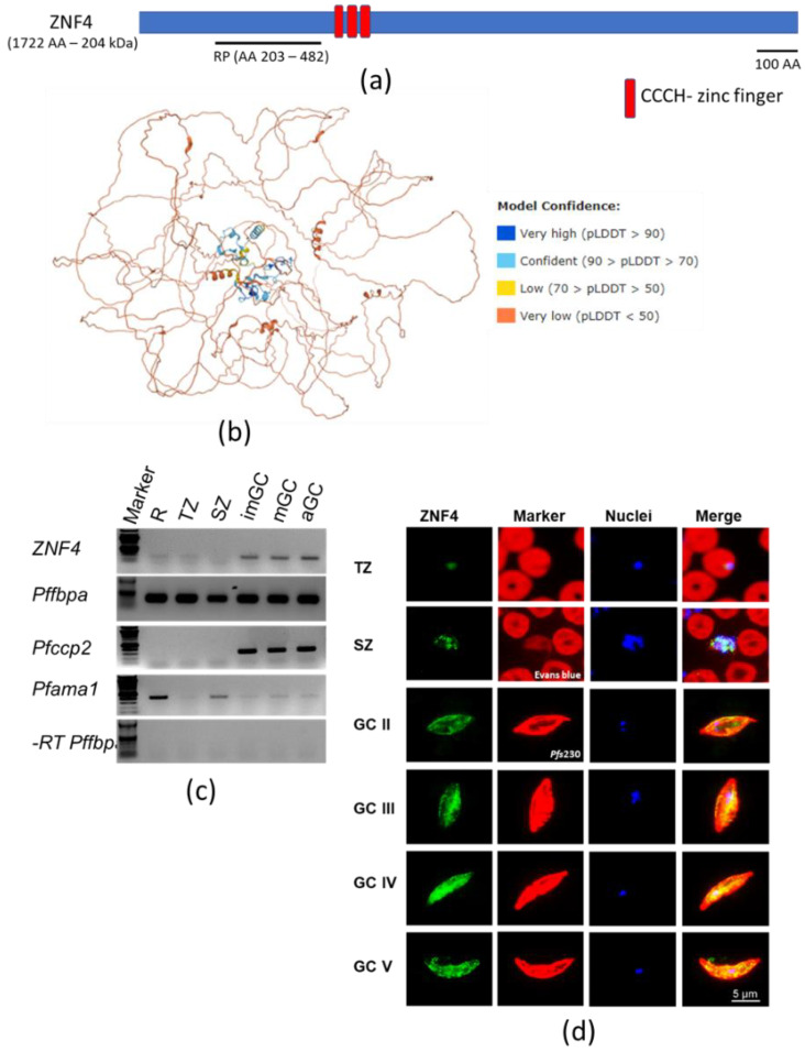
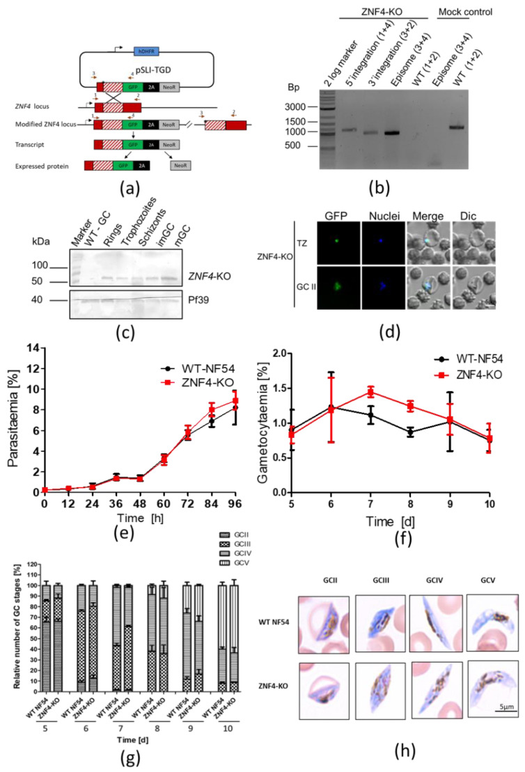
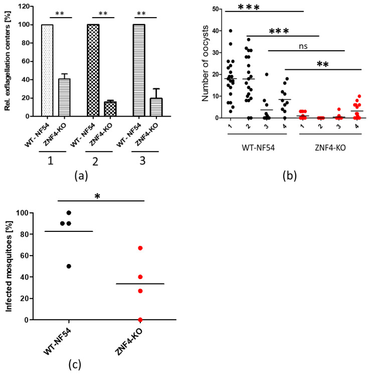
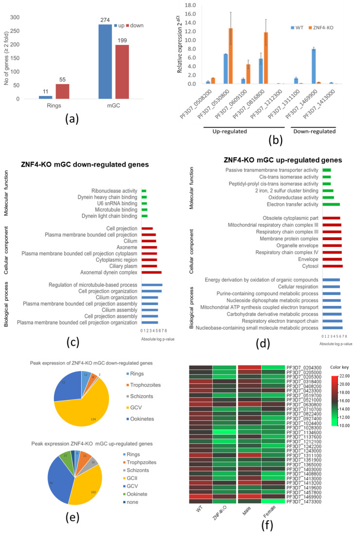
CCCH zinc finger proteins (ZFPs) function mainly as RNA-binding proteins (RBPs) and play a central role in the mRNA metabolism. Over twenty seven CCCH-ZFPs are encoded in the genome of the human malaria parasite Plasmodium falciparum, the causative agent of malaria tropica. However, little is known about their functions. In this study, we characterize one member of the PfCCCH-ZFP named ZNF4. We show that ZNF4 is highly expressed in mature gametocytes, where it predominantly localizes to the cytoplasm. Targeted gene disruption of ZNF4 showed no significant effect in asexual blood stage replication and gametocyte development while male gametocyte exflagellation was significantly impaired, leading to reduced malaria transmission in the mosquito. Comparative transcriptomics between wildtype (WT) and the ZNF4-deficient line (ZNF4-KO) demonstrated the deregulation of about 473 genes (274 upregulated and 199 downregulated) in mature gametocytes. Most of the downregulated genes show peak expression in mature gametocyte with male enriched genes associated to the axonemal dynein complex formation, and cell projection organization is highly affected, pointing to the phenotype in male gametocyte exflagellation. Upregulated genes are associated to ATP synthesis. Our combined data therefore indicate that ZNF4 is a CCCH zinc finger protein which plays an important role in male gametocyte exflagellation through the regulation of male gametocyte-enriched genes.
Overview
The claims and statistics of this paper haven’t been checked yet.
Run the check — free, no account needed. It reads the open-access text and checks the sources it cites.
Integrity signals
Registry
Cites
Statistics
Figures
Verification
Claims the paper makes
The claims and statistics of this paper haven’t been checked yet.
Run the check — free, no account needed. It reads the open-access text and checks the sources it cites.
Self-check
Percentages, sums and reported differences re-computed from the paper’s own numbers.
The self-check needs the paper’s full text.
Citations
Cites
Highly accurate protein structure prediction with AlphaFold
Jumper, J., Evans, R., Pritzel, A., Green, T., Figurnov, M., Ronneberger, O., Tunyasuvunakool, K., Bates, R., Žídek, A., Potapenko, A., et al. (2021) · Nature · doi:10.1038/s41586-021-03819-2 · Cited by 43,764
Genome sequence of the human malaria parasite Plasmodium falciparum
Gardner, M. J., Hall, N., Fung, E., White, O., Berriman, M., Hyman, R. W., Carlton, J. M., Pain, A., Nelson, K. E., Bowman, S., et al. (2002) · Nature · doi:10.1038/nature01097 · Cited by 4,407 · cited 2×
“falciparum 3D7 EPr1 [31]) with default parameters.”
A compendium of RNA-binding motifs for decoding gene regulation
Ray, D., Kazan, H., Cook, K. B., Weirauch, M. T., Najafabadi, H. S., Li, X., Gueroussov, S., Albu, M., Zheng, H., Yang, A., et al. (2013) · Nature · doi:10.1038/nature12311 · Cited by 1,480
Research works printed but not linked: 58
- Kuehn, 2010, The coming-out of malaria gametocytes, J. Biomed. Biotechnol., 2010, 976827, 10.1155/2010/976827 · DOI
- Bennink, 2016, The development of malaria parasites in the mosquito midgut, Cell. Microbiol., 18, 905, 10.1111/cmi.12604 · DOI
- Josling, 2015, Sexual development in Plasmodium parasites: Knowing when it’s time to commit, Nat. Rev. Microbiol., 13, 573, 10.1038/nrmicro3519 · DOI
- Kafsack, 2014, A transcriptional switch underlies commitment to sexual development in malaria parasites, Nature, 507, 248, 10.1038/nature12920 · DOI
- Sinha, 2014, A cascade of DNA-binding proteins for sexual commitment and development in Plasmodium, Nature, 507, 253, 10.1038/nature12970 · DOI
- Shang, 2021, A cascade of transcriptional repression determines sexual commitment and development in Plasmodium falciparum, Nucleic Acids Res., 49, 9264, 10.1093/nar/gkab683 · DOI
- Yuda, 2020, Female-specific gene regulation in malaria parasites by an AP2-family transcription factor, Mol. Microbiol., 113, 40, 10.1111/mmi.14334 · DOI
- Li, 2021, Plasmodium transcription repressor AP2-O3 regulates sex-specific identity of gene expression in female gametocytes, EMBO Rep., 22, e51660, 10.15252/embr.202051660 · DOI
- Kaneko, I., Iwanaga, S., Kato, T., Kobayashi, I., and Yuda, M. (2015). Genome-Wide Identification of the Target Genes of AP2-O, a Plasmodium AP2-Family Transcription Factor. PLoS Pathog., 11. · DOI
- Modrzynska, 2017, A Knockout Screen of ApiAP2 Genes Reveals Networks of Interacting Transcriptional Regulators Controlling the Plasmodium Life Cycle, Cell Host Microbe, 21, 11, 10.1016/j.chom.2016.12.003 · DOI
- Bennink, 2019, The molecular machinery of translational control in malaria parasites, Mol. Microbiol., 112, 1658, 10.1111/mmi.14388 · DOI
- Bennink, S., von Bohl, A., Ngwa, C.J., Henschel, L., Kuehn, A., Pilch, N., Weißbach, T., Rosinski, A.N., Scheuermayer, M., and Repnik, U. (2018). A seven-helix protein constitutes stress granules crucial for regulating translation during human-to-mosquito transmission of Plasmodium falciparum. PLoS Pathog., 14. · DOI
- Mair, G.R., Lasonder, E., Garver, L.S., Franke-Fayard, B.M.D., Carret, C.K., Wiegant, J.C.A.G., Dirks, R.W., Dimopoulos, G., Janse, C.J., and Waters, A.P. (2010). Universal Features of Post-Transcriptional Gene Regulation Are Critical for Plasmodium Zygote Development. PLoS Pathog., 6. · DOI
- Mair, 2006, Regulation of sexual development of Plasmodium by translational repression, Science, 313, 667, 10.1126/science.1125129 · DOI
- Miao, 2010, The Puf-family RNA-binding protein PfPuf2 regulates sexual development and sex differentiation in the malaria parasite Plasmodium falciparum, J. Cell Sci., 123, 1039, 10.1242/jcs.059824 · DOI
- Shrestha, 2016, The RNA-binding protein Puf1 functions in the maintenance of gametocytes in Plasmodium falciparum, J. Cell Sci., 129, 3144
- Ngwa, 2017, Transcriptional Profiling Defines Histone Acetylation as a Regulator of Gene Expression during Human-to-Mosquito Transmission of the Malaria Parasite Plasmodium falciparum, Front. Cell. Infect. Microbiol., 7, 320, 10.3389/fcimb.2017.00320 · DOI
- Ngwa, C.J., Kiesow, M.J., Orchard, L.M., Farrukh, A., Llinás, M., and Pradel, G. (2019). The g9a histone methyltransferase inhibitor BIX-01294 modulates gene expression during plasmodium falciparum gametocyte development and transmission. Int. J. Mol. Sci., 20. · DOI
- Ngwa, 2021, Zinc finger proteins of Plasmodium falciparum, Cell. Microbiol., 23, e13387, 10.1111/cmi.13387 · DOI
- Hajikhezri, Z., Darweesh, M., Akusjärvi, G., and Punga, T. (2020). Role of CCCH-Type Zinc Finger Proteins in Human Adenovirus Infections. Viruses, 12. · DOI
- Taylor, 1996, Mitogens stimulate the rapid nuclear to cytosolic translocation of tristetraprolin, a potential zinc-finger transcription factor, Mol. Endocrinol., 10, 140
- Pomeranz, 2010, The Arabidopsis tandem zinc finger protein AtTZF1 traffics between the nucleus and cytoplasmic foci and binds both DNA and RNA, Plant Physiol., 152, 151, 10.1104/pp.109.145656 · DOI
- Pomeranz, 2011, Putative molecular mechanisms underlying tandem CCCH zinc finger protein mediated plant growth, stress, and gene expression responses, Plant Signal. Behav., 6, 647, 10.4161/psb.6.5.15105 · DOI
- Ngwa, C.J., Scheuermayer, M., Mair, G.R., Kern, S., Brügl, T., Wirth, C.C., Aminake, M.N., Wiesner, J., Fischer, R., and Vilcinskas, A. (2013). Changes in the transcriptome of the malaria parasite Plasmodium falciparum during the initial phase of transmission from the human to the mosquito. BMC Genom., 14. · DOI
- Ifediba, 1981, Complete in vitro maturation of Plasmodium falciparum gametocytes, Nature, 294, 364, 10.1038/294364a0 · DOI
- Lambros, 1979, Synchronization of Plasmodium falciparum erythrocytic stages in culture, J. Parasitol., 65, 418, 10.2307/3280287 · DOI
- Birnbaum, 2017, A genetic system to study Plasmodium falciparum protein function, Nat. Methods, 14, 450, 10.1038/nmeth.4223 · DOI
- Wagle, P., Nikolić, M., and Frommolt, P. (2015). QuickNGS elevates Next-Generation Sequencing data analysis to a new level of automation. BMC Genom., 16. · DOI
- Dobin, 2013, STAR: Ultrafast universal RNA-seq aligner, Bioinformatics, 29, 15, 10.1093/bioinformatics/bts635 · DOI · Paper page
- Pertea, 2015, StringTie enables improved reconstruction of a transcriptome from RNA-seq reads, Nat. Biotechnol., 33, 290, 10.1038/nbt.3122 · DOI
- Liao, 2014, featureCounts: An efficient general purpose program for assigning sequence reads to genomic features, Bioinformatics, 30, 923, 10.1093/bioinformatics/btt656 · DOI · Paper page
- Love, 2014, Moderated estimation of fold change and dispersion for RNA-seq data with DESeq2, Genome Biol., 15, 1, 10.1186/s13059-014-0550-8 · DOI · Paper page
- Livak, 2001, Analysis of Relative Gene Expression Data Using Real-Time Quantitative PCR and the 2−ΔΔCT Method, Methods, 25, 402, 10.1006/meth.2001.1262 · DOI · Paper page
- Salanti, 2003, Selective upregulation of a single distinctly structured var gene in chondroitin sulphate A-adhering Plasmodium falciparum involved in pregnancy-associated malaria, Mol. Microbiol., 49, 179, 10.1046/j.1365-2958.2003.03570.x · DOI · Paper page
- Wang, 2010, Identification of a major rif transcript common to gametocytes and sporozoites of Plasmodium falciparum, Malar. J., 9, 147, 10.1186/1475-2875-9-147 · DOI
- Kariuki, 1998, Plasmodium falciparum: Purification of the various gametocyte developmental stages from in vitro-cultivated parasites, Am. J. Trop. Med. Hyg., 59, 505, 10.4269/ajtmh.1998.59.505 · DOI
- Fivelman, 2007, Improved synchronous production of Plasmodium falciparum gametocytes in vitro, Mol. Biochem. Parasitol., 154, 119, 10.1016/j.molbiopara.2007.04.008 · DOI
- Varadi, 2022, AlphaFold Protein Structure Database: Massively expanding the structural coverage of protein-sequence space with high-accuracy models, Nucleic Acids Res., 50, D439, 10.1093/nar/gkab1061 · DOI · Paper page
- Chen, 2020, TBtools: An Integrative Toolkit Developed for Interactive Analyses of Big Biological Data, Mol. Plant, 13, 1194, 10.1016/j.molp.2020.06.009 · DOI
- Grant, C.E., and Bailey, T.L. (2021). XSTREME: Comprehensive motif analysis of biological sequence datasets. bioRxiv. · DOI
- López-Barragán, M.J., Lemieux, J., Quiñones, M., Williamson, K.C., Molina-Cruz, A., Cui, K., Barillas-Mury, C., Zhao, K., and Su, X. (2011). Directional gene expression and antisense transcripts in sexual and asexual stages of Plasmodium falciparum. BMC Genom., 12. · DOI
- Lasonder, 2016, Integrated transcriptomic and proteomic analyses of P. falciparum gametocytes: Molecular insight into sex-specific processes and translational repression, Nucleic Acids Res., 44, 6087, 10.1093/nar/gkw536 · DOI
- Zhang, 2018, Uncovering the essential genome of the human malaria parasitePlasmodium falciparum by saturationmutagenesis, Science, 360, eaap7847, 10.1126/science.aap7847 · DOI
- Silvie, 2021, Pleiotropic Roles for the Plasmodium berghei RNA Binding Protein UIS12 in Transmission and Oocyst Maturation, Front. Cell. Infect. Microbiol., 11, 624945, 10.3389/fcimb.2021.624945 · DOI
- Aprea, I., Raidt, J., Höben, I.M., Loges, N.T., Nöthe-Menchen, T., Pennekamp, P., Olbrich, H., Kaiser, T., Biebach, L., and Tüttelmann, F. (2021). Defects in the Cytoplasmic Assembly of Axonemal Dynein Arms Cause Morphological Abnormalities and Dysmotility in Sperm Cells Leading to male Infertility. PLoS Genet., 17. · DOI
- Lindemann, 2010, Flagellar and ciliary beating: The proven and the possible, J. Cell Sci., 123, 519, 10.1242/jcs.051326 · DOI
- Stone, 2017, A Molecular Assay to Quantify Male and Female Plasmodium falciparum Gametocytes: Results From 2 Randomized Controlled Trials Using Primaquine for Gametocyte Clearance, J. Infect. Dis., 216, 457, 10.1093/infdis/jix237 · DOI
- Bradley, 2018, Predicting the likelihood and intensity of mosquito infection from sex specific plasmodium falciparum gametocyte density, Elife, 7, e34463, 10.7554/eLife.34463 · DOI
- Leslie, 2020, MNS1 variant associated with situs inversus and male infertility, Eur. J. Hum. Genet., 28, 50, 10.1038/s41431-019-0489-z · DOI
- Ta-Shma, A., Hjeij, R., Perles, Z., Dougherty, G.W., Abu Zahira, I., Letteboer, S.J.F., Antony, D., Darwish, A., Mans, D.A., and Spittler, S. (2018). Homozygous loss-of-function mutations in MNS1 cause laterality defects and likely male infertility. PLoS Genet., 14. · DOI
- Parkhurst, 1994, Sex determination and dosage compensation: Lessons from flies and worms, Science, 264, 924, 10.1126/science.8178152 · DOI
- Wang, 1994, The sex-lethal amino terminus mediates cooperative interactions in RNA binding and is essential for splicing regulation, Genes Dev., 8, 2072, 10.1101/gad.8.17.2072 · DOI
- Schmid, 2015, The Nuclear PolyA-Binding Protein Nab2p Is Essential for mRNA Production, Cell Rep., 12, 128, 10.1016/j.celrep.2015.06.008 · DOI
- Green, 2002, Nab2p is required for poly(A) RNA export in Saccharomyces cerevisiae and is regulated by arginine methylation via Hmt1p, J. Biol. Chem., 277, 7752, 10.1074/jbc.M110053200 · DOI
- Kini, 2014, Cytoplasmic poly(A) binding protein C4 serves a critical role in erythroid differentiation, Mol. Cell. Biol., 34, 1300, 10.1128/MCB.01683-13 · DOI
- Miao, J., Fan, Q., Parker, D., Li, X., Li, J., and Cui, L. (2013). Puf mediates translation repression of transmission-blocking vaccine candidates in malaria parasites. PLoS Pathog., 9. · DOI
- Hirai, M., Maeta, A., Mori, T., and Mita, T. (2021). Pb103 Regulates Zygote/Ookinete Development in Plasmodium berghei via Double Zinc Finger Domains. Pathogens, 10. · DOI
- Fu, 2017, RNA-binding proteins in immune regulation: A focus on CCCH zinc finger proteins, Nat. Rev. Immunol., 17, 130, 10.1038/nri.2016.129 · DOI
Metadata
- Authors
- Hanhsen, B., Farrukh, A., Pradel, G., & Ngwa, C. J.
- Affiliations
- RWTH Aachen University (DE)
- Corresponding author
- Ngwa, Che Julius
- Venue
- Cells
- Publisher
- MDPI AG
- Published
- 2022-05-17
- Volume
- 11
- Issue
- 10
- Pages
- 1666
- ISSN
- 2073-4409 (ISSN-L)
- PMC
- PMC9139750
- OpenAlex
- W4280578208
- Cited by
- 27
- References
- 3
- Citations by year
- 2026: 4 · 2025: 4 · 2024: 5 · 2023: 11 · 2022: 2
- FWCI
- 4.86
- Type
- journal article
- Language
- en
- Open access
- gold
- doi.org
- Licence
- Locations
- Cells (publishedVersion, cc-by)
- bioRxiv (Cold Spring Harbor Laboratory) (acceptedVersion, cc-by)
- PubMed Central (publishedVersion, cc-by)
- PubMed (publishedVersion)
- DOAJ (DOAJ: Directory of Open Access Journals) (submittedVersion, cc-by-sa)
- RWTH Publications (RWTH Aachen) (submittedVersion, other-oa)
- Funders
- the Deutsche Forschungsgemeinschaft (NG170/1-1, PR905/20-1, No 731060)
- the German Academic Exchange Service (DAAD) (NG170/1-1, PR905/20-1, No 731060)
- the European Union’s Horizon 2020 research and innovation program (NG170/1-1, PR905/20-1, No 731060)
- Topics
- Health Sciences › Medicine › Public Health, Environmental and Occupational Health › Malaria Research and Control
- Life Sciences › Immunology and Microbiology › Parasitology › Bird parasitology and diseases
- Life Sciences › Biochemistry, Genetics and Molecular Biology › Molecular Biology › RNA Research and Splicing
- Keywords
- Plasmodium falciparum, CCCH zinc finger proteins, male gametocytes, ATP production, mosquito-borne transmission, gene knockout, transcriptomics, RNA-binding proteins
- MeSH terms
- Animals, Cytoplasm, Malaria, Male, Culicidae, Plasmodium falciparum, Zinc Fingers
About these checks
Registry, retraction, Cites and statistics results are computed, not judged, with one exception: a printed reference that no index lists is read once by a model before it counts as a citation error. Claims are extracted by a model and kept only when their quote appears verbatim in the text; each cited source is then checked, with a model’s help, for whether it says what the claim relies on. Those verdicts can be wrong — read the claim’s quote and the cited source before relying on one.
Statistics and arithmetic are recomputed, not judged.
Confidence bands are shown only for fields calibrated on our annotated test set. None is calibrated yet, so no band is shown.
What counts as a claim ›How we measure confidence ›How we check this ›
Are you an author, or do you see a problem with this page? Request a review
