

The DOI matches its Crossref record; 42 of the 42 references it cites were found in a registry or an index.
Explainable Artificial Intelligence in Radiological Cardiovascular Imaging—A Systematic Review
Open accessAbstract
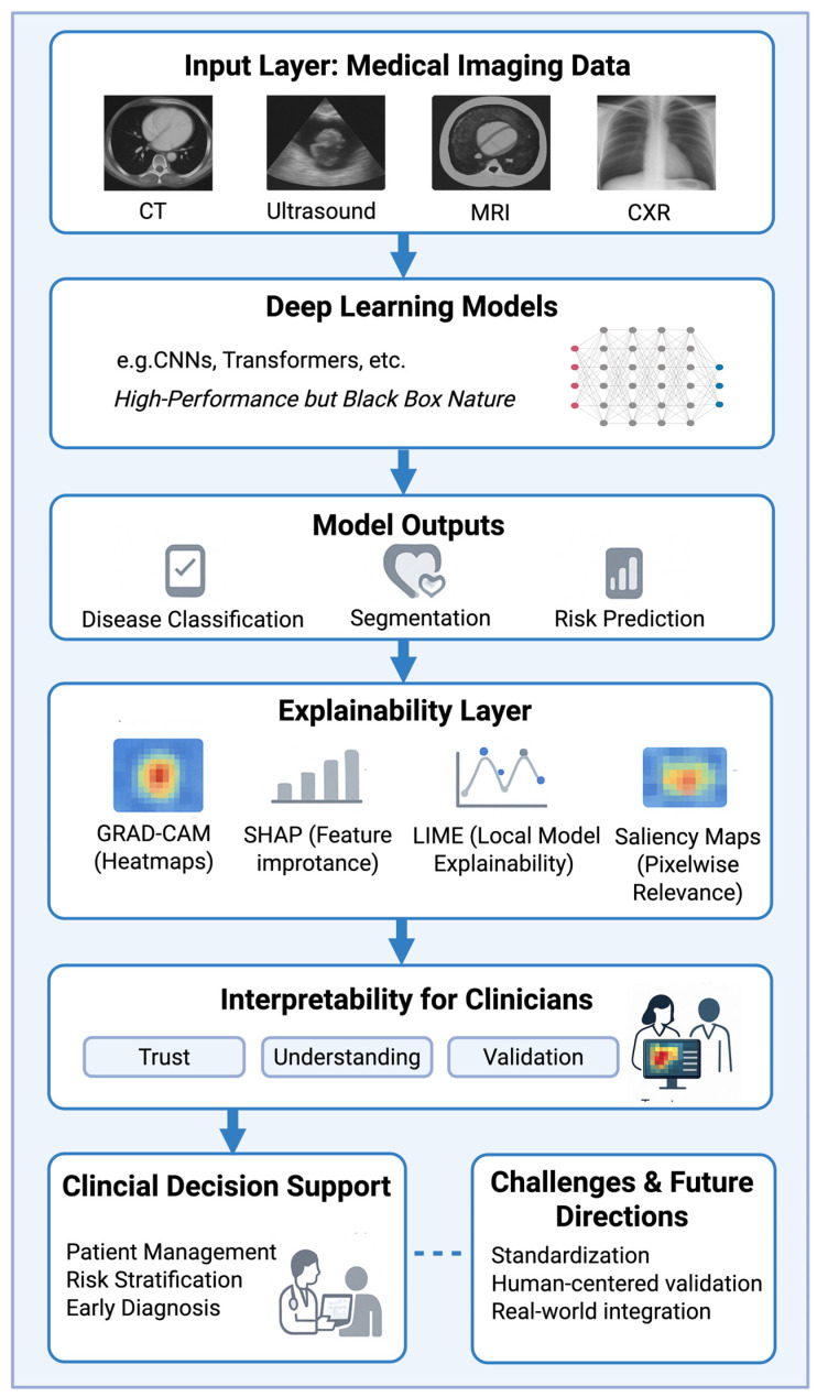
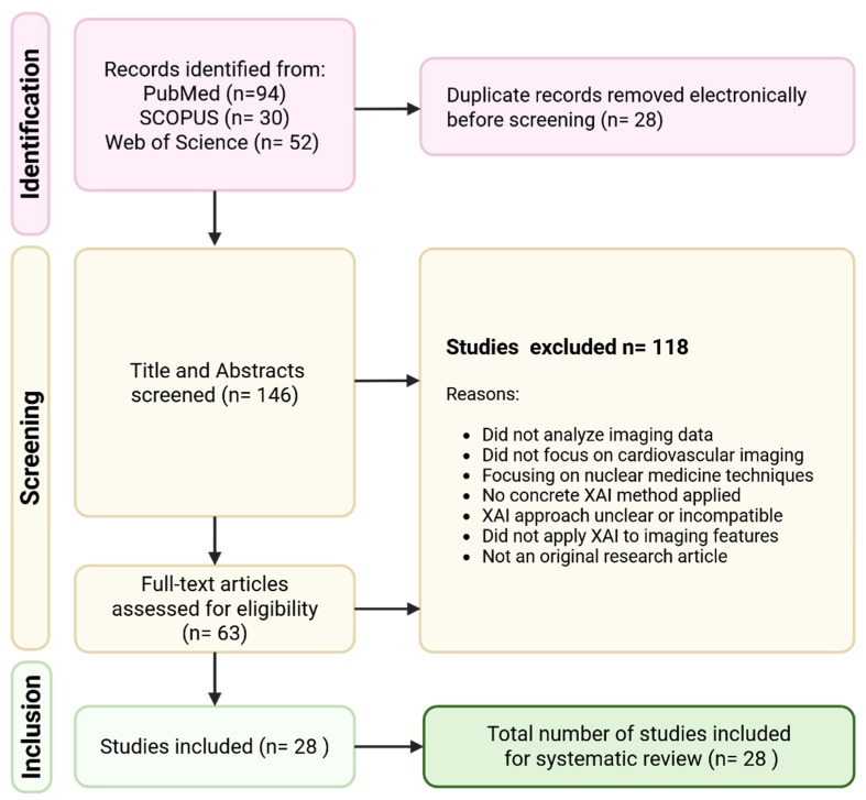
Background: Artificial intelligence (AI) and deep learning are increasingly applied in cardiovascular imaging. However, the “black box” nature of these models raises challenges for clinical trust and integration. Explainable Artificial Intelligence (XAI) seeks to address these concerns by providing insights into model decision-making. This systematic review synthesizes current research on the use of XAI methods in radiological cardiovascular imaging. Methods: A systematic literature search was conducted in PubMed, Scopus, and Web of Science to identify peer-reviewed original research articles published between January 2015 and March 2025. Studies were included if they applied XAI techniques—such as Gradient-Weighted Class Activation Mapping (Grad-CAM), Shapley Additive Explanations (SHAPs), Local Interpretable Model-Agnostic Explanations (LIMEs), or saliency maps—to cardiovascular imaging modalities, including cardiac computed tomography (CT), magnetic resonance imaging (MRI), echocardiography and other ultrasound examinations, and chest X-ray (CXR). Studies focusing on nuclear medicine, structured/tabular data without imaging, or lacking concrete explainability features were excluded. Screening and data extraction followed PRISMA guidelines. Results: A total of 28 studies met the inclusion criteria. Ultrasound examinations (n = 9) and CT (n = 9) were the most common imaging modalities, followed by MRI (n = 6) and chest X-rays (n = 4). Clinical applications included disease classification (e.g., coronary artery disease and valvular heart disease) and the detection of myocardial or congenital abnormalities. Grad-CAM was the most frequently employed XAI method, followed by SHAP. Most studies used saliency-based techniques to generate visual explanations of model predictions. Conclusions: XAI holds considerable promise for improving the transparency and clinical acceptance of deep learning models in cardiovascular imaging. However, the evaluation of XAI methods remains largely qualitative, and standardization is lacking. Future research should focus on the robust, quantitative assessment of explainability, prospective clinical validation, and the development of more advanced XAI techniques beyond saliency-based methods. Strengthening the interpretability of AI models will be crucial to ensuring their safe, ethical, and effective integration into cardiovascular care.
Overview
The claims and statistics of this paper haven’t been checked yet.
Free: it reads the open-access text and checks the first 5 claims that cite a source we can check. Create a free account to check every claim.
Integrity signals
Registry
Cites
Statistics
Figures
Verification
Claims the paper makes
The claims and statistics of this paper haven’t been checked yet.
Free: it reads the open-access text and checks the first 5 claims that cite a source we can check. Create a free account to check every claim.
Self-check
Percentages, sums and reported differences re-computed from the paper’s own numbers.
The self-check needs the paper’s full text.
Citations
Cites
Explainable AI: a review of machine learning interpretability methods
Linardatos, P., Papastefanopoulos, V., & Kotsiantis, S. (2020) · Entropy · doi:10.3390/e23010018 · Cited by 2,922 · cited 3×
“The general XAI literature underscores why this is the case: building inherently inter-pretable yet accurate models for high-dimensional image data is exceedingly difficult, so the community has gravitated toward complex models plus XAI as a compromise [5].”
InfluentialInterpreting Black-Box Models: A Review on Explainable Artificial Intelligence
Hassija, V., Chamola, V., Mahapatra, A., Singal, A., Goel, D., Huang, K., Scardapane, S., Spinelli, I., Mahmud, M., & Hussain, A. (2024) · Cognitive Computation · doi:10.1007/s12559-023-10179-8 · Cited by 2,495 · cited 3×
“The prevalence of such approaches is unsurprising—they offer intuitive, image-overlay explanations that align with radiologists’ visual reasoning and are readily supported by open-source tools, factors which have likely spurred their widespread adoption [8].”
InfluentialGlobal burden of cardiovascular diseases: projections from 2025 to 2050
Chong, B., Jayabaskaran, J., Jauhari, S. M., Chan, S. P., Goh, R., Kueh, M. T. W., Li, H., Chin, Y. H., Kong, G., Anand, V. V., et al. (2025) · European Journal of Preventive Cardiology · doi:10.1093/eurjpc/zwae281 · Cited by 793
Salahuddin, Z., Woodruff, H. C., Chatterjee, A., & Lambin, P. (2022) · Computers in Biology and Medicine · doi:10.1016/j.compbiomed.2021.105111 · Cited by 508
The role of explainable AI in the context of the AI Act
Panigutti, C., Hamon, R., Hupont, I., Llorca, D. F., Yela, D. F., Junklewitz, H., Scalzo, S., Mazzini, G., Sánchez, I., Garrido, J. S., et al. (2023) · ACM Conference on Fairness, Accountability, and Transparency (FAccT) · doi:10.1145/3593013.3594069 · Cited by 270
Muhammad, D., & Bendechache, M. (2024) · Computational and Structural Biotechnology Journal · doi:10.1016/j.csbj.2024.08.005 · Cited by 258
Borys, K., Schmitt, Y. A., Nauta, M., Seifert, C., Krämer, N., Friedrich, C. M., & Nensa, F. (2023) · European Journal of Radiology · doi:10.1016/j.ejrad.2023.110786 · Cited by 233 · cited 4×
“AI systems can assist in various imaging-based tasks, including disease classification, the segmentation of anatomical structures, and even image reconstruction [3].”
InfluentialMarey, A., Arjmand, P., Alerab, A. D. S., Eslami, M. J., Saad, A. M., Sanchez, N., & Umair, M. (2024) · Egyptian Journal of Radiology and Nuclear Medicine · doi:10.1186/s43055-024-01356-2 · Cited by 196
Wang, Y.-R., Yang, K., Wen, Y., Wang, P., Hu, Y., Lai, Y., Wang, Y., Zhao, K., Tang, S., Zhang, A., et al. (2024) · Nature Medicine · doi:10.1038/s41591-024-02971-2 · Cited by 188
Gala, D., Behl, H., Shah, M., & Makaryus, A. N. (2024) · Healthcare · doi:10.3390/healthcare12040481 · Cited by 183
Severe aortic stenosis detection by deep learning applied to echocardiography
Holste, G., Oikonomou, E. K., Mortazavi, B. J., Coppi, A. C., Faridi, K. F., Miller, E. J., Forrest, J. K., McNamara, R. L., Ohno-Machado, L., Yuan, N., et al. (2023) · European Heart Journal · doi:10.1093/eurheartj/ehad456 · Cited by 169
Hassan, S. U., Abdulkadir, S. J., Zahid, M. S. M., & Al-Selwi, S. M. (2024) · Computers in Biology and Medicine · doi:10.1016/j.compbiomed.2024.109569 · Cited by 138
A review of evaluation approaches for explainable AI with applications in cardiology
Salih, A. M., Galazzo, I. B., Gkontra, P., Rauseo, E., Lee, A. M., Lekadir, K., Radeva, P. I., Petersen, S. E., & Menegaz, G. (2024) · Artificial Intelligence Review · doi:10.1007/s10462-024-10852-w · Cited by 104
Ueda, D., Matsumoto, T., Ehara, S., Yamamoto, A., Walston, S. L., Ito, A., Shimono, T., Shiba, M., Takeshita, T., Fukuda, D., et al. (2023) · The Lancet Digital Health · doi:10.1016/s2589-7500(23)00107-3 · Cited by 84
Candemir, S., White, R. D., Demirer, M., Gupta, V., Bigelow, M. T., Prevedello, L. M. S., & Erdal, B. S. (2020) · Computerized Medical Imaging and Graphics · doi:10.1016/j.compmedimag.2020.101721 · Cited by 74
Sakai, A., Komatsu, M., Komatsu, R., Matsuoka, R., Yasutomi, S., Dozen, A., Shozu, K., Arakaki, T., Machino, H., Asada, K., et al. (2022) · Biomedicines · doi:10.3390/biomedicines10030551 · Cited by 68
Mouchès, P., Wilms, M., Rajashekar, D., Langner, S., & Forkert, N. D. (2022) · Human Brain Mapping · doi:10.1002/hbm.25805 · Cited by 67
Nurmaini, S., Partan, R. U., Bernolian, N., Sapitri, A. I., Tutuko, B., Rachmatullah, M. N., Darmawahyuni, A., Firdaus, F., & Mose, J. C. (2022) · Journal of Clinical Medicine · doi:10.3390/jcm11216454 · Cited by 61
McNamara, S. L., Yi, P. H., & Lotter, W. (2024) · npj Digital Medicine · doi:10.1038/s41746-024-01080-1 · Cited by 60
Chao, C., Jeong, J., Arsanjani, R., Kim, K. H., Tsai, Y., YU, W., Farina, J., Mahmoud, A., Ayoub, C., Grogan, M. A., et al. (2023) · JACC. Cardiovascular imaging · doi:10.1016/j.jcmg.2023.09.011 · Cited by 50
Mitigating Algorithmic Bias in AI-Driven Cardiovascular Imaging for Fairer Diagnostics
Sufian, M. A., Alsadder, L., Hamzi, W., Zaman, S., Sagar, A. S. M. S., & Hamzi, B. (2024) · Diagnostics · doi:10.3390/diagnostics14232675 · Cited by 49
Clinician's guide to trustworthy and responsible artificial intelligence in cardiovascular imaging
Szabó, L. E., Raisi‐Estabragh, Z., Salih, A. M., McCracken, C., Pujadas, E. R., Gkontra, P., Kiss, M., Maurovich‐Horvat, P., Vágó, H., Merkely, B. P., et al. (2022) · Frontiers in Cardiovascular Medicine · doi:10.3389/fcvm.2022.1016032 · Cited by 45
Wang, S., Wu, K., Chu, T., Fernandes, S. L., Zhou, Q., Zhang, Y., & Sun, J. (2021) · Wireless Communications and Mobile Computing · doi:10.1155/2021/5792975 · Cited by 45
Lee, H., Eun, Y., Hwang, J. Y., & Eun, L. Y. (2022) · Computer Methods and Programs in Biomedicine · doi:10.1016/j.cmpb.2022.106970 · Cited by 39
Deep learning to detect left ventricular structural abnormalities in chest X-rays
Bhave, S., Rodriguez, V., Poterucha, T., Mutasa, S., Aberle, D., Capaccione, K. M., Chen, Y., Dsouza, B., Dumeer, S., Goldstein, J., et al. (2024) · European Heart Journal · doi:10.1093/eurheartj/ehad782 · Cited by 38
Self-citationVafaeezadeh, M., Behnam, H., Hosseinsabet, A., & Gifani, P. (2021) · International Journal of Computer Assisted Radiology and Surgery · doi:10.1007/s11548-021-02542-7 · Cited by 31
Day, T. G., Matthew, J., Budd, S., Venturini, L., Wright, R., Farruggia, A., Vigneswaran, T. V., Zidere, V., Hajnal, J. V., Razavi, R., et al. (2024) · Ultrasound in Obstetrics and Gynecology · doi:10.1002/uog.27577 · Cited by 31
Deep Learning Based Prediction of Pulmonary Hypertension in Newborns Using Echocardiograms
Ragnarsdóttir, H., Özkan, E., Michel, H., Chin-Cheong, K., Manduchi, L., Wellmann, S. M., & Vogt, J. E. (2024) · International Journal of Computer Vision · doi:10.1007/s11263-024-01996-x · Cited by 27
Penso, M., Moccia, S., Caiani, E. G., Caredda, G., Lampus, M., Carerj, M. L., Babbaro, M., Pepi, M., Chiesa, M., & Pontone, G. (2022) · Computers in Biology and Medicine · doi:10.1016/j.compbiomed.2022.106484 · Cited by 25
Iacono, F. L., Maragna, R., Pontone, G., & Corino, V. (2023) · Frontiers in Radiology · doi:10.3389/fradi.2023.1193046 · Cited by 23
Gerbasi, A., Dagliati, A., Albi, G., Chiesa, M., Andreini, D., Baggiano, A., Mushtaq, S., Pontone, G., Bellazzi, R., & Colombo, G. I. (2023) · Computer Methods and Programs in Biomedicine · doi:10.1016/j.cmpb.2023.107989 · Cited by 23
Huo, Y., Terry, J. G., Wang, J., Nath, V., Bermudez, C., Bao, S., Parvathaneni, P., Carr, J. J., & Landman, B. A. (2019) · Medical Imaging 2019: Image Processing · doi:10.1117/12.2512541 · Cited by 22
Cai, L., Zhao, E., Niu, H., Liu, Y., Zhang, T., Liu, D., Zhang, Z., Li, J., Qiao, P., Lv, H., et al. (2023) · Computers in Biology and Medicine · doi:10.1016/j.compbiomed.2023.107264 · Cited by 22
Artificial intelligence-based detection of atrial fibrillation from chest radiographs
Matsumoto, T., Ehara, S., Walston, S. L., Mitsuyama, Y., Miki, Y., & Ueda, D. (2022) · European Radiology · doi:10.1007/s00330-022-08752-0 · Cited by 20
Paciorek, A. M., von Schacky, C. E., Foreman, S. C., Gassert, F. G., Gassert, F. T., Kirschke, J. S., Laugwitz, K., Geith, T., Hadamitzky, M., & Nadjiri, J. (2024) · BMC Medical Imaging · doi:10.1186/s12880-024-01217-4 · Cited by 20
Deep Learning for Detection of Exercise-Induced Pulmonary Hypertension Using Chest X-Ray Images
Kusunose, K., Hirata, Y., Yamaguchi, N., et al. (2022) · Frontiers in Cardiovascular Medicine · doi:10.3389/fcvm.2022.891703 · Cited by 18
Lo, S., & Yin, Y. (2021) · Discover Artificial Intelligence · doi:10.1007/s44163-021-00015-z · Cited by 15
Cardiac Segmentation Method Based on Domain Knowledge
Wang, Y., Chen, W., Tang, T., Xie, W., Jiang, Y., Zhang, H., Zhou, X., & Yuan, K. (2022) · Ultrasonic Imaging · doi:10.1177/01617346221099435 · Cited by 12
Zhang, Z., Dai, Y., Peng, X., et al. (2025) · Scientific Reports · doi:10.1038/s41598-025-87828-5 · Cited by 7
Fu, J., Fang, M., Lin, Z., Qiu, J., Yang, M., Tian, J., Dong, D., & Zou, Y. (2024) · Visual Computing for Industry Biomedicine and Art · doi:10.1186/s42492-023-00152-5 · Cited by 5
Lopes, R. R., van den Boogert, T. P. W., Lobé, N. H. J., et al. (2022) · European Radiology · doi:10.1007/s00330-022-08901-5 · Cited by 3
Sakai, Y., Kim, J., Phi, H. Q., Hu, A. C., Balali, P., Guggenberger, K. V., Woo, J. H., Bos, D., Kasner, S. E., Cucchiara, B. L., et al. (2024) · medRxiv · doi:10.1101/2024.10.25.24316081 · Cited by 3
Cited by
Abbas, Q., Jeong, W., & Lee, S. W. (2025) · Healthcare · doi:10.3390/healthcare13172154 · Cited by 236
“For example, in diagnostic imaging, XAI can highlight specific regions of interest on radiographs or MRIs that contribute to a diagnosis, allowing radiologists to verify and validate the model’s conclusions [22,23].”
Metadata
- Authors
- Haupt, M., Maurer, M. H., & Thomas, R. P.
- Affiliations
- Carl von Ossietzky Universität Oldenburg (DE)
- Corresponding author
- Haupt, Matteo
- Venue
- Diagnostics
- Publisher
- MDPI AG
- Published
- 2025-05-31
- Volume
- 15
- Issue
- 11
- Pages
- 1399
- ISSN
- 2075-4418 (ISSN-L)
- PMC
- PMC12155260
- OpenAlex
- W4410970602
- Semantic Scholar
- f2875a6425b0c0db7a41f7efd17d773aef16e9f2
- Cited by
- 58
- References
- 42
- Citations by year
- 2026: 46 · 2025: 12
- FWCI
- 5.57
- Type
- journal article
- Language
- en
- Open access
- gold
- doi.org
- Licence
- Locations
- Diagnostics (publishedVersion, cc-by)
- PubMed Central (publishedVersion, cc-by)
- PubMed (publishedVersion)
- DOAJ (DOAJ: Directory of Open Access Journals) (submittedVersion, cc-by-sa)
- Topics
- Physical Sciences › Computer Science › Artificial Intelligence › Explainable Artificial Intelligence (XAI)
- Health Sciences › Medicine › Health Informatics › Artificial Intelligence in Healthcare and Education
- Health Sciences › Medicine › Radiology, Nuclear Medicine and Imaging › COVID-19 diagnosis using AI
- Keywords
- explainable AI, cardiovascular imaging, Grad-CAM, SHAP, lime, saliency maps, cardiac CT, cardiac magnetic resonance imaging, echocardiography, chest radiography, coronary artery disease, valvular heart disease, congenital heart disease, myocardial abnormalities, model interpretability, clinical trust in AI
About these checks
Registry, retraction, Cites and statistics results are computed, not judged, with one exception: a printed reference that no index lists is read once by a model before it counts as a citation error. Claims are extracted by a model and kept only when their quote appears verbatim in the text; each cited source is then checked, with a model’s help, for whether it says what the claim relies on. Those verdicts can be wrong — read the claim’s quote and the cited source before relying on one.
Statistics and arithmetic are recomputed, not judged.
Confidence bands are shown only for fields calibrated on our annotated test set. None is calibrated yet, so no band is shown.
What counts as a claim ›How we measure confidence ›How we check this ›
Are you an author, or do you see a problem with this page? Request a review
Data sources
Bibliographic data: Semantic Scholar, OpenAlex, Crossref.
Contains information from the Semantic Scholar Academic Graph Datasets, made available under the ODC Attribution License (ODC-BY 1.0).
Semantic Scholar’s data is described in: Kinney, R., et al. (2023). The Semantic Scholar Open Data Platform. arXiv:2301.10140.
