

The DOI matches its Crossref record; 1 of the 1 references it cites were found in a registry or an index.
Anti-Inflammation and Protective Effects of Anethum graveolens L. (Dill Seeds) on Esophageal Mucosa Damages in Reflux Esophagitis-Induced Rats
Abstract
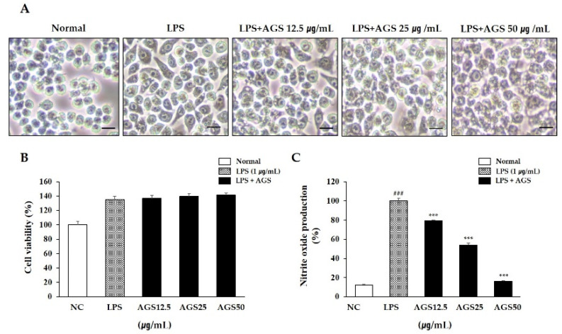
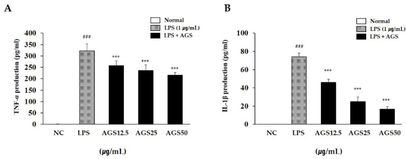
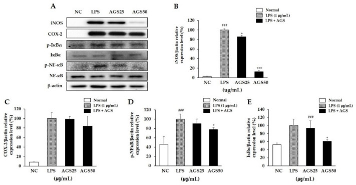
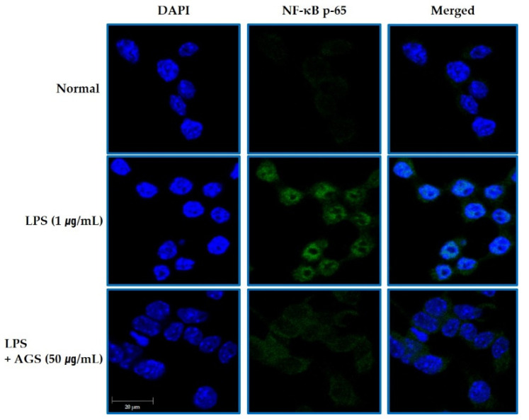
Anethum graveolens L. (dill seeds) are important medicinal and functional foods in Europe and central and south Asia, often used as a seasoning in daily diets. Anethum graveolens L. seeds (AGS) are used to treat indigestion and have shown physiological activities such as those against hypoglycemia and gastroesophageal disease. This study explored the protective effects of AGS extract on mucosal damages and inflammation in reflux esophagitis rats. AGS inhibited cellular inflammation including NO production and the expression of inflammatory proteins (iNOS and COX2 etc.), cytokines (IL-1β and TNF-α) and nuclear transfer factor related to NF-κB signaling caused by LPS stimulation in vitro. Furthermore, reflux esophagitis-induced rats were used to observe the anti-inflammatory effect of AGS. Tissue staining and inflammation-related protein expression of rats with acute reflux esophagitis indicated that AGS improved this inflammatory response, such as COX-2 and TNF-α in mucosa. In conclusion, AGS have good physiological activity and the possibility of being used as a medicinal food and a functional resource for the prevention and therapy of gastroesophageal diseases.
Overview
The claims and statistics of this paper haven’t been checked yet.
Run the check — free, no account needed. It reads the open-access text and checks the sources it cites.
Integrity signals
Registry
Cites
Statistics
Figures
Verification
Claims the paper makes
The claims and statistics of this paper haven’t been checked yet.
Run the check — free, no account needed. It reads the open-access text and checks the sources it cites.
Claims other papers make about this one
Each is checked against this paper’s own text: does it say what the citing paper says it does?
In the citing paper
“At the scientific level, numerous previous studies have highlighted the biological potential of Anethum graveolens, such as anti-inflammatory [10], antidiabetic [11], antimicrobial [12], diuretic [13], insecticidal [14], hepatoprotective [15], anticancer, and antioxidant [16].”
In the citing paper
“It contains a variety of natural products, like EO, flavonoids, phenolic and fatty acids, cardiac glycosides, saponins and tannins [2,3,4].”
From Maceration, Soxhlet Extraction, and Steam Hydrodistillation of Anethum graveolens Seeds: Phytochemical Profiles and Cytotoxic Effects (2026) · cites this paper as [3]
Self-check
Percentages, sums and reported differences re-computed from the paper’s own numbers.
The self-check needs the paper’s full text.
Citations
Cites
Cancer-related inflammation and treatment effectiveness
Diakos, C. I., Charles, K. A., McMillan, D. C., & Clarke, S. J. (2014) · The Lancet Oncology · doi:10.1016/s1470-2045(14)70263-3 · Cited by 2,193
Research works printed but not linked: 43
- Mohammed, 2018, Anethum graveolens (dill)—A medicinal herb induces apoptosis and cell cycle arrest in HepG2 cell line, J. Ethnopharmacol., 219, 15, 10.1016/j.jep.2018.03.008 · DOI · Paper page
- Talebi, 2020, Effect of dill (Anethum graveolens Linn) seed on the duration of labor: A systematic review, Complement Ther. Clin. Pract., 41, 101251, 10.1016/j.ctcp.2020.101251 · DOI
- Badar, 2008, Characteristics of Anethum graveolens (umbelliferae) seed oil: Extraction, composition and antimicrobial activity, Int. J. Agri. Biol., 10, 329
- Li, 2018, The antioxidation of different fractions of Dill (Anethum graveolens) and their influences on cytokines in macrophages RAW264.7, J. Oleo. Sci., 67, 1535, 10.5650/jos.ess18134 · DOI
- Chen, 2013, Antifungal mechanism of essential oil from Anethum graveolens seeds against Candida albicans, J. Med. Microbiol., 62, 1175, 10.1099/jmm.0.055467-0 · DOI
- Hosseinzadeh, H., Karimi, G.R., and Ameri, M. (2002). Effects of Anethum graveolens L. seed extracts on experimental gastric irritation models in mice. BMC Pharmacol., 2. · DOI · Paper page
- Eshwar, 2016, Comparison of Dill seed oil mouth rinse and chlorhexidine mouth rinse on plaque levels and gingivitis—A double blind randomized clinical trial, Open Dent. J., 10, 207, 10.2174/1874210601610010207 · DOI
- Kaur, 2019, Antioxidant activity of Anethum graveolens L. essential oil constituents and their chemical analogues, J. Food Biochem., 43, e12782, 10.1111/jfbc.12782 · DOI · Paper page
- Velioglu, 1998, Antioxidant activity and total phenolics in selected fruits, vegetables, and grain products, J. Agric. Food Chem., 46, 4113, 10.1021/jf9801973 · DOI
- Zeng, H., Tian, J., Zheng, Y., Ban, X., Zeng, J., Mao, Y., and Wang, Y. (2011). In vitro and in vivo activities of essential oil from the seed of Anethum graveolens L. against Candida spp.. Evid. Base Complement Alternat. Med., 659704. · DOI
- Naseri, 2012, The study of anti-inflammatory activity of oil-based Dill (Anethum graveolens L.) extract used topically in formalin-induced inflammation male rat paw, Iran J. Pharm. Res., 11, 1169
- Schepp, 1988, Modulation of rat gastric mucosal prostaglandin E2 release by dietary linoleic acid: Effects on gastric acid secretion and stress-induced mucosal damage, Gastroenterology, 95, 18, 10.1016/0016-5085(88)90285-5 · DOI
- Shimoyama, 2013, Antiulcerogenic activity of chlorogenic acid in different models of gastric ulcer, Naunyn. Schmiedebergs. Arch. Pharmacol., 386, 5, 10.1007/s00210-012-0807-2 · DOI
- Vieira, 2019, Gastroprotective effect of limonene in rats: Influence on oxidative stress, inflammation and gene expression, Phytomedicine, 53, 37, 10.1016/j.phymed.2018.09.027 · DOI
- Goodarzi, 2016, The role of Anethum graveolens L. (Dill) in the management of diabetes, J. Trop. Med., 2016, 1098916, 10.1155/2016/1098916 · DOI · Paper page
- Fullerton, 2016, Resolution of inflammation: A new therapeutic frontier, Nat. Rev. Drug Discov., 15, 551, 10.1038/nrd.2016.39 · DOI · Paper page
- Hu, 2019, Anti-inflammation action of xanthones from Swertia chirayita by regulating COX-2/NF-κB/MAPKs/Akt signaling pathways in RAW 264.7 macrophage cells, Phytomedicine, 55, 214, 10.1016/j.phymed.2018.08.001 · DOI
- Kellerman, 2017, Gastroesophageal Reflux Disease, Prim. Care, 44, 561, 10.1016/j.pop.2017.07.001 · DOI
- Negrutiu, 2019, Food and gastroesophageal reflux disease, Curr. Med. Chem., 26, 3497, 10.2174/0929867324666170515123807 · DOI
- Mikami, 2015, Physiology and pathogenesis of gastroesophageal reflux disease, Surg. Clin. N. Am., 95, 515, 10.1016/j.suc.2015.02.006 · DOI
- Chen, 2019, Gastroesophageal reflux disease pathophysiology, diagnosis, and treatment, Gastroenterol. Nurs., 42, 20, 10.1097/SGA.0000000000000359 · DOI
- 2015, NSAIDs: Learning new tricks from old drugs, Eur. J. Immunol., 45, 679, 10.1002/eji.201445222 · DOI
- Haley, 2019, Localized and targeted delivery of NSAIDs for treatment of inflammation: A review, Exp. Biol. Med., 244, 433, 10.1177/1535370218787770 · DOI
- Tsai, 2013, Therapeutic potential of traditional chinese medicine on inflammatory diseases, J. Tradit. Complement Med., 3, 142, 10.4103/2225-4110.114898 · DOI
- Kim, 2019, Determination of fatty acids in the seeds of Lepidium apetalum Willdenow, Descurainia sophia (L.) Webb ex Prantl, and Draba nemorosa L. by ultra-high-performance liquid chromatography equipped with a charged aerosol detector, J. Liq. Chromatogr. Relat. Technol., 42, 128, 10.1080/10826076.2019.1571509 · DOI
- Sandhu, 2018, Current Trends in the Management of Gastroesophageal Reflux Disease, Gut Liver, 15, 7, 10.5009/gnl16615 · DOI
- Baskaran, A., Chua, K.H., Sabaratnam, V., Ram, M.R., and Kuppusamy, U.R. (2017). Pleurotus giganteus (Berk. Karun & Hyde), the giant oyster mushroom inhibits NO production in LPS/H2O2 stimulated RAW 264.7 cells via STAT 3 and COX-2 pathways. BMC Complement Altern. Med., 17. · DOI
- Wei, 2020, A highly efficient hybrid peptide ameliorates intestinal inflammation and mucosal barrier damage by neutralizing lipopolysaccharides and antagonizing the lipopolysaccharide-receptor interaction, FASEB J., 34, 16049, 10.1096/fj.201903263RRR · DOI
- Sun, 2019, Inhibition of dextran sodium sulfate-induced colitis in mice by baker’s yeast polysaccharides, Carbohydr. Polym., 207, 371, 10.1016/j.carbpol.2018.11.087 · DOI
- Hwang, 2020, Anti-Inflammatory effect of immature sword bean pod (Canavalia gladiata) in lipopolysaccharide-induced RAW264.7 Cells, J. Med. Food, 23, 1183, 10.1089/jmf.2020.4733 · DOI
- Li, 2018, Ulinastatin attenuates LPS-induced inflammation in mouse macrophage RAW264.7 cells by inhibiting the JNK/NF-κB signaling pathway and activating the PI3K/Akt/Nrf2 pathway, Acta. Pharmacol. Sin., 39, 1294, 10.1038/aps.2017.143 · DOI
- Yang, C., You, L., Yin, X., Liu, Y., Leng, X., Wang, W., Sai, N., and Ni, J. (2018). Heterophyllin B ameliorates lipopolysaccharide-induced inflammation and oxidative stress in RAW 264.7 macrophages by suppressing the PI3K/Akt pathways. Molecules, 23. · DOI
- Giridharan, 2018, Mechanisms of NF-κB p65 and strategies for therapeutic manipulation, J. Inflamm. Res., 11, 407, 10.2147/JIR.S140188 · DOI · Paper page
- Xu, 2021, Astragalus polysaccharides attenuate ovalbumin-induced allergic rhinitis in rats by inhibiting NLRP3 inflammasome activation and NOD2-mediated NF- κ B activation, J. Med. Food, 24, 1, 10.1089/jmf.2020.4750 · DOI
- Kawai, 2007, Signaling to NF-κB by Toll-like receptors, Trends Mol. Med., 13, 460, 10.1016/j.molmed.2007.09.002 · DOI
- Souza, 2009, Gastroesophageal reflux might cause esophagitis through a cytokine-mediated mechanism rather than caustic acid injury, Gastroenterology, 137, 1776, 10.1053/j.gastro.2009.07.055 · DOI
- Huo, 2017, Hypoxia-inducible factor-2alpha plays a role in mediating oesophagitis in GORD, Gut, 66, 1542, 10.1136/gutjnl-2016-312595 · DOI
- Jabri, 2016, Ameliorative and antioxidant effects of myrtle berry seed (Myrtus communis) extract during reflux-induced esophagitis in rats, Pharm. Biol., 54, 1575, 10.3109/13880209.2015.1107748 · DOI
- Che, 2017, Macrophages induce EMT to promote invasion of lung cancer cells through the IL-6-mediated COX-2/PGE 2/β-catenin signaling pathway, Mol. Immunol., 90, 197, 10.1016/j.molimm.2017.06.018 · DOI
- Zhang, 2020, Menthol relieves acid reflux inflammation by regulating TRPV1 in esophageal epithelial cells, Biochem. Biophys. Res. Commun., 525, 113, 10.1016/j.bbrc.2020.02.050 · DOI
- Akshay, 2020, Chlorogenic Acid Potentiates the Anti-Inflammatory Activity of Curcumin in LPS-Stimulated THP-1 Cells, Nutrients, 12, 2706, 10.3390/nu12092706 · DOI
- Dipasquale, 2018, Anti-inflammatory effects of conjugated linoleic acid isomers and essential fatty acids in bovine mammary epithelial cells, Animal, 12, 2108, 10.1017/S1751731117003676 · DOI
- Saiki, 2017, The anti-inflammatory effect of Agaricus brasiliensis is partly due to its linoleic acid content, Food Funct., 8, 4150, 10.1039/C7FO01172E · DOI
Cited by
Hadi, N., Drioiche, A., Bouchra, E. M., Baammi, S., Abdelaziz Shahat, A., Tagnaout, I., Radi, M., Remok, F., Bouzoubaa, A., & Zair, T. (2024) · Pharmaceuticals · doi:10.3390/ph17070862 · Cited by 30
Goldiș, C., Racoviceanu, R., Negrea-Ghiulai, R., Mioc, A., Prodea, A., Atyim, E., Maksimovic, T., Lukinich-Gruia, A. T., Pricop, M.-A., & Șoica, C. (2026) · Molecules · doi:10.3390/molecules31183337
Metadata
- Authors
- Nam, H.-H., Nan, L., & Choo, B.-K.
- Venue
- Foods
- Publisher
- MDPI AG
- Published
- 2021-10-18
- Volume
- 10
- Issue
- 10
- Pages
- 2500
- ISSN
- 2304-8158
- PMC
- PMC8535990
- Cited by
- 22
- References
- 1
- Language
- en
- Funders
- Rural Development Administration (PJ0142272020)
- Jeonbuk National University (2020)
About these checks
Registry, retraction, Cites and statistics results are computed, not judged, with one exception: a printed reference that no index lists is read once by a model before it counts as a citation error. Claims are extracted by a model and kept only when their quote appears verbatim in the text; each cited source is then checked, with a model’s help, for whether it says what the claim relies on. Those verdicts can be wrong — read the claim’s quote and the cited source before relying on one.
Statistics and arithmetic are recomputed, not judged.
Confidence bands are shown only for fields calibrated on our annotated test set. None is calibrated yet, so no band is shown.
What counts as a claim ›How we measure confidence ›How we check this ›
Are you an author, or do you see a problem with this page? Request a review
Data sources
Bibliographic data: Semantic Scholar, OpenAlex, Crossref.
Contains information from the Semantic Scholar Academic Graph Datasets, made available under the ODC Attribution License (ODC-BY 1.0).
Semantic Scholar’s data is described in: Kinney, R., et al. (2023). The Semantic Scholar Open Data Platform. arXiv:2301.10140.
