

The DOI matches its Crossref record; 4 of the 4 references it cites were found in a registry or an index.
Suppression Effect of Astaxanthin on Osteoclast Formation In Vitro and Bone Loss In Vivo
Abstract
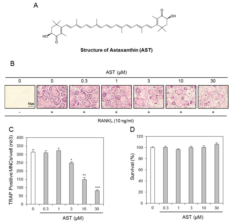
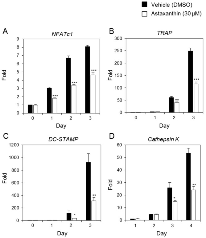
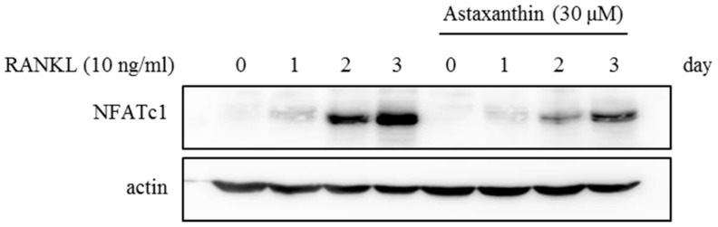
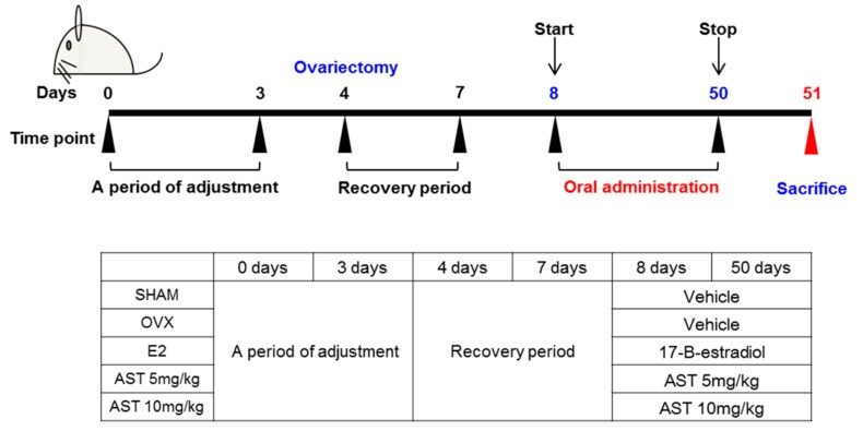
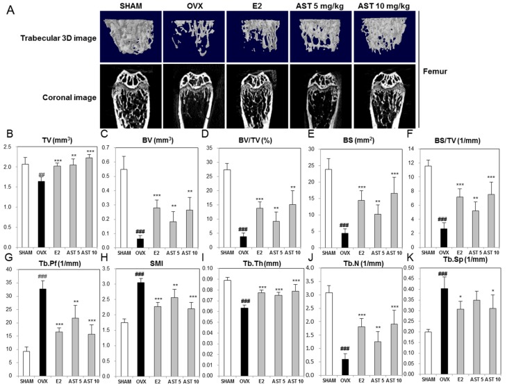
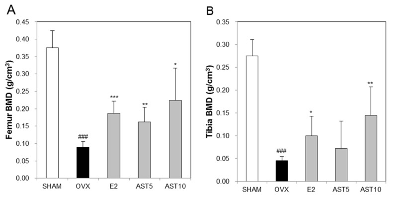
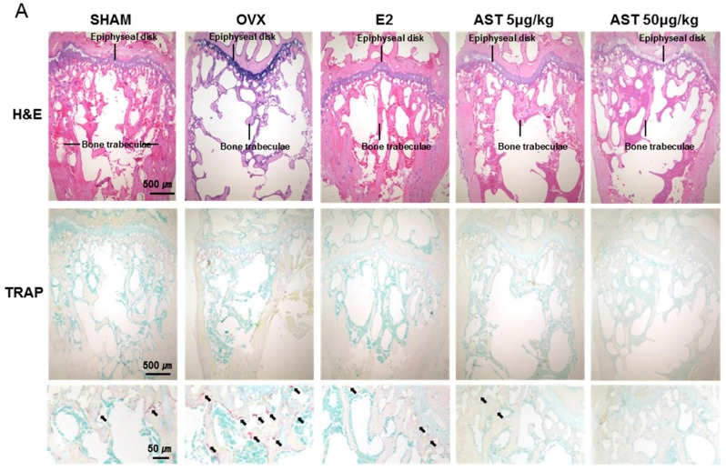
Osteoporosis is characterized by a reduction of the bone mineral density (BMD) and microarchitectural deterioration of the bone, which lead to bone fragility and susceptibility to fracture. Astaxanthin (AST) has a variety of biological activities, such as a protective effect against asthma or neuroinflammation, antioxidant effect, and decrease of the osteoclast number in the right mandibles in the periodontitis model. Although treatment with AST is known to have an effect on inflammation, no studies on the effect of AST exposure on bone loss have been performed. Thus, in the present study, we examined the antiosteoporotic effect of AST on bone mass in ovariectomized (OVX) mice and its possible mechanism of action. The administration of AST (5, 10 mg/kg) for 6 weeks suppressed the enhancement of serum calcium, inorganic phosphorus, alkaline phosphatase, total cholesterol, and tartrate-resistant acid phosphatase (TRAP) activity. The bone mineral density (BMD) and bone microarchitecture of the trabecular bone in the tibia and femur were recovered by AST exposure. Moreover, in the in vitro experiment, we demonstrated that AST inhibits osteoclast formation through the expression of the nuclear factor of activated T cells (NFAT) c1, dendritic cell-specific transmembrane protein (DC-STAMP), TRAP, and cathepsin K without any cytotoxic effects on bone marrow-derived macrophages (BMMs). Therefore, we suggest that AST may have therapeutic potential for the treatment of postmenopausal osteoporosis.
Overview
The claims and statistics of this paper haven’t been checked yet.
Run the check — free, no account needed. It reads the open-access text and checks the sources it cites.
Integrity signals
Registry
Cites
Statistics
Figures
Verification
Claims the paper makes
The claims and statistics of this paper haven’t been checked yet.
Run the check — free, no account needed. It reads the open-access text and checks the sources it cites.
Claims other papers make about this one
Each is checked against this paper’s own text: does it say what the citing paper says it does?
In the citing paper
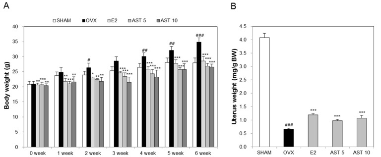
“During the progression of osteoporosis induced by OVX surgery, uterine weight notably decreases while body weight increases [36].”
From Anti-Osteoporotic Potential of Water Extract of Anethum graveolens L. Seeds (2023) · cites this paper as [36]
Self-check
Percentages, sums and reported differences re-computed from the paper’s own numbers.
The self-check needs the paper’s full text.
Citations
Cites
Takayanagi, H., Kim, S., Koga, T., Nishina, H., Isshiki, M., Yoshida, H., Saiura, A., Isobe, M., Yokochi, T., Inoue, J.-I., et al. (2002) · Developmental Cell · doi:10.1016/s1534-5807(02)00369-6 · Cited by 2,701
Self-citationEstrogen deficiency and bone loss: an inflammatory tale
Weitzmann, M. N. (2006) · Journal of Clinical Investigation · doi:10.1172/jci28550 · Cited by 990 · cited 11×
“The BMCs were cultured with M-CSF (10 ng/mL) for 1 day, and the nonadherent cells were cultured for a further 3 days in the presence of M-CSF (30 ng/mL).”
InfluentialThe role of NFAT in osteoclast formation
TAKAYANAGI, H. (2007) · Annals of the New York Academy of Sciences · doi:10.1196/annals.1402.071 · Cited by 442
GALLAGHER, J. C., RIGGS, B. L., & DELUCA, H. F. (1980) · The Journal of Clinical Endocrinology & Metabolism · doi:10.1210/jcem-51-6-1359 · Cited by 428 · cited 1×
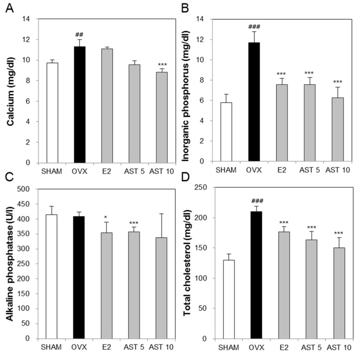
“Previous research showed that estrogen treatment increases calcium absorption in postmenopausal osteoporosis [42].”
Background
Research works printed but not linked: 54
- Kanis, 1994, Assessment of fracture risk and its application to screening for postmenopausal osteoporosis: Synopsis of a WHO report, Osteoporos. Int., 4, 368, 10.1007/BF01622200 · DOI
- Weycker (2016). D.; Li, X.; Barron, R.; Bornheimer, R.; Chandler, D. Hospitalizations for osteoporosis-related fractures: Economic costs and clinical outcomes. Bone Rep., 5, 186–191. · DOI
- Fitzpatrick, 2006, Estrogen therapy for postmenopausal osteoporosis, Arq. Bras. Endocrinol. Metab., 50, 705, 10.1590/S0004-27302006000400016 · DOI
- Zhao, 2012, Immune regulation of osteoclast function in postmenopausal osteoporosis: A critical interdisciplinary perspective, Int. J. Med. Sci., 9, 825, 10.7150/ijms.5180 · DOI
- Kim, 2015, Decursin inhibits osteoclastogenesis by downregulating NFATc1 and blocking fusion of pre-osteoclasts, Bone, 81, 208, 10.1016/j.bone.2015.07.023 · DOI
- Rossouw, 2002, Writing Group for the Women’s Health Initiative Investigators. Risks and benefits of estrogen plus progestin in healthy postmenopausal women: Principal results From the Women’s Health Initiative randomized controlled trial, JAMA, 288, 321, 10.1001/jama.288.3.321 · DOI · Paper page
- Lobo, 1995, Benefits and risks of estrogen replacement therapy, Am. J. Obstet. Gynecol., 173, 982, 10.1016/0002-9378(95)90247-3 · DOI
- An, 2016, Natural products for treatment of osteoporosis: The effects and mechanisms on promoting osteoblast-mediated bone formation, Life Sci., 147, 46, 10.1016/j.lfs.2016.01.024 · DOI
- Yamaguchi, 2017, Combination of alendronate and genistein synergistically suppresses osteoclastic differentiation of RAW267.4 cells in vitro, Exp. Ther. Med., 14, 1769, 10.3892/etm.2017.4695 · DOI
- Ishimi, 1999, Selective effects of genistein, a soybean isoflavone, on B-lymphopoiesis and bone loss caused by estrogen deficiency, Endocrinology, 140, 1893, 10.1210/endo.140.4.6663 · DOI
- Atteritano, 2014, Genistein effects on quality of life and depression symptoms in osteopenic postmenopausal women: A 2-year randomized, double-blind, controlled study, Osteoporos. Int., 25, 1123, 10.1007/s00198-013-2512-5 · DOI
- Arcoraci, V., Atteritano, M., Squadrito, F., D’Anna, R., Marini, H., Santoro, D., Minutoli, L., Messina, S., Altavilla, D., and Bitto, A. (2017). Antiosteoporotic Activity of Genistein Aglycone in Postmenopausal Women: Evidence from a Post-Hoc Analysis of a Multicenter Randomized Controlled Trial. Nutrients, 9. · DOI
- Lorenz, 2000, Commercial potential for Haematococcus microalgae as a natural source of astaxanthin, Trends Biotechnol., 18, 160, 10.1016/S0167-7799(00)01433-5 · DOI
- Guerin, 2003, Haematococcus astaxanthin: Applications for human health and nutrition, Trends Biotechnol., 21, 210, 10.1016/S0167-7799(03)00078-7 · DOI · Paper page
- Kurashige, 1990, Inhibition of oxidative injury of biological membranes by astaxanthin, Physiol. Chem. Phys. Med. NMR, 22, 27
- Hwang, Y.H., Hong, S.G., Mun, S.K., Kim, S.J., Lee, S.J., Kim, J.J., Kang, K.Y., and Yee, S.T. (2017). The Protective Effects of Astaxanthin on the OVA-Induced Asthma Mice Model. Molecules. · DOI
- Li, S., Takahara, T., Fujino, M., Fukuhara, Y., Sugiyama, T., Li, X.K., and Takahara, S. (2017). Astaxanthin prevents ischemia-reperfusion injury of the steatotic liver in mice. PLoS ONE. · DOI
- Zhang, 2017, Astaxanthin pretreatment attenuates acetaminophen-induced liver injury in mice, Int. Immunopharmacol., 45, 26, 10.1016/j.intimp.2017.01.028 · DOI
- Wu, 2016, Astaxanthin inhibits proliferation and promotes apoptosis of A549 lung cancer cells via blocking JAK1/STAT3 pathway, Xi Bao Yu Fen Zi Mian Yi Xue Za Zhi, 32, 784
- Wen, 2017, Astaxanthin acts via LRP-1 to inhibit inflammation and reverse lipopolysaccharide-induced M1/M2 polarization of microglial cells, Oncotarget, 8, 69370, 10.18632/oncotarget.20628 · DOI
- Gevrek, 2018, Investigation of the effect of astaxanthin on alveolar bone loss in experimental periodontitis, J. Periodontal Res., 53, 131, 10.1111/jre.12497 · DOI
- Blume, 2011, Medical costs of osteoporosis in the elderly Medicare population, Osteoporos. Int., 22, 1835, 10.1007/s00198-010-1419-7 · DOI
- Boyle, 2003, Osteoclast differentiation and activation, Nature, 423, 337, 10.1038/nature01658 · DOI · Paper page
- Kim, 2008, NFATc1 induces osteoclast fusion via up-regulation of Atp6v0d2 and the dendritic cell-specific transmembrane protein (DC-STAMP), Mol. Endocrinol., 22, 176, 10.1210/me.2007-0237 · DOI · Paper page
- Lange, 2006, NFATc1 expression in the developing heart valves is responsive to the RANKL pathway and is required for endocardial expression of cathepsin K, Dev. Biol., 292, 407, 10.1016/j.ydbio.2006.01.017 · DOI
- Lelovas, 2008, The laboratory rat as an animal model for osteoporosis research, Comp. Med., 58, 424
- Xu, 2016, Salvia miltiorrhiza bunge increases estrogen level without side effects on reproductive tissues in immature/ovariectomized mice, Aging, 9, 156, 10.18632/aging.101145 · DOI
- Wang, X., He, Y., Guo, B., Tsang, M.C., Tu, F., Dai, Y., Yao, Z., Zheng, L., Xie, X., and Wang, N. (2015). In Vivo Screening for Anti-Osteoporotic Fraction from Extract of Herbal Formula Xianlinggubao in Ovariectomized Mice. PLoS ONE. · DOI
- Kanaya, N., Kubo, M., Liu, Z., Chu, P., Wang, C., Yuan, Y.C., and Chen, S. (2011). Protective Effects of White Button Mushroom (Agaricus bisporus) against Hepatic Steatosis in Ovariectomized Mice as a Model of Postmenopausal Women. PLoS ONE. · DOI
- Hwang, Y.H., Kang, K.Y., Kim, J.J., Lee, S.J., Son, Y.J., Paik, S.H., and Yee, S.T. (2016). Effects of Hot Water Extracts from Polygonum multiflorum on Ovariectomy Induced Osteopenia in Mice. Evid Based Complement. Altern. Med. · DOI
- Kim, 2016, The effects of Lycii Radicis Cortex on RANKL-induced osteoclast differentiation and activation in RAW 264.7 cells, Int. J. Mol. Med., 37, 649, 10.3892/ijmm.2016.2477 · DOI
- Dang, 2002, Exposure of KS483 cells to estrogen enhances osteogenesis and inhibits adipogenesis, J. Bone Miner. Res., 17, 394, 10.1359/jbmr.2002.17.3.394 · DOI
- Wehrle, 2015, The impact of low-magnitude high-frequency vibration on fracture healing is profoundly influenced by the oestrogen status in mice, Dis. Model. Mech., 8, 93
- Chung, H.J., Cho, L., Shin, J.S., Lee, J., Ha, I.H., Park, H.J., and Lee, S.K. (2014). Effects of JSOG-6 on protection against bone loss in ovariectomized mice through regulation of osteoblast differentiation and osteoclast formation. BMC Complement. Altern. Med., 14. · DOI
- Wang, 2015, Bu-Shen-Ning-Xin Decoction ameliorated the osteoporotic phenotype of ovariectomized mice without affecting the serum estrogen concentration or uterus, Drug. Des. Develop. Ther., 9, 5019, 10.2147/DDDT.S89505 · DOI
- Han, J., and Wang, W. (2017). Effects of tanshinol on markers of bone turnover in ovariectomized rats and osteoblast cultures. PLoS ONE, 12. · DOI
- Nordin, 1997, Calcium and osteoporosis, Nutrition, 13, 664, 10.1016/S0899-9007(97)83011-0 · DOI
- Penido, 2012, Phosphate homeostasis and its role in bone health, Pediatr. Nephrol., 27, 2039, 10.1007/s00467-012-2175-z · DOI
- Zhou, 2016, Polydatin possesses notable anti‑osteoporotic activity via regulation of OPG, RANKL and β-catenin, Mol. Med. Rep., 14, 1865, 10.3892/mmr.2016.5432 · DOI
- Wu, 2017, Evaluation of Osteogenesis and Angiogenesis of Icariin in Local Controlled Release and Systemic Delivery for Calvarial Defect in Ovariectomized Rats, Sci. Rep., 7, 5077, 10.1038/s41598-017-05392-z · DOI
- Pasquali, 1997, Influence of menopause on blood cholesterol levels in women: The role of body composition, fat distribution and hormonal milieu, J. Int. Med., 241, 195, 10.1046/j.1365-2796.1997.96114000.x · DOI
- Trimpou, 2011, High serum total cholesterol is a long-term cause of osteoporotic fracture, Osteoporos. Int., 22, 1615, 10.1007/s00198-010-1367-2 · DOI
- Liu, 1998, An investigation of the relationship between estrogen, estrogen metabolites and blood cholesterol levels in ovariectomized rats, J. Pharmacol. Exp. Ther., 286, 561, 10.1016/S0022-3565(24)37618-9 · DOI
- Nuttall, 1998, Idoxifene: A novel selective estrogen receptor modulator prevents bone loss and lowers cholesterol levels in ovariectomized rats and decreases uterine weight in intact rats, Endocrinology, 139, 5224, 10.1210/endo.139.12.6343 · DOI
- Kim, 2013, Effects of dietary isoflavones from Puerariae radix on lipid and bone metabolism in ovariectomized rats, Nutrients, 5, 2734, 10.3390/nu5072734 · DOI
- Brandi, M.L. (2009). Microarchitecture, the key to bone quality. Rheumatology. · DOI
- Kulak, 2010, Bone histomorphometry: A concise review for endocrinologists and clinicians, Arq. Bras. Endocrinol. Metabol., 54, 87, 10.1590/S0004-27302010000200002 · DOI
- Vidal, 2012, Bone histomorphometry revisited, Acta Reumatol. Port., 37, 294
- Chappard, 2008, Interindividual and intraspecimen variability of 3-D bone microarchitectural parameters in iliac crest biopsies imaged by conventional micro-computed tomography, J. Bone Miner. Metab., 26, 506, 10.1007/s00774-008-0856-2 · DOI
- Xiang, 2007, Changes in micro-CT 3D bone parameters reflect effects of a potent cathepsin K inhibitor (SB-553484) on bone resorption and cortical bone formation in ovariectomized mice, Bone, 40, 1231, 10.1016/j.bone.2007.01.010 · DOI
- Spilmont, 2013, Pomegranate seed oil prevents bone loss in a mice model of osteoporosis, through osteoblastic stimulation, osteoclastic inhibition and decreased inflammatory status, J. Nutr. Biochem., 24, 1840, 10.1016/j.jnutbio.2013.04.005 · DOI
- Ryan, 1997, Overview of role of BMD measurements in managing osteoporosis, Semin. Nucl. Med., 27, 197, 10.1016/S0001-2998(97)80024-4 · DOI
- Livak, 2001, Analysis of relative gene expression data using real-time quantitative PCR and the 2(-Delta Delta C(T)) Method, Methods, 25, 402, 10.1006/meth.2001.1262 · DOI · Paper page
- Rozen, 2000, Primer3 on the WWW for general users and for biologist programmers, Methods Mol. Biol., 132, 365
Cited by
Zhang, J., Yang, H., Sun, Y., Yan, B., Chen, W., & Fan, D. (2024) · Comprehensive Reviews in Food Science and Food Safety · doi:10.1111/1541-4337.13418 · Cited by 20
Anti-Osteoporotic Potential of Water Extract of Anethum graveolens L. Seeds
Jang, S.-A., Lee, S.-J., Hwang, Y.-H., & Ha, H. (2023) · Nutrients · doi:10.3390/nu15194302 · Cited by 2
Self-citation
Metadata
- Authors
- Hwang, Y.-H., Kim, K.-J., Kim, S.-J., Mun, S.-K., Hong, S.-G., Son, Y.-J., & Yee, S.-T.
- Venue
- International Journal of Molecular Sciences
- Publisher
- MDPI AG
- Published
- 2018-03-19
- Volume
- 19
- Issue
- 3
- Pages
- 912
- ISSN
- 1422-0067
- PMC
- PMC5877773
- Cited by
- 48
- References
- 4
- Language
- en
About these checks
Registry, retraction, Cites and statistics results are computed, not judged, with one exception: a printed reference that no index lists is read once by a model before it counts as a citation error. Claims are extracted by a model and kept only when their quote appears verbatim in the text; each cited source is then checked, with a model’s help, for whether it says what the claim relies on. Those verdicts can be wrong — read the claim’s quote and the cited source before relying on one.
Statistics and arithmetic are recomputed, not judged.
Confidence bands are shown only for fields calibrated on our annotated test set. None is calibrated yet, so no band is shown.
What counts as a claim ›How we measure confidence ›How we check this ›
Are you an author, or do you see a problem with this page? Request a review
How to cite
Cite CoChat for the checks on this page, and the paper for its own findings.
- This page
- CoChat. (2026). Citation check: Suppression Effect of Astaxanthin on Osteoclast Formation In Vitro and Bone Loss In Vivo. https://app.cochat.ai/papers/10.3390/ijms19030912
- The paper
- Hwang, Y.-H., Kim, K.-J., Kim, S.-J., Mun, S.-K., Hong, S.-G., Son, Y.-J., & Yee, S.-T. (2018). Suppression Effect of Astaxanthin on Osteoclast Formation In Vitro and Bone Loss In Vivo. International Journal of Molecular Sciences. https://doi.org/10.3390/ijms19030912
Data sources
Bibliographic data: Semantic Scholar, OpenAlex, Crossref.
Contains information from the Semantic Scholar Academic Graph Datasets, made available under the ODC Attribution License (ODC-BY 1.0).
Semantic Scholar’s data is described in: Kinney, R., et al. (2023). The Semantic Scholar Open Data Platform. arXiv:2301.10140.
