

The DOI matches its Crossref record.
Novel Triterpenic Acid—Benzotriazole Esters Act as Pro-Apoptotic Antimelanoma Agents
Abstract
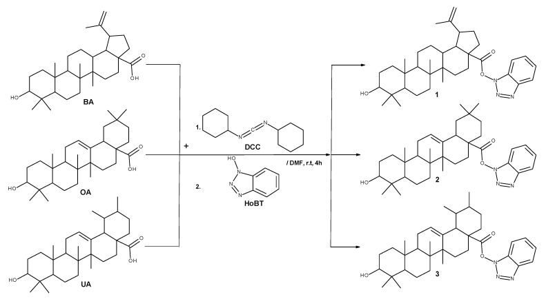
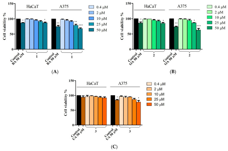
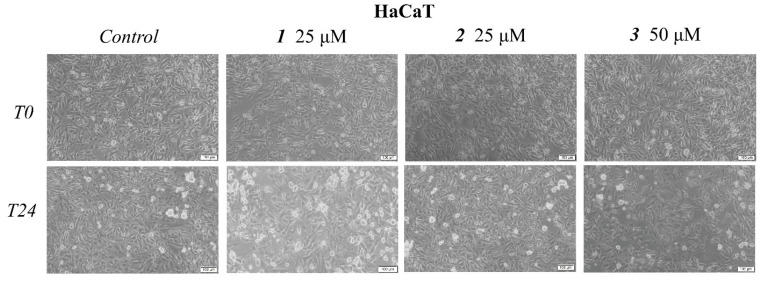
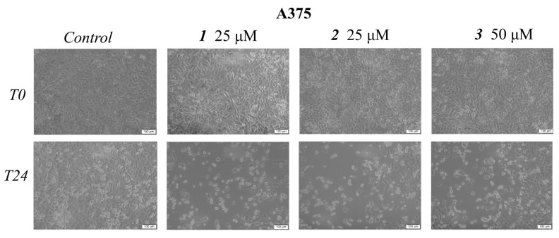
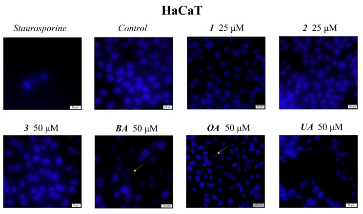
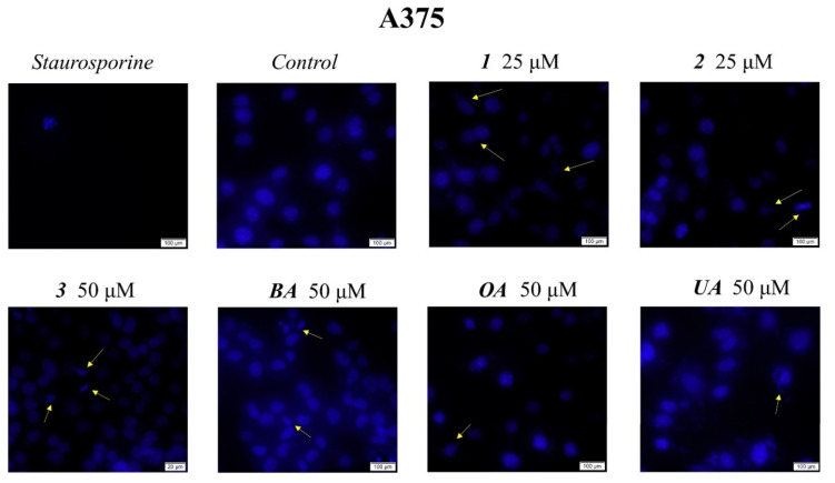
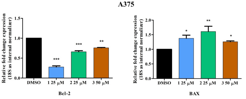
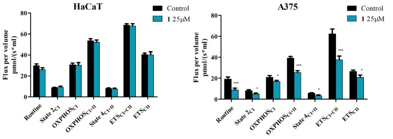
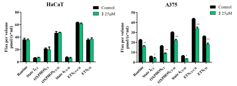
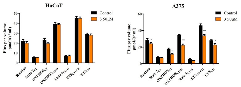
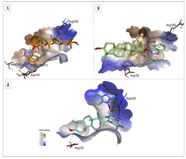
Pentacyclic triterpenes, such as betulinic, ursolic, and oleanolic acids are efficient and selective anticancer agents whose underlying mechanisms of action have been widely investigated. The introduction of N-bearing heterocycles (e.g., triazoles) into the structures of natural compounds (particularly pentacyclic triterpenes) has yielded semisynthetic derivatives with increased antiproliferative potential as opposed to unmodified starting compounds. In this work, we report the synthesis and biological assessment of benzotriazole esters of betulinic acid (BA), oleanolic acid (OA), and ursolic acid (UA) (compounds 1–3). The esters were obtained in moderate yields (28–42%). All three compounds showed dose-dependent reductions in cell viability against A375 melanoma cells and no cytotoxic effects against healthy human keratinocytes. The morphology analysis of treated cells showed characteristic apoptotic changes consisting of nuclear shrinkage, condensation, fragmentation, and cellular membrane disruption. rtPCR analysis reinforced the proapoptotic evidence, showing a reduction in anti-apoptotic Bcl-2 expression and upregulation of the pro-apoptotic Bax. High-resolution respirometry studies showed that all three compounds were able to significantly inhibit mitochondrial function. Molecular docking showed that compounds 1–3 showed an increase in binding affinity against Bcl-2 as opposed to BA, OA, and UA and similar binding patterns compared to known Bcl-2 inhibitors.
Overview
The claims and statistics of this paper haven’t been checked yet.
Run the check — free, no account needed. It reads the open-access text and checks the sources it cites.
Integrity signals
Registry
Cites
Statistics
Figures
Verification
Claims the paper makes
The claims and statistics of this paper haven’t been checked yet.
Run the check — free, no account needed. It reads the open-access text and checks the sources it cites.
Claims other papers make about this one
Each is checked against this paper’s own text: does it say what the citing paper says it does?
In the citing paper
“The process is linked to an increase in proton leak across the inner mitochondrial membrane [55], leading to an increase of State 2 and State 4 [51].”
From Maceration, Soxhlet Extraction, and Steam Hydrodistillation of Anethum graveolens Seeds: Phytochemical Profiles and Cytotoxic Effects (2026) · cites this paper as [55]
Self-check
Percentages, sums and reported differences re-computed from the paper’s own numbers.
The self-check needs the paper’s full text.
Citations
Cites
No references could be resolved yet.
Research works printed but not linked: 60
- Hughes, 2011, Principles of early drug discovery, Br. J. Pharmacol., 162, 1239, 10.1111/j.1476-5381.2010.01127.x · DOI · Paper page
- Ghante, 2019, Role of Pentacyclic Triterpenoids in Chemoprevention and Anticancer Treatment: An Overview on Targets and Underling Mechanisms, J. Pharmacopunct., 22, 55, 10.3831/KPI.201.22.007 · DOI
- Sung, 2021, Global Cancer Statistics 2020: GLOBOCAN Estimates of Incidence and Mortality Worldwide for 36 Cancers in 185 Countries. CA, Cancer J. Clin., 71, 209, 10.3322/caac.21660 · DOI · Paper page
- Zhong, 2021, Recent progress on betulinic acid and its derivatives as antitumor agents: A mini review, Chin. J. Nat. Med., 19, 641
- Ayeleso, T., Matumba, M., and Mukwevho, E. (2017). Oleanolic Acid and Its Derivatives: Biological Activities and Therapeutic Potential in Chronic Diseases. Molecules, 22. · DOI
- Mlala, S., Oyedeji, A.O., Gondwe, M., and Oyedeji, O.O. (2019). Ursolic Acid and Its Derivatives as Bioactive Agents. Molecules, 24. · DOI
- Dangroo, 2017, A convergent synthesis of novel alkyne–azide cycloaddition congeners of betulinic acid as potent cytotoxic agent, Steroids, 123, 1, 10.1016/j.steroids.2017.04.002 · DOI
- Wei, 2015, A library of 1,2,3-triazole-substituted oleanolic acid derivatives as anticancer agents: Design, synthesis, and biological evaluation, Org. Biomol. Chem., 13, 1507, 10.1039/C4OB01605J · DOI
- Suman, 2017, Synthesis and cytotoxicity of Baylis-Hillman template derived betulinic acid-triazole conjugates, Tetrahedron, 73, 4214, 10.1016/j.tet.2016.11.056 · DOI
- Banerji, 2018, Synthesis of Triazole-Substituted Quinazoline Hybrids for Anticancer Activity and a Lead Compound as the EGFR Blocker and ROS Inducer Agent, ACS Omega, 3, 16134, 10.1021/acsomega.8b01960 · DOI
- Chan, 2007, Kinetics of Amide Formation through Carbodiimide/N-Hydroxybenzotriazole (HOBt) Couplings, J. Org. Chem., 72, 8863, 10.1021/jo701558y · DOI
- Sheikh, 2010, Mechanistic studies of DCC/HOBt-mediated reaction of 3-phenylpropionic acid with benzyl alcohol and studies on the reactivities of ‘active ester’ and the related derivatives with nucleophiles, Tetrahedron, 66, 7272, 10.1016/j.tet.2010.07.011 · DOI
- Demuner, 2015, Hederagenin as a triterpene template for the development of new antitumor compounds, Eur. J. Med. Chem., 105, 57, 10.1016/j.ejmech.2015.10.006 · DOI
- Wang, 2019, Synthesis and biological evaluation of novel H6 analogues as drug resistance reversal agents, Eur. J. Med. Chem., 161, 364, 10.1016/j.ejmech.2018.10.033 · DOI
- He, 2020, Synthesis and biological evaluation of celastrol derivatives as potential immunosuppressive agents, J. Nat. Prod., 83, 2578, 10.1021/acs.jnatprod.0c00067 · DOI
- Wu, 2007, Synthesis, in vitro inhibitory activity towards COX-2 and haemolytic activity of derivatives of Esculentoside, A, Bioorg. Med. Chem. Lett., 17, 6430, 10.1016/j.bmcl.2007.10.006 · DOI
- Schwarz, 2014, Amino derivatives of glycyrrhetinic acid as potential inhibitors of cholinesterases, Bioorg. Med. Chem., 22, 3370, 10.1016/j.bmc.2014.04.046 · DOI
- Ghosh, P., Vidal, C., Dey, S., and Zhang, L. (2020). Mitochondria Targeting as an Effective Strategy for Cancer Therapy. Int. J. Mol. Sci., 21. · DOI
- Porporato, 2018, Mitochondrial metabolism and cancer, Cell Res., 28, 265, 10.1038/cr.2017.155 · DOI
- Ren, 2019, Natural Product Triterpenoids and Their Semi-Synthetic Derivatives with Potential Anticancer Activity, Planta Med., 85, 802, 10.1055/a-0832-2383 · DOI
- Guo, 2021, Application of triazoles in the structural modification of natural products, J. Enzym. Inhib. Med. Chem., 36, 1115, 10.1080/14756366.2021.1890066 · DOI
- Zlatskiy, 2020, Comparative Analysis of the Different Dyes’ Potential to Assess Human Normal and Cancer Cell Viability In Vitro under Different D/H Ratios in a Culture Medium, Sci. World J., 2020, 2373021, 10.1155/2020/2373021 · DOI
- Knez, 2021, Antiproliferative Activity of Selected Triterpene Acids from Rosemary on Metastatic Melanoma Cell Line WM-266-4, Open Access Maced. J. Med. Sci., 9, 515, 10.3889/oamjms.2021.6176 · DOI
- Coricovac, D., Dehelean, C.A., Pinzaru, I., Mioc, A., Aburel, O.-M., Macasoi, I., Draghici, G.A., Petean, C., Soica, C., and Boruga, M. (2021). Assessment of Betulinic Acid Cytotoxicity and Mitochondrial Metabolism Impairment in a Human Melanoma Cell Line. Int. J. Mol. Sci., 22. · DOI · Paper page
- Oprean, 2018, Selective in vitro anti-melanoma activity of ursolic and oleanolic acids, Toxicol. Mech. Methods, 28, 148, 10.1080/15376516.2017.1373881 · DOI
- Khan, 2016, A novel triazole derivative of betulinic acid induces extrinsic and intrinsic apoptosis in human leukemia HL-60 cells, Eur. J. Med. Chem., 108, 104, 10.1016/j.ejmech.2015.11.018 · DOI
- Wang, W., Lei, L., Liu, Z., Wang, H., and Meng, Q. (2019). Design, Synthesis, and Biological Evaluation of Novel Nitrogen Heterocycle-Containing Ursolic Acid Analogs as Antitumor Agents. Molecules, 24. · DOI
- Singh, V., Khurana, A., Navik, U., Allawadhi, P., Bharani, K.K., and Weiskirchen, R. (2022). Apoptosis and Pharmacological Therapies for Targeting Thereof for Cancer Therapeutics. Science, 4. · DOI
- Vianna, 2021, Natural and Semisynthetic Pentacyclic Triterpenes for Chronic Myeloid Leukemia Therapy: Reality, Challenges and Perspectives, ChemMedChem, 16, 1835, 10.1002/cmdc.202100038 · DOI
- Borska, 2017, Anticancer properties of ester derivatives of betulin in human metastatic melanoma cells (Me-45), Cancer Cell Int., 17, 4, 10.1186/s12935-016-0369-3 · DOI
- Zhuo, 2017, Novel betulin derivative induces anti-proliferative activity by G2/M phase cell cycle arrest and apoptosis in Huh7 cells, Oncol. Lett., 15, 2097
- Gou, 2022, Ursolic Acid Derivative UA232 Promotes Tumor Cell Apoptosis by Inducing Endoplasmic Reticulum Stress and Lysosomal Dysfunction, Int. J. Biol. Sci., 18, 2639, 10.7150/ijbs.67166 · DOI
- Rivas, 2016, The oleanolic acid derivative, 3-O-succinyl-28-O-benzyl oleanolate, induces apoptosis in B16–F10 melanoma cells via the mitochondrial apoptotic pathway, RSC Adv., 6, 93590, 10.1039/C6RA18879F · DOI
- Park, 2014, Relationships between chemical structures and functions of triterpene glycosides isolated from sea cucumbers, Front. Chem., 2, 77, 10.3389/fchem.2014.00077 · DOI
- Malyarenko, T.V., Kicha, A.A., Kalinovsky, A.I., Dmitrenok, P.S., Malyarenko, O.S., Kuzmich, A.S., Stonik, V.A., and Ivanchina, N.V. (2021). New Triterpene Glycosides from the Far Eastern Starfish Solaster pacificus and Their Biological Activity. Biomolecules, 11. · DOI
- Dubinin, 2020, Effect of betulin and betulonic acid on isolated rat liver mitochondria and liposomes, Biochim. Biophys. Acta-Biomembr., 1862, 183383, 10.1016/j.bbamem.2020.183383 · DOI
- George, 2012, Apoptosis-Induced Cell Death due to Oleanolic Acid in HaCaT Keratinocyte Cells—A Proof-of-Principle Approach for Chemopreventive Drug Development, Asian Pac. J. Cancer Prev., 13, 2015, 10.7314/APJCP.2012.13.5.2015 · DOI
- Harmand, 2003, Ursolic acid induces apoptosis through caspase-3 activation and cell cycle arrest in HaCat cells, Int. J. Oncol., 23, 105
- Guo, Y., Zhu, H., Weng, M., Wang, C., and Sun, L. (2020). Chemopreventive effect of Betulinic acid via mTOR -Caspases/Bcl2/Bax apoptotic signaling in pancreatic cancer. BMC Complement. Med. Ther., 20. · DOI
- Pratheeshkumar, 2011, Oleanolic acid induces Apoptosis by modulating p53, Bax, Bcl-2 and caspase-3 gene expression and regulates the activation of transcription factors and cytokine profile in B16F, J. Environ. Pathol. Toxicol. Oncol., 30, 21, 10.1615/JEnvironPatholToxicolOncol.v30.i1.30 · DOI
- Kim, 2011, Induction of apoptotic cell death by ursolic acid through mitochondrial death pathway and extrinsic death receptor pathway in MDA-MB-231 cells, Arch. Pharmacal Res., 34, 1363, 10.1007/s12272-011-0817-5 · DOI
- Mioc, M., Milan, A., Malița, D., Mioc, A., Prodea, A., Racoviceanu, R., Ghiulai, R., Cristea, A., Căruntu, F., and Șoica, C. (2022). Recent Advances Regarding the Molecular Mechanisms of Triterpenic Acids: A Review (Part I). Int. J. Mol. Sci., 23. · DOI
- Klein, 2020, Role of Mitochondria in Cancer Immune Evasion and Potential Therapeutic Approaches, Front. Immunol., 11, 573326, 10.3389/fimmu.2020.573326 · DOI
- Viale, 2015, Tumors and Mitochondrial Respiration: A Neglected Connection, Cancer Res., 75, 3687, 10.1158/0008-5472.CAN-15-0491 · DOI
- Prestegarden, 2019, Inhibition of mitochondrial respiration prevents BRAF-mutant melanoma brain metastasis, Acta Neuropathol. Commun., 7, 55, 10.1186/s40478-019-0712-8 · DOI
- Kumar, 2021, Mitochondrial oxidative phosphorylation in cutaneous melanoma, Br. J. Cancer, 124, 115, 10.1038/s41416-020-01159-y · DOI
- Danese, 2017, Calcium regulates cell death in cancer: Roles of the mitochondria and mitochondria-associated membranes (MAMs), Biochim. Biophys. Acta-Bioenerg., 1858, 615, 10.1016/j.bbabio.2017.01.003 · DOI
- Konopleva, 2013, Mitochondrial Uncoupling and the Reprograming of Intermediary Metabolism in Leukemia Cells, Front. Oncol., 3, 67
- Brookes, 2005, Mitochondrial H+ leak and ROS generation: An odd couple, Free Radic. Biol. Med., 38, 12, 10.1016/j.freeradbiomed.2004.10.016 · DOI · Paper page
- Ling, 2022, Triterpenoids as Reactive Oxygen Species Modulators of Cell Fate, Chem. Res. Toxicol., 35, 569, 10.1021/acs.chemrestox.1c00428 · DOI
- Brand, 2011, Assessing mitochondrial dysfunction in cells, Biochem. J., 435, 297, 10.1042/BJ20110162 · DOI
- Pinzi, L., and Rastelli, G. (2019). Molecular Docking: Shifting Paradigms in Drug Discovery. Int. J. Mol. Sci., 20. · DOI
- Bruncko, 2007, Studies Leading to Potent, Dual Inhibitors of Bcl-2 and Bcl-xL, J. Med. Chem., 50, 641, 10.1021/jm061152t · DOI
- Porter, 2009, Tetrahydroisoquinoline amide substituted phenyl pyrazoles as selective Bcl-2 inhibitors, Bioorg. Med. Chem. Lett., 19, 230, 10.1016/j.bmcl.2008.10.113 · DOI
- Petrus, 2017, Assessment of Mitochondrial Respiration in Human Platelets, Rev. Chim., 68, 768, 10.37358/RC.17.4.5549 · DOI
- Pesta, D., and Gnaiger, E. (2012). High-Resolution Respirometry: OXPHOS Protocols for Human Cells and Permeabilized Fibers from Small Biopsies of Human Muscle. Mitochondrial Bioenergetic, Humana Press. · DOI
- Kazakova, O., Mioc, A., Smirnova, I., Baikova, I., Voicu, A., Vlaia, L., Macașoi, I., Mioc, M., Drăghici, G., and Avram, Ş. (2021). Novel Synthesized N-Ethyl-Piperazinyl-Amides of C2-Substituted Oleanonic and Ursonic Acids Exhibit Cytotoxic Effects through Apoptotic Cell Death Regulation. Int. J. Mol. Sci., 22. · DOI
- Kazakova, O., Șoica, C., Babaev, M., Petrova, A., Khusnutdinova, E., Poptsov, A., Macașoi, I., Drăghici, G., Avram, Ș., and Vlaia, L. (2021). 3-Pyridinylidene Derivatives of Chemically Modified Lupane and Ursane Triterpenes as Promising Anticancer Agents by Targeting Apoptosis. Int. J. Mol. Sci., 22. · DOI
- Berman, 2000, The Protein Data Bank, Nucleic Acids Res., 28, 235, 10.1093/nar/28.1.235 · DOI · Paper page
- Trott, 2009, AutoDock Vina: Improving the speed and accuracy of docking with a new scoring function, efficient optimization, and multithreading, J. Comput. Chem., 31, 455, 10.1002/jcc.21334 · DOI · Paper page
Cited by
Goldiș, C., Racoviceanu, R., Negrea-Ghiulai, R., Mioc, A., Prodea, A., Atyim, E., Maksimovic, T., Lukinich-Gruia, A. T., Pricop, M.-A., & Șoica, C. (2026) · Molecules · doi:10.3390/molecules31183337
Self-citation
Metadata
- Authors
- Mioc, M., Mioc, A., Prodea, A., Milan, A., Balan-Porcarasu, M., Racoviceanu, R., Ghiulai, R., Iovanescu, G., Macasoi, I., Draghici, G., Dehelean, C., & Soica, C.
- Venue
- International Journal of Molecular Sciences
- Publisher
- MDPI AG
- Published
- 2022-09-01
- Volume
- 23
- Issue
- 17
- Pages
- 9992
- ISSN
- 1422-0067
- PMC
- PMC9456456
- Cited by
- 19
- References
- 0
- Language
- en
- Funders
- Romanian UEFISCDI (PN-III-P1-1.1-PD-2019-1078)
About these checks
Registry, retraction, Cites and statistics results are computed, not judged, with one exception: a printed reference that no index lists is read once by a model before it counts as a citation error. Claims are extracted by a model and kept only when their quote appears verbatim in the text; each cited source is then checked, with a model’s help, for whether it says what the claim relies on. Those verdicts can be wrong — read the claim’s quote and the cited source before relying on one.
Statistics and arithmetic are recomputed, not judged.
Confidence bands are shown only for fields calibrated on our annotated test set. None is calibrated yet, so no band is shown.
What counts as a claim ›How we measure confidence ›How we check this ›
Are you an author, or do you see a problem with this page? Request a review
Data sources
Bibliographic data: Semantic Scholar, OpenAlex, Crossref.
Contains information from the Semantic Scholar Academic Graph Datasets, made available under the ODC Attribution License (ODC-BY 1.0).
Semantic Scholar’s data is described in: Kinney, R., et al. (2023). The Semantic Scholar Open Data Platform. arXiv:2301.10140.
