

The DOI matches its Crossref record; 5 of the 5 references it cites were found in a registry or an index.
Targeting Key Signaling Pathways in Glioblastoma Stem Cells for the Development of Efficient Chemo- and Immunotherapy
Abstract
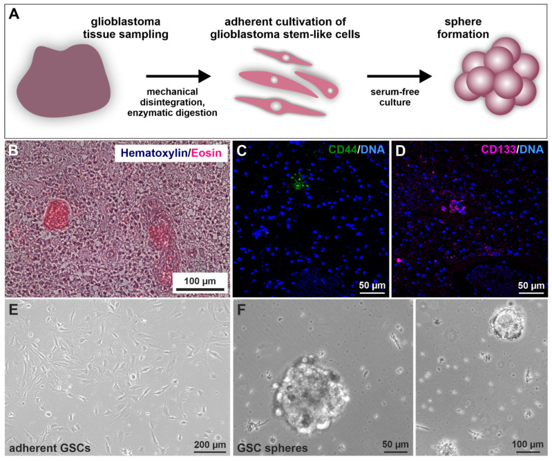
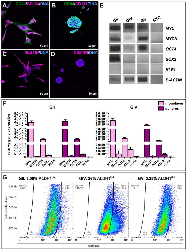
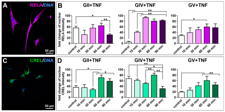
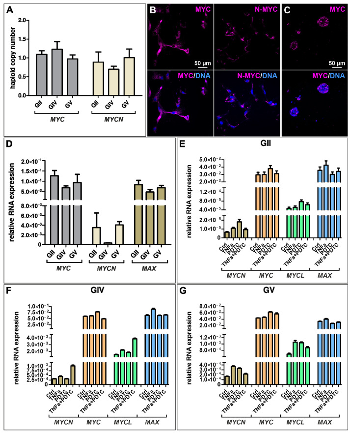
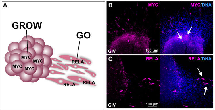
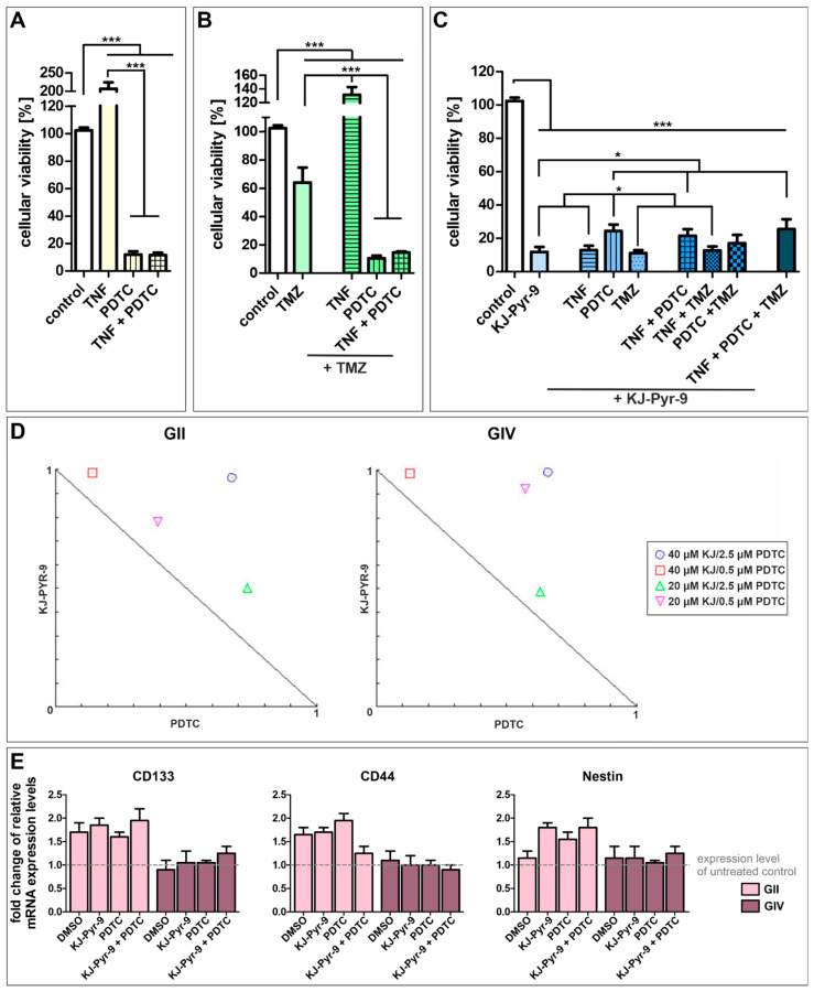
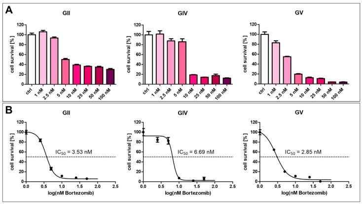
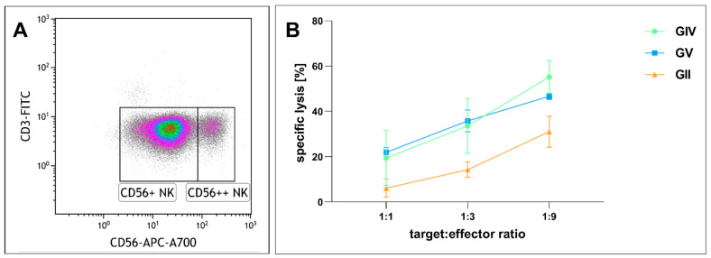
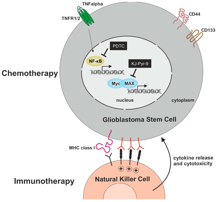
Glioblastoma multiforme (GBM) is the most aggressive and most common malignant brain tumor with poor patient survival despite therapeutic intervention. On the cellular level, GBM comprises a rare population of glioblastoma stem cells (GSCs), driving therapeutic resistance, invasion, and recurrence. GSCs have thus come into the focus of therapeutic strategies, although their targeting remains challenging. In the present study, we took advantage of three GSCs-populations recently established in our lab to investigate key signaling pathways and subsequent therapeutic strategies targeting GSCs. We observed that NF-κB, a crucial transcription factor in GBM progression, was expressed in all CD44+/CD133+/Nestin+-GSC-populations. Exposure to TNFα led to activation of NF-κB-RELA and/or NF-κB-c-REL, depending on the GBM type. GSCs further expressed the proto-oncogene MYC family, with MYChigh GSCs being predominantly located in the tumor spheres (“GROW”-state) while NF-κB-RELAhigh GSCs were migrating out of the sphere (“GO”-state). We efficiently targeted GSCs by the pharmacologic inhibition of NF-κB using PTDC/Bortezomib or inhibition of MYC by KJ-Pyr-9, which significantly reduced GSC-viability, even in comparison to the standard chemotherapeutic drug temozolomide. As an additional cell-therapeutic strategy, we showed that NK cells could kill GSCs. Our findings offer new perspectives for developing efficient patient-specific chemo- and immunotherapy against GBM.
Overview
The claims and statistics of this paper haven’t been checked yet.
Run the check — free, no account needed. It reads the open-access text and checks the sources it cites.
Integrity signals
Registry
Cites
Statistics
Figures
Verification
Claims the paper makes
The claims and statistics of this paper haven’t been checked yet.
Run the check — free, no account needed. It reads the open-access text and checks the sources it cites.
Claims other papers make about this one
Each is checked against this paper’s own text: does it say what the citing paper says it does?
The source backs part of the claim, not all of it — or backs it for a different population, condition or comparison.
In the citing paper
“GB tumors are related to neural stem cells and Myc signaling, but effective targeting of Myc in clinical practice is yet to be achieved despite efforts, and there are various challenges in translating related strategies to clinical settings [202, 203].”
From Emerging role of exosomes in cancer therapy: progress and challenges (2025) · cites this paper as [203]
Experimental — accuracy not yet measured
Self-check
Percentages, sums and reported differences re-computed from the paper’s own numbers.
The self-check needs the paper’s full text.
Citations
Cites
Natural killer cells in cancer biology and therapy
Wu, S.-Y., Fu, T., Jiang, Y.-Z., & Shao, Z.-M. (2020) · Molecular Cancer · doi:10.1186/s12943-020-01238-x · Cited by 863
In vivo Switching of Human Melanoma Cells between Proliferative and Invasive States
Hoek, K. S., Eichhoff, O. M., Schlegel, N. C., Döbbeling, U., Kobert, N., Schaerer, L., Hemmi, S., & Dummer, R. (2008) · Cancer Research · doi:10.1158/0008-5472.can-07-2491 · Cited by 743
Myc-Driven glycolysis is a therapeutic target in glioblastoma
Tateishi, K., Iafrate, A. J., Ho, Q., Curry, W. T., Batchelor, T. T., Flaherty, K. T., Onozato, M. L., Lelic, N., Sundaram, S., Cahill, D. P., et al. (2016) · Clinical Cancer Research · doi:10.1158/1078-0432.ccr-15-2274 · Cited by 160
Wiewrodt, D., Nagel, G., Dreimüller, N., Hundsberger, T., Perneczky, A., & Kaina, B. (2008) · International Journal of Cancer · doi:10.1002/ijc.23219 · Cited by 114
A Role for NF-κB in Organ Specific Cancer and Cancer Stem Cells
Kaltschmidt, C., Banz-Jansen, C., Benhidjeb, T., Beshay, M., Förster, C., Greiner, J., Hamelmann, E., Jorch, N., Mertzlufft, F., Pfitzenmaier, J., et al. (2019) · Cancers · doi:10.3390/cancers11050655 · Cited by 102
Self-citation
Research works printed but not linked: 81
- Ostrom, 2013, CBTRUS statistical report: Primary brain and central nervous system tumors diagnosed in the United States in 2006–2010, Neuro Oncol., 15, ii1, 10.1093/neuonc/not151 · DOI
- Medina, 2020, Advances in the Knowledge of the Molecular Biology of Glioblastoma and Its Impact in Patient Diagnosis, Stratification, and Treatment, Adv. Sci., 7, 1902971, 10.1002/advs.201902971 · DOI
- Davis, 2016, Glioblastoma: Overview of Disease and Treatment, Clin. J. Oncol. Nurs., 20, S2, 10.1188/16.CJON.S1.2-8 · DOI
- Ohgaki, 2013, The definition of primary and secondary glioblastoma, Clin. Cancer Res., 19, 764, 10.1158/1078-0432.CCR-12-3002 · DOI
- Chen, 2016, Isocitrate Dehydrogenase (IDH)1/2 Mutations as Prognostic Markers in Patients With Glioblastomas, Medicine (Baltimore), 95, e2583, 10.1097/MD.0000000000002583 · DOI
- Wee, 2017, Novel recursive partitioning analysis classification for newly diagnosed glioblastoma: A multi-institutional study highlighting the MGMT promoter methylation and IDH1 gene mutation status, Radiother. Oncol., 123, 106, 10.1016/j.radonc.2017.02.014 · DOI
- Kitange, 2009, Induction of MGMT expression is associated with temozolomide resistance in glioblastoma xenografts, Neuro Oncol., 11, 281, 10.1215/15228517-2008-090 · DOI
- Happold, 2018, Transcriptional control of O(6) -methylguanine DNA methyltransferase expression and temozolomide resistance in glioblastoma, J. Neurochem., 144, 780, 10.1111/jnc.14326 · DOI
- Xia, 2014, NF-κB, an active player in human cancers, Cancer Immunol. Res., 2, 823, 10.1158/2326-6066.CIR-14-0112 · DOI · Paper page
- Taniguchi, 2018, NF-κB, inflammation, immunity and cancer: Coming of age, Nat. Rev. Immunol., 18, 309, 10.1038/nri.2017.142 · DOI · Paper page
- Kaltschmidt, B., Greiner, J.F.W., Kadhim, H.M., and Kaltschmidt, C. (2018). Subunit-Specific Role of NF-κB in Cancer. Biomedicines, 6. · DOI
- Soubannier, V., and Stifani, S. (2017). NF-κB Signalling in Glioblastoma. Biomedicines, 5. · DOI
- Eilers, 2008, Myc’s broad reach, Genes Dev., 22, 2755, 10.1101/gad.1712408 · DOI · Paper page
- Zaytseva, O., Kim, N.-h., and Quinn, L.M. (2020). MYC in Brain Development and Cancer. Int. J. Mol. Sci., 21. · DOI
- Vita, 2006, The Myc oncoprotein as a therapeutic target for human cancer, Semin. Cancer Biol., 16, 318, 10.1016/j.semcancer.2006.07.015 · DOI
- Herms, 1999, c-myc oncogene family expression in glioblastoma and survival, Surg. Neurol., 51, 536, 10.1016/S0090-3019(98)00028-7 · DOI
- Hodgson, 2009, Comparative analyses of gene copy number and mRNA expression in glioblastoma multiforme tumors and xenografts, Neuro-Oncology, 11, 477, 10.1215/15228517-2008-113 · DOI
- Orian, 1992, Overexpression of multiple oncogenes related to histological grade of astrocytic glioma, Br. J. Cancer, 66, 106, 10.1038/bjc.1992.225 · DOI
- Duyao, 1990, Interaction of an NF-kappa B-like factor with a site upstream of the c-myc promoter, Proc. Natl. Acad. Sci. USA, 87, 4727, 10.1073/pnas.87.12.4727 · DOI
- Kessler, 1992, NF-kappa B-like factors mediate interleukin 1 induction of c-myc gene transcription in fibroblasts, J. Exp. Med., 176, 787, 10.1084/jem.176.3.787 · DOI
- Pierce, 1994, Differential regulation of the c-myc oncogene promoter by the NF-kappa B rel family of transcription factors, Mol. Cell Biol., 14, 1039
- Moser, 2021, The inflammatory kinase IKKalpha phosphorylates and stabilizes c-Myc and enhances its activity, Mol. Cancer, 20, 16, 10.1186/s12943-021-01308-8 · DOI
- Grumont, 2004, The mitogen-induced increase in T cell size involves PKC and NFAT activation of Rel/NF-kappaB-dependent c-myc expression, Immunity, 21, 19, 10.1016/j.immuni.2004.06.004 · DOI
- Grumont, 2002, B cell growth is controlled by phosphatidylinosotol 3-kinase-dependent induction of Rel/NF-kappaB regulated c-myc transcription, Mol. Cell, 10, 1283, 10.1016/S1097-2765(02)00779-7 · DOI
- Hayes, 2021, Cellular immunotherapies for cancer, Ir. J. Med. Sci., 190, 41, 10.1007/s11845-020-02264-w · DOI
- Jones, A.B., Rocco, A., Lamb, L.S., Friedman, G.K., and Hjelmeland, A.B. (2022). Regulation of NKG2D Stress Ligands and Its Relevance in Cancer Progression. Cancers, 14. · DOI
- Chitadze, 2022, Immune surveillance in glioblastoma: Role of the NKG2D system and novel cell-based therapeutic approaches, Scand. J. Immunol., 96, e13201, 10.1111/sji.13201 · DOI
- Brandetti, E., Focaccetti, C., Pezzolo, A., Ognibene, M., Folgiero, V., Cotugno, N., Benvenuto, M., Palma, P., Manzari, V., and Rossi, P. (2021). Enhancement of Neuroblastoma NK-Cell-Mediated Lysis through NF-kB p65 Subunit-Induced Expression of FAS and PVR, the Loss of Which Is Associated with Poor Patient Outcome. Cancers, 13. · DOI
- Wang, 2021, Aneuploid senescent cells activate NF-kappaB to promote their immune clearance by NK cells, EMBO Rep., 22, e52032, 10.15252/embr.202052032 · DOI
- Guegan, 2021, CD95/Fas suppresses NF-kappaB activation through recruitment of KPC2 in a CD95L/FasL-independent mechanism, iScience, 24, 103538, 10.1016/j.isci.2021.103538 · DOI
- Gimple, 2019, Glioblastoma stem cells: Lessons from the tumor hierarchy in a lethal cancer, Genes Dev., 33, 591, 10.1101/gad.324301.119 · DOI · Paper page
- Lathia, 2015, Cancer stem cells in glioblastoma, Genes Dev., 29, 1203, 10.1101/gad.261982.115 · DOI · Paper page
- Yan, 2013, The evolving landscape of glioblastoma stem cells, Curr. Opin. Neurol., 26, 701, 10.1097/WCO.0000000000000032 · DOI
- Prager, 2020, Glioblastoma Stem Cells: Driving Resilience through Chaos, Trends. Cancer, 6, 223, 10.1016/j.trecan.2020.01.009 · DOI · Paper page
- Glas, 2010, Residual tumor cells are unique cellular targets in glioblastoma, Ann. Neurol., 68, 264, 10.1002/ana.22036 · DOI
- Seymour, 2015, Targeting Aggressive Cancer Stem Cells in Glioblastoma, Front. Oncol., 5, 159, 10.3389/fonc.2015.00159 · DOI
- Witte, K.E., Hertel, O., Windmöller, B.A., Helweg, L.P., Höving, A.L., Knabbe, C., Busche, T., Greiner, J.F.W., Kalinowski, J., and Noll, T. (2021). Nanopore Sequencing Reveals Global Transcriptome Signatures of Mitochondrial and Ribosomal Gene Expressions in Various Human Cancer Stem-like Cell Populations. Cancers, 13. · DOI
- Schulte Am Esch, J.S.A., Windmöller, B.A., Hanewinkel, J., Storm, J., Förster, C., Wilkens, L., Krüger, M., Kaltschmidt, B., and Kaltschmidt, C. (2020). Isolation and Characterization of Two Novel Colorectal Cancer Cell Lines, Containing a Subpopulation with Potential Stem-Like Properties: Treatment Options by MYC/NMYC Inhibition. Cancers, 12. · DOI
- Entrena, 2016, Low adherent cancer cell subpopulations are enriched in tumorigenic and metastatic epithelial-to-mesenchymal transition-induced cancer stem-like cells, Sci. Rep., 6, 18772, 10.1038/srep18772 · DOI
- Walia, 2010, Enrichment for breast cancer cells with stem/progenitor properties by differential adhesion, Stem. Cells Dev., 19, 1175, 10.1089/scd.2009.0430 · DOI
- Li, Y., Sharma, A., Maciaczyk, J., and Schmidt-Wolf, I.G.H. (2022). Recent Development in NKT-Based Immunotherapy of Glioblastoma: From Bench to Bedside. Int. J. Mol. Sci., 23. · DOI
- Golán, I., Rodríguez de la Fuente, L., and Costoya, J.A. (2018). NK Cell-Based Glioblastoma Immunotherapy. Cancers, 10. · DOI
- Schreck, 1992, Dithiocarbamates as potent inhibitors of nuclear factor kappa B activation in intact cells, J. Exp. Med., 175, 1181, 10.1084/jem.175.5.1181 · DOI · Paper page
- Witte, K.E., Pfitzenmaier, J., Storm, J., Lütkemeyer, M., Wimmer, C., Schulten, W., Czaniera, N., Geisler, M., Förster, C., and Wilkens, L. (2021). Analysis of Several Pathways for Efficient Killing of Prostate Cancer Stem Cells: A Central Role of NF-κB RELA. Int. J. Mol. Sci., 22. · DOI
- Windmöller, B.A., Beshay, M., Helweg, L.P., Flottmann, C., Beermann, M., Förster, C., Wilkens, L., Greiner, J.F.W., Kaltschmidt, C., and Kaltschmidt, B. (2021). Novel Primary Human Cancer Stem-Like Cell Populations from Non-Small Cell Lung Cancer: Inhibition of Cell Survival by Targeting NF-κB and MYC Signaling. Cells, 10. · DOI
- Miraglia, 1997, A novel five-transmembrane hematopoietic stem cell antigen: Isolation, characterization, and molecular cloning, Blood, 90, 5013, 10.1182/blood.V90.12.5013 · DOI
- Galli, 2004, Isolation and characterization of tumorigenic, stem-like neural precursors from human glioblastoma, Cancer Res., 64, 7011, 10.1158/0008-5472.CAN-04-1364 · DOI
- Singh, 2004, Identification of human brain tumour initiating cells, Nature, 432, 396, 10.1038/nature03128 · DOI
- Brescia, 2013, CD133 is essential for glioblastoma stem cell maintenance, Stem. Cells, 31, 857, 10.1002/stem.1317 · DOI
- Li, 2012, Cancer Stem Cells: Distinct Entities or Dynamically Regulated Phenotypes?, Cancer Res., 72, 576, 10.1158/0008-5472.CAN-11-3070 · DOI
- Du, 2009, Oct4 is expressed in human gliomas and promotes colony formation in glioma cells, Glia, 57, 724, 10.1002/glia.20800 · DOI
- Johnson, 2022, Sox2 induces glioblastoma cell stemness and tumor propagation by repressing TET2 and deregulating 5hmC and 5mC DNA modifications, Signal. Transduct. Target. Ther., 7, 37, 10.1038/s41392-021-00857-0 · DOI
- Ostrom, 2018, CBTRUS Statistical Report: Primary Brain and Other Central Nervous System Tumors Diagnosed in the United States in 2011–2015, Neuro Oncol., 20, iv1, 10.1093/neuonc/noy131 · DOI
- Alves, 2021, Role of glioblastoma stem cells in cancer therapeutic resistance: A perspective on antineoplastic agents from natural sources and chemical derivatives, Stem. Cell Res. Ther., 12, 206, 10.1186/s13287-021-02231-x · DOI
- Hanif, 2017, Glioblastoma Multiforme: A Review of its Epidemiology and Pathogenesis through Clinical Presentation and Treatment, Asian. Pac. J. Cancer Prev., 18, 3
- Schafer, 2012, Aldehyde dehydrogenase 1A1--a new mediator of resistance to temozolomide in glioblastoma, Neuro Oncol., 14, 1452, 10.1093/neuonc/nos270 · DOI
- Kaltschmidt, B., Witte, K.E., Greiner, J.F.W., Weissinger, F., and Kaltschmidt, C. (2022). Targeting NF-kappaB Signaling in Cancer Stem Cells: A Narrative Review. Biomedicines, 10. · DOI
- Bredel, 2011, NFKBIA deletion in glioblastomas, N. Engl. J. Med., 364, 627, 10.1056/NEJMoa1006312 · DOI
- Wang, J., Wang, H., Li, Z., Wu, Q., Lathia, J.D., McLendon, R.E., Hjelmeland, A.B., and Rich, J.N. (2008). c-Myc is required for maintenance of glioma cancer stem cells. PLoS ONE, 3. · DOI
- Zheng, 2008, Pten and p53 converge on c-Myc to control differentiation, self-renewal, and transformation of normal and neoplastic stem cells in glioblastoma, Cold Spring Harb. Symp. Quant. Biol., 73, 427, 10.1101/sqb.2008.73.047 · DOI
- Hoek, 2006, Metastatic potential of melanomas defined by specific gene expression profiles with no BRAF signature, Pigment. Cell Res., 19, 290, 10.1111/j.1600-0749.2006.00322.x · DOI
- Hatzikirou, 2012, ‘Go or grow’: The key to the emergence of invasion in tumour progression?, Math. Med. Biol., 29, 49, 10.1093/imammb/dqq011 · DOI
- Bhat, 2013, Mesenchymal differentiation mediated by NF-kappaB promotes radiation resistance in glioblastoma, Cancer Cell, 24, 331, 10.1016/j.ccr.2013.08.001 · DOI
- Rinkenbaugh, 2016, IKK/NF-kappaB signaling contributes to glioblastoma stem cell maintenance, Oncotarget, 7, 69173, 10.18632/oncotarget.12507 · DOI
- Avci, 2020, NF-κB inhibitor with Temozolomide results in significant apoptosis in glioblastoma via the NF-κB(p65) and actin cytoskeleton regulatory pathways, Sci. Rep., 10, 13352, 10.1038/s41598-020-70392-5 · DOI
- Bellas, 1997, Inhibition of NF-kappa B activity induces apoptosis in murine hepatocytes, Am. J. Pathol., 151, 891
- Yamaki, 2013, Temozolomide suppresses MYC via activation of TAp63 to inhibit progression of human glioblastoma, Sci. Rep., 3, 1160, 10.1038/srep01160 · DOI
- Pang, 2007, Sodium diethyldithiocarbamate, an AIDS progression inhibitor and a copper-binding compound, has proteasome-inhibitory and apoptosis-inducing activities in cancer cells, Int. J. Mol. Med., 19, 809
- Bota, 2013, Proteasome inhibition with bortezomib induces cell death in GBM stem-like cells and temozolomide-resistant glioma cell lines, but stimulates GBM stem-like cells’ VEGF production and angiogenesis, J. Neurosurg., 119, 1415, 10.3171/2013.7.JNS1323 · DOI
- Tang, 2019, Bortezomib inhibits growth and sensitizes glioma to temozolomide (TMZ) via down-regulating the FOXM1-Survivin axis, Cancer Commun. (Lond.), 39, 81, 10.1186/s40880-019-0424-2 · DOI
- Curran, 2009, Bortezomib: A review of its use in patients with multiple myeloma, Drugs, 69, 859, 10.2165/00003495-200969070-00006 · DOI
- Ishikawa, 2004, Autologous natural killer cell therapy for human recurrent malignant glioma, Anticancer Res., 24, 1861
- Castriconi, 2009, NK cells recognize and kill human glioblastoma cells with stem cell-like properties, J. Immunol., 182, 3530, 10.4049/jimmunol.0802845 · DOI
- Avril, 2012, Human glioblastoma stem-like cells are more sensitive to allogeneic NK and T cell-mediated killing compared with serum-cultured glioblastoma cells, Brain Pathol., 22, 159, 10.1111/j.1750-3639.2011.00515.x · DOI
- Haspels, 2018, Glioblastoma Stem-Like Cells Are More Susceptible Than Differentiated Cells to Natural Killer Cell Lysis Mediated Through Killer Immunoglobulin-Like Receptors-Human Leukocyte Antigen Ligand Mismatch and Activation Receptor-Ligand Interactions, Front. Immunol., 9, 1345, 10.3389/fimmu.2018.01345 · DOI
- Friebel, 2020, Single-Cell Mapping of Human Brain Cancer Reveals Tumor-Specific Instruction of Tissue-Invading Leukocytes, Cell, 181, 1626, 10.1016/j.cell.2020.04.055 · DOI
- Romee, 2013, NK cell CD16 surface expression and function is regulated by a disintegrin and metalloprotease-17 (ADAM17), Blood, 121, 3599, 10.1182/blood-2012-04-425397 · DOI
- Ramakers, 2003, Assumption-free analysis of quantitative real-time polymerase chain reaction (PCR) data, Neurosci. Lett., 339, 62, 10.1016/S0304-3940(02)01423-4 · DOI
- Speleman, 2002, Quantification of MYCN, DDX1, and NAG gene copy number in neuroblastoma using a real-time quantitative PCR assay, Mod. Pathol., 15, 159, 10.1038/modpathol.3880508 · DOI
- Witte, 2020, PLEKHG5 regulates autophagy, survival and MGMT expression in U251-MG glioblastoma cells, Sci. Rep., 10, 21858, 10.1038/s41598-020-77958-3 · DOI
- Chou, 2010, Drug combination studies and their synergy quantification using the Chou-Talalay method, Cancer Res., 70, 440, 10.1158/0008-5472.CAN-09-1947 · DOI · Paper page
Cited by
Sorted by citations
Emerging role of exosomes in cancer therapy: progress and challenges
Li, J., Wang, J., & Chen, Z. (2025) · Molecular Cancer · doi:10.1186/s12943-024-02215-4 · Cited by 297
Metadata
- Authors
- Helweg, L. P., Storm, J., Witte, K. E., Schulten, W., Wrachtrup, L., Janotte, T., Kitke, A., Greiner, J. F. W., Knabbe, C., Kaltschmidt, B., Simon, M., & Kaltschmidt, C.
- Venue
- International Journal of Molecular Sciences
- Publisher
- MDPI AG
- Published
- 2022-10-26
- Volume
- 23
- Issue
- 21
- Pages
- 12919
- ISSN
- 1422-0067
- PMC
- PMC9659205
- Cited by
- 10
- References
- 5
- Language
- en
- Funders
- University of Bielefeld
- Deutsche Forschungsgemeinschaft and the Open Access Publication Fund of Bielefeld University
- Bethel Foundation, Bielefeld, Germany
About these checks
Registry, retraction, Cites and statistics results are computed, not judged, with one exception: a printed reference that no index lists is read once by a model before it counts as a citation error. Claims are extracted by a model and kept only when their quote appears verbatim in the text; each cited source is then checked, with a model’s help, for whether it says what the claim relies on. Those verdicts can be wrong — read the claim’s quote and the cited source before relying on one.
Statistics and arithmetic are recomputed, not judged.
Confidence bands are shown only for fields calibrated on our annotated test set. None is calibrated yet, so no band is shown.
What counts as a claim ›How we measure confidence ›How we check this ›
Are you an author, or do you see a problem with this page? Request a review
