

The DOI matches its Crossref record; 4 of the 4 references it cites were found in a registry or an index.
Advancements in Therapeutic Approaches for Degenerative Tendinopathy: Evaluating Efficacy and Challenges
Open accessAbstract
Degenerative tendinopathy results from the accumulation of minor injuries following unsuccessful tendon repair during acute tendon injuries. The process of tendon repair is prolonged and varies between individuals, making it susceptible to reinjury. Moreover, treating chronic tendinopathy often requires expensive and extensive rehabilitation, along with a variety of combined therapies to facilitate recovery. This condition significantly affects the quality of life of affected individuals, underscoring the urgent need for more efficient and cost-effective treatment options. Although traditional treatments have improved significantly and are being used as substitutes for surgical interventions, the findings have been inconsistent and conflicting. This review aims to clarify these issues by exploring the strengths and limitations of current treatments as well as recent innovations in managing various forms of degenerative tendinopathy.
Overview
The claims and statistics of this paper haven’t been checked yet.
Run the check — free, no account needed. It reads the open-access text and checks the sources it cites.
Integrity signals
Registry
Cites
Statistics
Figures
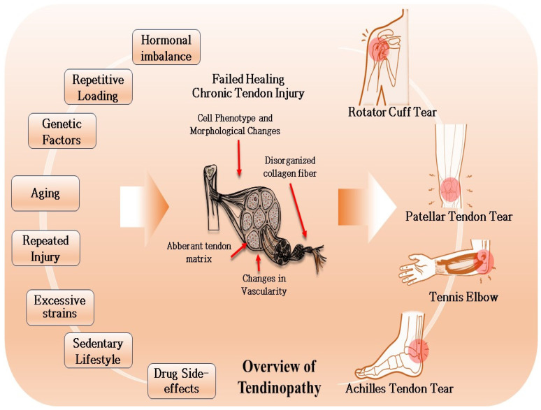
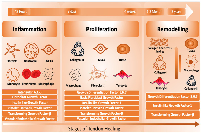
Verification
Claims the paper makes
The claims and statistics of this paper haven’t been checked yet.
Run the check — free, no account needed. It reads the open-access text and checks the sources it cites.
Claims other papers make about this one
Each is checked against this paper’s own text: does it say what the citing paper says it does?
The source backs part of the claim, not all of it — or backs it for a different population, condition or comparison.
In the citing paper
“Available data suggest that MSC-based treatments provide more durable long-term benefits in cartilage repair, whereas PRP offers a more accessible but shorter-lived alternative for pain relief [106,107].”
From Advancements in Regenerative Therapies for Orthopedics: A Comprehensive Review of Platelet-Rich Plasma, Mesenchymal Stem Cells, Peptide Therapies, and Biomimetic Applications (2025) · cites this paper as [106]
Experimental — accuracy not yet measured
Self-check
Percentages, sums and reported differences re-computed from the paper’s own numbers.
The self-check needs the paper’s full text.
Citations
Cites
Wernbom, M., Augustsson, J., & Thome??, R. (2007) · Sports Medicine · doi:10.2165/00007256-200737030-00004 · Cited by 583
Karimi, M. M., Goyal, P., Maksakova, I. A., Bilenky, M., Leung, D., Tang, J. X., Shinkai, Y., Mager, D. L., Jones, S., Hirst, M., et al. (2011) · Cell Stem Cell · doi:10.1016/j.stem.2011.04.004 · Cited by 535 · cited 1×
“Identifying epigenetic marker is difficult because of large gene lists, few comparative studies, and conflicting studies [219].”
Resistance training modes: specificity and effectiveness
MORRISSEY, M. C., HARMAN, E. A., & JOHNSON, M. J. (1995) · Medicine & Science in Sports & Exercise · doi:10.1249/00005768-199505000-00006 · Cited by 195
Sex differences and considerations for female specific nutritional strategies: a narrative review
Wohlgemuth, K. J., Arieta, L. R., Brewer, G. J., Hoselton, A. L., Gould, L. M., & Smith-Ryan, A. E. (2021) · Journal of the International Society of Sports Nutrition · doi:10.1186/s12970-021-00422-8 · Cited by 111
Research works printed but not linked: 265
- Loiacono, C., Palermi, S., Massa, B., Belviso, I., Romano, V., Gregorio, A.D., Sirico, F., and Sacco, A.M. (2019). Tendinopathy: Pathophysiology, Therapeutic Options, and Role of Nutraceutics. A Narrative Literature Review. Medicina, 55. · DOI
- 2022, Current understanding of the diagnosis and management of the tendinopathy: An update from the lab to the clinical practice, Dis. Mon., 68, 101314, 10.1016/j.disamonth.2021.101314 · DOI
- Jaworski, Ł., Zabrzyńska, M., Klimaszewska-Wiśniewska, A., Zielińska, W., Grzanka, D., and Gagat, M. (2022). Advances in Microscopic Studies of Tendinopathy: Literature Review and Current Trends, with Special Reference to Neovascularization Process. J. Clin. Med., 11. · DOI
- Sciarretta, 2023, Current trends in rehabilitation of rotator cuff injuries, Sicot J., 9, 14, 10.1051/sicotj/2023011 · DOI
- Challoumas, 2020, Recent advances in tendinopathy, Fac. Rev., 9, 16, 10.12703/b/9-16 · DOI
- Luo, 2023, Animal model for tendinopathy, J. Orthop. Translat., 42, 43, 10.1016/j.jot.2023.06.005 · DOI
- Millar, 2021, Tendinopathy, Nat. Rev. Dis. Primers, 7, 1, 10.1038/s41572-020-00234-1 · DOI
- Hopkins, 2016, Critical review on the socio-economic impact of tendinopathy, Asia Pac. J. Sports Med. Arthrosc. Rehabil. Technol., 4, 9
- Lui, P.P.Y., Zhang, X., Yao, S., Sun, H., and Huang, C. (2022). Roles of Oxidative Stress in Acute Tendon Injury and Degenerative Tendinopathy-A Target for Intervention. Int. J. Mol. Sci., 23. · DOI
- Docheva, 2015, Biologics for tendon repair, Adv. Drug Deliv. Rev., 84, 222, 10.1016/j.addr.2014.11.015 · DOI
- Dimitrios, 2015, Exercise for tendinopathy, World J. Methodol., 5, 51, 10.5662/wjm.v5.i2.51 · DOI
- Ragni, 2022, The effects of orthobiologics in the treatment of tendon pathologies: A systematic review of preclinical evidence, J. Exp. Orthop., 9, 31, 10.1186/s40634-022-00468-w · DOI
- Lee, 2021, Orthobiologics: Diagnosis and Treatment of Common Tendinopathies, Semin. Musculoskelet. Radiol., 25, 735, 10.1055/s-0041-1735475 · DOI
- Spargoli, 2018, Supraspinatus tendon pathomechanics: A current concepts review, Int. J. Sports Phys. Ther., 13, 1083, 10.26603/ijspt20181083 · DOI
- Worsfold, 2024, Rotator Cuff Tendinopathy: Pathways of Apoptosis, Sports Med. Arthrosc. Rev., 32, 12, 10.1097/JSA.0000000000000387 · DOI
- Caballero, 2024, Effectiveness of non-surgical management in rotator cuff calcific tendinopathy (the effect trial): Protocol for a randomised clinical trial, BMJ Open, 14, e074949, 10.1136/bmjopen-2023-074949 · DOI
- French, 2022, Imaging of Tendinopathies in Advancing Age, Radiol. Clin. N. Am., 60, 583, 10.1016/j.rcl.2022.03.002 · DOI
- Plachel, 2020, Risk Factors for Rotator Cuff Disease: An Experimental Study on Intact Human Subscapularis Tendons, J. Orthop. Res., 38, 182, 10.1002/jor.24385 · DOI
- Leong, 2019, Risk factors for rotator cuff tendinopathy: A systematic review and meta-analysis, J. Rehabil. Med., 51, 627, 10.2340/16501977-2598 · DOI
- Pasculli, 2022, Evidence-based Management of Rotator Cuff Tears (Acute and Chronic), Curr. Phys. Med. Rehabil. Rep., 10, 239, 10.1007/s40141-022-00363-6 · DOI
- Jensen, 2020, Factors Influencing the Reparability and Healing Rates of Rotator Cuff Tears, Curr. Rev. Musculoskelet. Med., 13, 572, 10.1007/s12178-020-09660-w · DOI
- Larsson, R., Bernhardsson, S., and Nordeman, L. (2019). Effects of eccentric exercise in patients with subacromial impingement syndrome: A systematic review and meta-analysis. BMC Musculoskelet. Disord., 20. · DOI
- 2018, Health-related quality of life of patients with rotator cuff injuries, Cofee Triangle, Colombia, 2013, Rev. Bras. Ortop., 53, 364
- Feeney, 2022, The Effectiveness of Extracorporeal Shockwave Therapy for Midportion Achilles Tendinopathy: A Systematic Review, Cureus, 14, e26960
- Chen, 2024, Epidemiology of insertional and midportion Achilles tendinopathy in runners: A prospective cohort study, J. Sport. Health Sci., 13, 256, 10.1016/j.jshs.2023.03.007 · DOI
- Chen, 2022, Management of Insertional Achilles Tendinopathy, J. Am. Acad. Orthop. Surg., 30, e751
- Knapik, 2020, Achilles Tendinopathy: Pathophysiology, Epidemiology, Diagnosis, Treatment, Prevention, and Screening, J. Spec. Oper. Med., 20, 125, 10.55460/QXTX-A72P · DOI
- Li, H.Y., and Hua, Y.H. (2016). Achilles Tendinopathy: Current Concepts about the Basic Science and Clinical Treatments. Biomed. Res. Int., 2016. · DOI
- Heitkamp, 2021, Das Management der Mid-Portions-Achillessehnentendinopathie in der Physiotherapie–eine systematische Literaturrecherche, Sportverletz. Sportschaden, 35, 24, 10.1055/a-0877-3407 · DOI
- Bah, I., Fernandes, N.R.J., Chimenti, R.L., Ketz, J., Flemister, A.S., and Buckley, M.R. (2020). Tensile mechanical changes in the Achilles tendon due to Insertional Achilles tendinopathy. J. Mech. Behav. Biomed. Mater., 112. · DOI
- Corrigan, 2020, Tendon Morphology and Mechanical Properties Are Associated with the Recovery of Symptoms and Function in Patients with Achilles Tendinopathy, Orthop. J. Sports Med., 8, 2325967120917271, 10.1177/2325967120917271 · DOI
- Chen, 2021, FGF gene expression in injured tendons as a prognostic biomarker of 1-year patient outcome after Achilles tendon repair, J. Exp. Orthop., 8, 20, 10.1186/s40634-021-00335-0 · DOI
- Pavone, V., Vescio, A., Mobilia, G., Dimartino, S., Di Stefano, G., Culmone, A., and Testa, G. (2019). Conservative Treatment of Chronic Achilles Tendinopathy: A Systematic Review. J. Funct. Morphol. Kinesiol., 4. · DOI
- Miller, 2013, The patellar tendon, Semin. Musculoskelet. Radiol., 17, 56, 10.1055/s-0033-1333915 · DOI
- Theodorou, 2023, Patellar tendinopathy: An overview of prevalence, risk factors, screening, diagnosis, treatment and prevention, Arch. Orthop. Trauma. Surg., 143, 6695, 10.1007/s00402-023-04998-5 · DOI
- Hutchison, 2019, Prevalence of Patellar Tendinopathy and Patellar Tendon Abnormality in Male Collegiate Basketball Players: A Cross-Sectional Study, J. Athl. Train., 54, 953, 10.4085/1062-6050-70-18 · DOI
- Walton, 2023, A Review of Patellar Tendinopathy in Athletes Involved in Jumping Sports, Cureus, 15, e47459
- Zhang, Z.J., Ng, G.Y., Lee, W.C., and Fu, S.N. (2014). Changes in morphological and elastic properties of patellar tendon in athletes with unilateral patellar tendinopathy and their relationships with pain and functional disability. PLoS ONE, 9. · DOI
- Samiric, 2009, Changes in the composition of the extracellular matrix in patellar tendinopathy, Matrix Biol., 28, 230, 10.1016/j.matbio.2009.04.001 · DOI
- Parkinson, 2010, Change in proteoglycan metabolism is a characteristic of human patellar tendinopathy, Arthritis Rheum., 62, 3028, 10.1002/art.27587 · DOI
- Llombart, R., Mariscal, G., Barrios, C., and Llombart-Ais, R. (2024). The Best Current Research on Patellar Tendinopathy: A Review of Published Meta-Analyses. Sports, 12. · DOI
- Cenni, 2015, Patellar tendinopathy: Late-stage results from surgical treatment, Rev. Bras. Ortop., 50, 550, 10.1016/j.rbo.2014.09.004 · DOI
- Maffulli, 2021, Failed Surgery for Patellar Tendinopathy in Athletes: Midterm Results of Further Surgical Management, Orthop. J. Sports Med., 9, 2325967121994550, 10.1177/2325967121994550 · DOI
- Morgan, 2016, Causative factors and rehabilitation of patellar tendinopathy: A systematic review, S. Afr. J. Physiother., 72, 338, 10.4102/sajp.v72i1.338 · DOI
- Crossland, 2023, Metabolic and molecular responses of human patellar tendon to concentric- and eccentric-type exercise in youth and older age, Geroscience, 45, 331, 10.1007/s11357-022-00636-x · DOI
- Han, S.H., Kim, H.K., Jang, Y., Lee, H.H., Rhie, J., Han, D., Oh, J., and Lee, S. (2021). The expression of substance P and calcitonin gene-related peptide is associated with the severity of tendon degeneration in lateral epicondylitis. BMC Musculoskelet. Disord., 22. · DOI
- Alakhdar Mohmara, Y., Cook, J., Benítez-Martínez, J.C., McPeek, E.R., Aguilar, A.A., Olivas, E.S., and Hernandez-Sanchez, S. (2020). Influence of genetic factors in elbow tendon pathology: A case-control study. Sci. Rep., 10. · DOI
- Day, 2019, A comprehensive rehabilitation program for treating lateral elbow tendinopathy, Int. J. Sports Phys. Ther., 14, 818, 10.26603/ijspt20190818 · DOI
- Screen, 2015, Tendon functional extracellular matrix, J. Orthop. Res., 33, 793, 10.1002/jor.22818 · DOI
- Li, Y., Wu, T., and Liu, S. (2021). Identification and Distinction of Tenocytes and Tendon-Derived Stem Cells. Front. Cell Dev. Biol., 9. · DOI
- Oliva, 2013, Metalloproteases and tendinopathy, Muscles Ligaments Tendons J., 3, 51
- Minkwitz, S., Schmock, A., Kurtoglu, A., Tsitsilonis, S., Manegold, S., Wildemann, B., and Klatte-Schulz, F. (2017). Time-Dependent Alterations of MMPs, TIMPs and Tendon Structure in Human Achilles Tendons after Acute Rupture. Int. J. Mol. Sci., 18. · DOI
- Jones, 2006, Expression profiling of metalloproteinases and tissue inhibitors of metalloproteinases in normal and degenerate human achilles tendon, Arthritis Rheum., 54, 832, 10.1002/art.21672 · DOI
- Yu, 2013, Aging is associated with increased activities of matrix metalloproteinase-2 and-9 in tenocytes, BMC Musculoskelet. Disord., 14, 2, 10.1186/1471-2474-14-2 · DOI
- Tsai, 2013, High glucose concentration up-regulates the expression of matrix metalloproteinase-9 and-13 in tendon cells, BMC Musculoskelet. Disord., 14, 1, 10.1186/1471-2474-14-255 · DOI
- Chatterjee, 2022, The role of the tendon ECM in mechanotransduction: Disruption and repair following overuse, Connect. Tissue Res., 63, 28, 10.1080/03008207.2021.1925663 · DOI
- Dean, 2017, Review: Emerging concepts in the pathogenesis of tendinopathy, Surgeon, 15, 349, 10.1016/j.surge.2017.05.005 · DOI
- McBeath, 2019, Tendinosis develops from age- and oxygen tension-dependent modulation of Rac1 activity, Aging Cell, 18, e12934, 10.1111/acel.12934 · DOI
- Sunwoo, 2020, The role of the macrophage in tendinopathy and tendon healing, J. Orthop. Res., 38, 1666, 10.1002/jor.24667 · DOI
- Zhang, 2020, Expression of alarmins in a murine rotator cuff tendinopathy model, J. Orthop. Res., 38, 2513, 10.1002/jor.24690 · DOI
- Thorpe, 2015, Tendon overload results in alterations in cell shape and increased markers of inflammation and matrix degradation, Scand. J. Med. Sci. Sports, 25, e381, 10.1111/sms.12333 · DOI
- Jayaseelan, D.J., Mischke, J.J., and Strazzulla, R.L. (2019). Eccentric Exercise for Achilles Tendinopathy: A Narrative Review and Clinical Decision-Making Considerations. J. Funct. Morphol. Kinesiol., 4. · DOI
- Challoumas, 2023, Effectiveness of Exercise Treatments with or without Adjuncts for Common Lower Limb Tendinopathies: A Living Systematic Review and Network Meta-analysis, Sports Med. Open, 9, 71, 10.1186/s40798-023-00616-1 · DOI
- 2022, The role of progressive, therapeutic exercise in the management of upper limb tendinopathies: A systematic review and meta-analysis, Musculoskelet. Sci. Pract., 62, 102645, 10.1016/j.msksp.2022.102645 · DOI
- 2015, Fortalecimiento excéntrico en tendinopatías del manguito de los rotadores asociadas a pinzamiento subacromial. Evidencia actual, CirugÍA Y Cir., 83, 74, 10.1016/j.circir.2015.04.029 · DOI
- Watson, 2015, Why are eccentric exercises effective for achilles tendinopathy?, Int. J. Sports Phys. Ther., 10, 552
- Langberg, 2007, Eccentric rehabilitation exercise increases peritendinous type I collagen synthesis in humans with Achilles tendinosis, Scand. J. Med. Sci. Sports, 17, 61, 10.1111/j.1600-0838.2006.00522.x · DOI
- Majorczyk, 2016, Effect of physical activity on IGF-1 and IGFBP levels in the context of civilization diseases prevention, Rocz. Panstw. Zakl. Hig., 67, 105
- Kraus, 2004, Circulating plasma VEGF response to exercise in sedentary and endurance-trained men, J. Appl. Physiol., 96, 1445, 10.1152/japplphysiol.01031.2003 · DOI
- Shalabi, 2004, Eccentric training of the gastrocnemius-soleus complex in chronic Achilles tendinopathy results in decreased tendon volume and intratendinous signal as evaluated by MRI, Am. J. Sports Med., 32, 1286, 10.1177/0363546504263148 · DOI
- Silbernagel, 2001, Eccentric overload training for patients with chronic Achilles tendon pain--a randomised controlled study with reliability testing of the evaluation methods, Scand. J. Med. Sci. Sports, 11, 197, 10.1034/j.1600-0838.2001.110402.x · DOI
- Habets, 2021, No Difference in Clinical Effects When Comparing Alfredson Eccentric and Silbernagel Combined Concentric-Eccentric Loading in Achilles Tendinopathy: A Randomized Controlled Trial, Orthop. J. Sports Med., 9, 23259671211031254, 10.1177/23259671211031254 · DOI
- Vetter, S., Schleichardt, A., Köhler, H.P., and Witt, M. (2022). The Effects of Eccentric Strength Training on Flexibility and Strength in Healthy Samples and Laboratory Settings: A Systematic Review. Front. Physiol., 13. · DOI
- Beyer, 2015, Heavy Slow Resistance Versus Eccentric Training as Treatment for Achilles Tendinopathy: A Randomized Controlled Trial, Am. J. Sports Med., 43, 1704, 10.1177/0363546515584760 · DOI
- Noriega-González, D.C., Drobnic, F., Caballero-García, A., Roche, E., Perez-Valdecantos, D., and Córdova, A. (2022). Effect of Vitamin C on Tendinopathy Recovery: A Scoping Review. Nutrients, 14. · DOI
- Shaw, 2017, Vitamin C-enriched gelatin supplementation before intermittent activity augments collagen synthesis, Am. J. Clin. Nutr., 105, 136, 10.3945/ajcn.116.138594 · DOI
- Choudhary, 2021, Comparing Effectiveness of Combination of Collagen Peptide Type-1, Low Molecular Weight Chondroitin Sulphate, Sodium Hyaluronate, and Vitamin-C Versus Oral Diclofenac Sodium in Achilles Tendinopathy: A Prospective Randomized Control Trial, Cureus, 13, e19737
- Kim, 2018, In vitro and in vivo anti-inflammatory and tendon-healing effects in Achilles tendinopathy of long-term curcumin delivery using porous microspheres, J. Ind. Eng. Chem., 58, 123, 10.1016/j.jiec.2017.09.016 · DOI
- Henrotin, 2021, Curcuminoids and Boswellia serrata extracts combination decreases tendinopathy symptoms: Findings from an open-label post-observational study, Curr. Med. Res. Opin., 37, 423, 10.1080/03007995.2020.1860923 · DOI
- Chen, Y., Lyu, K., Lu, J., Jiang, L., Zhu, B., Liu, X., Li, Y., Liu, X., Long, L., and Wang, X. (2022). Biological response of extracorporeal shock wave therapy to tendinopathy in vivo (review). Front. Vet. Sci., 9. · DOI
- Waugh, 2015, In vivo biological response to extracorporeal shockwave therapy in human tendinopathy, Eur. Cell Mater., 29, 268, 10.22203/eCM.v029a20 · DOI
- Chao, 2008, Effects of shock waves on tenocyte proliferation and extracellular matrix metabolism, Ultrasound Med. Biol., 34, 841, 10.1016/j.ultrasmedbio.2007.11.002 · DOI
- Vetrano, 2011, Extracorporeal shock wave therapy promotes cell proliferation and collagen synthesis of primary cultured human tenocytes, Knee Surg. Sports Traumatol. Arthrosc., 19, 2159, 10.1007/s00167-011-1534-9 · DOI
- Thiel, 2001, Application of shock waves in medicine, Clin. Orthop. Relat. Res., 387, 18, 10.1097/00003086-200106000-00004 · DOI
- Wang, 2012, Extracorporeal shockwave therapy in musculoskeletal disorders, J. Orthop. Surg. Res., 7, 11, 10.1186/1749-799X-7-11 · DOI
- Xue, X., Song, Q., Yang, X., Kuati, A., Fu, H., Liu, Y., and Cui, G. (2024). Effect of extracorporeal shockwave therapy for rotator cuff tendinopathy: A systematic review and meta-analysis. BMC Musculoskelet. Disord., 25. · DOI
- Li, 2023, Therapeutic effects following extracorporeal shock wave therapy for insertional and non-insertional Achilles tendinopathy, Asia Pac. J. Sports Med. Arthrosc. Rehabil. Technol., 34, 38
- Majidi, L., Khateri, S., Nikbakht, N., Moradi, Y., and Nikoo, M.R. (2024). The effect of extracorporeal shock-wave therapy on pain in patients with various tendinopathies: A systematic review and meta-analysis of randomized control trials. BMC Sports Sci. Med. Rehabil., 16. · DOI
- Punnoose, 2017, Extracorporeal shock wave therapy for Achilles and patellar tendinopathy: Meta-analysis and a systematic review of the literature, J. Physiother. Phys. Rehabil., 2, 124
- Yahata, 2016, Low-energy extracorporeal shock wave therapy for promotion of vascular endothelial growth factor expression and angiogenesis and improvement of locomotor and sensory functions after spinal cord injury, J. Neurosurg. Spine, 25, 745, 10.3171/2016.4.SPINE15923 · DOI
- Seabaugh, K.A., Thoresen, M., and Giguère, S. (2017). Extracorporeal Shockwave Therapy Increases Growth Factor Release from Equine Platelet-Rich Plasma In Vitro. Front. Vet. Sci., 4. · DOI
- Simplicio, 2020, Extracorporeal shock wave therapy mechanisms in musculoskeletal regenerative medicine, J. Clin. Orthop. Trauma., 11, S309, 10.1016/j.jcot.2020.02.004 · DOI
- Ziltener, 2010, Non-steroidal anti-inflammatory drugs for athletes: An update, Ann. Phys. Rehabil. Med., 53, 278, 10.1016/j.rehab.2010.03.001 · DOI
- Heinemeier, 2017, Effects of anti-inflammatory (NSAID) treatment on human tendinopathic tissue, J. Appl. Physiol., 123, 1397, 10.1152/japplphysiol.00281.2017 · DOI
- Andersen, 2021, No Additive Clinical or Physiological Effects of Short-term Anti-inflammatory Treatment to Physical Rehabilitation in the Early Phase of Human Achilles Tendinopathy: A Randomized Controlled Trial, Am. J. Sports Med., 49, 1711, 10.1177/0363546521991903 · DOI
- Bittermann, 2018, Oral Ibuprofen Interferes with Cellular Healing Responses in a Murine Model of Achilles Tendinopathy, J. Musculoskelet. Disord. Treat., 4, 49
- Dean, 2016, The Effects of Glucocorticoid on Tendon and Tendon Derived Cells, Adv. Exp. Med. Biol., 920, 239, 10.1007/978-3-319-33943-6_23 · DOI
- Abate, 2017, Clinical benefits and drawbacks of local corticosteroids injections in tendinopathies, Expert. Opin. Drug Saf., 16, 341, 10.1080/14740338.2017.1276561 · DOI
- Johannsen, 2022, Effect of Ultrasonography-Guided Corticosteroid Injection vs Placebo Added to Exercise Therapy for Achilles Tendinopathy: A Randomized Clinical Trial, JAMA Netw. Open., 5, e2219661, 10.1001/jamanetworkopen.2022.19661 · DOI
- Seah, 2023, Non-selective NSAIDs do not increase retear rates post-arthroscopic rotator cuff repair: A meta-analysis, J. ISAKOS, 8, 216, 10.1016/j.jisako.2023.04.001 · DOI
- Boudreault, 2014, The efficacy of oral non-steroidal anti-inflammatory drugs for rotator cuff tendinopathy: A systematic review and meta-analysis, J. Rehabil. Med., 46, 294, 10.2340/16501977-1800 · DOI
- Cooper, 2023, Exercise therapy for tendinopathy: A mixed-methods evidence synthesis exploring feasibility, acceptability and effectiveness, Health Technol. Assess., 27, 1, 10.3310/TFWS2748 · DOI
- Stania, 2023, The Efficacy of Extracorporeal Shock Wave Therapy as a Monotherapy for Achilles Tendinopathy: A Systematic Review and Meta-Analysis, J. Chiropr. Med., 22, 294, 10.1016/j.jcm.2023.04.003 · DOI
- Nuhmani, 2023, Dry needling in the management of tendinopathy: A systematic review of randomized control trials, J. Bodyw. Mov. Ther., 33, 128, 10.1016/j.jbmt.2022.09.021 · DOI
- Arthur Vithran, D.T., Xie, W., Opoku, M., Essien, A.E., He, M., and Li, Y. (2023). The Efficacy of Platelet-Rich Plasma Injection Therapy in the Treatment of Patients with Achilles Tendinopathy: A Systematic Review and Meta-Analysis. J. Clin. Med., 12. · DOI
- Jiang, 2023, Mesenchymal stem cells: An efficient cell therapy for tendon repair (Review), Int. J. Mol. Med., 52, 70, 10.3892/ijmm.2023.5273 · DOI
- Galang, 2015, Regenerative medicine techniques in the management of chronic tendinopathy, Tech. Reg. Anesth. Pain. Manag., 19, 73, 10.1053/j.trap.2016.09.013 · DOI
- Miller, 2017, Efficacy of platelet-rich plasma injections for symptomatic tendinopathy: Systematic review and meta-analysis of randomised injection-controlled trials, BMJ Open Sport. Exerc. Med., 3, e000237, 10.1136/bmjsem-2017-000237 · DOI
- Guelfi, 2015, Long-term beneficial effects of platelet-rich plasma for non-insertional Achilles tendinopathy, Foot Ankle Surg., 21, 178, 10.1016/j.fas.2014.11.005 · DOI
- Charousset, 2014, Are multiple platelet-rich plasma injections useful for treatment of chronic patellar tendinopathy in athletes? a prospective study, Am. J. Sports Med., 42, 906, 10.1177/0363546513519964 · DOI
- Kaux, 2014, Exuberant inflammatory reaction as a side effect of platelet-rich plasma injection in treating one case of tendinopathy, Clin. J. Sport. Med., 24, 150, 10.1097/JSM.0b013e31829aa410 · DOI
- Meijer, 2013, An exercise-based physical therapy program for patients with patellar tendinopathy after platelet-rich plasma injection, Phys. Ther. Sport., 14, 124, 10.1016/j.ptsp.2012.05.002 · DOI
- Filardo, 2013, Platelet-rich plasma for the treatment of patellar tendinopathy: Clinical and imaging findings at medium-term follow-up, Int. Orthop., 37, 1583, 10.1007/s00264-013-1972-8 · DOI
- 2022, An early-exercise-mediated adverse reaction associated with platelet-rich plasma therapy, TÜRk Fiz. Ve Rehabil. Derg., 33, 85, 10.21653/tjpr.954675 · DOI
- Pintaric, 2023, Assessment of short-term effect of platelet-rich plasma treatment of tendinosis using texture analysis of ultrasound images, Radiol. Oncol., 57, 465, 10.2478/raon-2023-0054 · DOI
- Kamble, 2023, Is Ultrasound (US)-Guided Platelet-Rich Plasma Injection More Efficacious as a Treatment Modality for Lateral Elbow Tendinopathy Than US-Guided Steroid Injection? A Prospective Triple-Blinded Study with Midterm Follow-up, Clin. Orthop. Surg., 15, 454, 10.4055/cios22128 · DOI
- Mallo, 2010, Exuberant synovitis after subacromial decompression and platelet rich growth factor (PRGF) injection, J. Shoulder Elb. Surg., 19, e6, 10.1016/j.jse.2010.01.020 · DOI
- Kwong, 2021, Platelet-Rich Plasma in Patients with Partial-Thickness Rotator Cuff Tears or Tendinopathy Leads to Significantly Improved Short-Term Pain Relief and Function Compared with Corticosteroid Injection: A Double-Blind Randomized Controlled Trial, Arthroscopy, 37, 510, 10.1016/j.arthro.2020.10.037 · DOI · Paper page
- Gupta, 2020, PRP versus steroids in a deadlock for efficacy: Long-term stability versus short-term intensity-results from a randomised trial, Musculoskelet. Surg., 104, 285, 10.1007/s12306-019-00619-w · DOI
- Kaux, 2016, Using platelet-rich plasma to treat jumper’s knees: Exploring the effect of a second closely-timed infiltration, J. Sci. Med. Sport., 19, 200, 10.1016/j.jsams.2015.03.006 · DOI
- Chun, S.W., Kim, W., Lee, S.Y., Lim, C.Y., Kim, K., Kim, J.G., Park, C.H., Hong, S.H., Yoo, H.J., and Chung, S.G. (2022). A randomized controlled trial of stem cell injection for tendon tear. Sci. Rep., 12. · DOI
- Usuelli, 2018, Intratendinous adipose-derived stromal vascular fraction (SVF) injection provides a safe, efficacious treatment for Achilles tendinopathy: Results of a randomized controlled clinical trial at a 6-month follow-up, Knee Surg. Sports Traumatol. Arthrosc., 26, 2000, 10.1007/s00167-017-4479-9 · DOI · Paper page
- Lamas, 2019, Adverse effects of xenogenic scaffolding in the context of a randomized double-blind placebo-controlled study for repairing full-thickness rotator cuff tears, Trials, 20, 387, 10.1186/s13063-019-3504-3 · DOI
- Rodas, 2021, Effect of Autologous Expanded Bone Marrow Mesenchymal Stem Cells or Leukocyte-Poor Platelet-Rich Plasma in Chronic Patellar Tendinopathy (with Gap >3 mm): Preliminary Outcomes After 6 Months of a Double-Blind, Randomized, Prospective Study, Am. J. Sports Med., 49, 1492, 10.1177/0363546521998725 · DOI
- Burk, J., Wittenberg-Voges, L., Schubert, S., Horstmeier, C., Brehm, W., and Geburek, F. (2023). Treatment of Naturally Occurring Tendon Disease with Allogeneic Multipotent Mesenchymal Stromal Cells: A Randomized, Controlled, Triple-Blinded Pilot Study in Horses. Cells, 12. · DOI
- Lee, 2015, Treatment of Lateral Epicondylosis by Using Allogeneic Adipose-Derived Mesenchymal Stem Cells: A Pilot Study, Stem Cells, 33, 2995, 10.1002/stem.2110 · DOI
- Geburek, 2017, Effect of single intralesional treatment of surgically induced equine superficial digital flexor tendon core lesions with adipose-derived mesenchymal stromal cells: A controlled experimental trial, Stem Cell Res. Ther., 8, 129, 10.1186/s13287-017-0564-8 · DOI
- Grassi, 2016, Treatment of Achilles Tendinopathy with Autologous Adipose-derived Stromal Vascular Fraction:Results of a Randomized Prospective Clinical Trial, Orthop. J. Sports Med., 4, 2325967116S2325900128
- Costa, 2021, Adipose-derived Stromal Vascular Fraction Injection in a Competitive High-level Athlete Affected by Insertional Achilles Tendinopathy, J. Foot Ankle Surg., 60, 626, 10.1053/j.jfas.2020.04.019 · DOI
- Roato, 2019, Concentrated adipose tissue infusion for the treatment of knee osteoarthritis: Clinical and histological observations, Int. Orthop., 43, 15, 10.1007/s00264-018-4192-4 · DOI
- Albano, 2017, Magnetic resonance and ultrasound in achilles tendinopathy: Predictive role and response assessment to platelet-rich plasma and adipose-derived stromal vascular fraction injection, Eur. J. Radiol., 95, 130, 10.1016/j.ejrad.2017.08.006 · DOI
- Khoury, 2021, Expanded adipose derived mesenchymal stromal cells are effective in treating chronic insertional patellar tendinopathy: Clinical and MRI evaluations of a pilot study, J. Exp. Orthop., 8, 49, 10.1186/s40634-021-00358-7 · DOI
- Pretorius, 2023, Current Status and Advancements in Platelet-Rich Plasma Therapy, Cureus, 15, e47176
- Pavlovic, 2016, Platelet Rich Plasma: A short overview of certain bioactive components, Open Med., 11, 242, 10.1515/med-2016-0048 · DOI
- Zhou, Y., and Wang, J.H. (2016). PRP Treatment Efficacy for Tendinopathy: A Review of Basic Science Studies. Biomed. Res. Int., 2016. · DOI
- Lee, J.M., Hwang, J.W., Kim, M.J., Jung, S.Y., Kim, K.S., Ahn, E.H., Min, K., and Choi, Y.S. (2021). Mitochondrial Transplantation Modulates Inflammation and Apoptosis, Alleviating Tendinopathy Both In Vivo and In Vitro. Antioxidants, 10. · DOI
- Rahman, 2022, The Effect of Platelet-Rich Plasma on Type I Collagen Production, VEGF Expression, and Neovascularization after Femoral Bone Implants: A Study on Rat Models, Orthop. Res. Rev., 14, 207
- Evrova, 2017, In vitro and in vivo effects of PDGF-BB delivery strategies on tendon healing: A review, Eur. Cell Mater., 34, 15, 10.22203/eCM.v034a02 · DOI
- Chen, Y., Jiang, L., Lyu, K., Lu, J., Long, L., Wang, X., Liu, T., and Li, S. (2022). A Promising Candidate in Tendon Healing Events-PDGF-BB. Biomolecules, 12. · DOI
- Liu, X., Zhu, B., Li, Y., Liu, X., Guo, S., Wang, C., Li, S., and Wang, D. (2021). The Role of Vascular Endothelial Growth Factor in Tendon Healing. Front. Physiol., 12. · DOI
- Shams, F., Moravvej, H., Hosseinzadeh, S., Mostafavi, E., Bayat, H., Kazemi, B., Bandehpour, M., Rostami, E., Rahimpour, A., and Moosavian, H. (2022). Overexpression of VEGF in dermal fibroblast cells accelerates the angiogenesis and wound healing function: In vitro and in vivo studies. Sci. Rep., 12. · DOI
- Lai, 2022, VEGF promotes tendon regeneration of aged rats by inhibiting adipogenic differentiation of tendon stem/progenitor cells and promoting vascularization, FASEB J., 36, e22433, 10.1096/fj.202200213R · DOI
- Tang, J.B., Wu, Y.F., Cao, Y., Chen, C.H., Zhou, Y.L., Avanessian, B., Shimada, M., Wang, X.T., and Liu, P.Y. (2016). Basic FGF or VEGF gene therapy corrects insufficiency in the intrinsic healing capacity of tendons. Sci. Rep., 6. · DOI
- Miescher, I., Rieber, J., Calcagni, M., and Buschmann, J. (2023). In Vitro and In Vivo Effects of IGF-1 Delivery Strategies on Tendon Healing: A Review. Int. J. Mol. Sci., 24. · DOI
- Wang, 2023, Current progress in growth factors and extracellular vesicles in tendon healing, Int. Wound J., 20, 3871, 10.1111/iwj.14261 · DOI
- Disser, 2019, Insulin-like growth factor 1 signaling in tenocytes is required for adult tendon growth, FASEB J., 33, 12680, 10.1096/fj.201901503R · DOI
- Chen, 2014, Tendon derived stem cells promote platelet-rich plasma healing in collagenase-induced rat achilles tendinopathy, Cell Physiol. Biochem., 34, 2153, 10.1159/000369659 · DOI
- Wesner, M., Defreitas, T., Bredy, H., Pothier, L., Qin, Z., McKillop, A.B., and Gross, D.P. (2016). A Pilot Study Evaluating the Effectiveness of Platelet-Rich Plasma Therapy for Treating Degenerative Tendinopathies: A Randomized Control Trial with Synchronous Observational Cohort. PLoS ONE, 11. · DOI
- Randelli, 2022, Platelet-Rich Plasma in Arthroscopic Rotator Cuff Repair: Clinical and Radiological Results of a Prospective Randomized Controlled Trial Study at 10-Year Follow-Up, Arthroscopy, 38, 51, 10.1016/j.arthro.2021.05.017 · DOI
- Annaniemi, J.A., Pere, J., and Giordano, S. (2022). Platelet-Rich Plasma Injections Decrease the Need for Any Surgical Procedure for Chronic Epicondylitis versus Conservative Treatment-A Comparative Study with Long-Term Follow-Up. J. Clin. Med., 12. · DOI
- Nguyen, 2016, Stromal vascular fraction: A regenerative reality? Part 1: Current concepts and review of the literature, J. Plast. Reconstr. Aesthet. Surg., 69, 170, 10.1016/j.bjps.2015.10.015 · DOI
- Behfar, 2011, Adipose-derived stromal vascular fraction improves tendon healing in rabbits, Chin. J. Traumatol., 14, 329
- Riccio, 2021, Adipose micro-grafts enhance tendinopathy healing in ovine model: An in vivo experimental perspective study, Stem Cells Transl. Med., 10, 1544, 10.1002/sctm.20-0496 · DOI
- Zhang, 2022, Mid-term prognosis of the stromal vascular fraction for knee osteoarthritis: A minimum 5-year follow-up study, Stem Cell Res. Ther., 13, 105, 10.1186/s13287-022-02788-1 · DOI
- Nguyen, 2017, Comparative Clinical Observation of Arthroscopic Microfracture in the Presence and Absence of a Stromal Vascular Fraction Injection for Osteoarthritis, Stem Cells Transl. Med., 6, 187, 10.5966/sctm.2016-0023 · DOI · Paper page
- Santoprete, 2021, Fresh autologous stromal tissue fraction for the treatment of knee osteoarthritis related pain and disability, Orthop. Rev., 13, 9161, 10.4081/or.2021.9161 · DOI
- Andia, 2021, Mesenchymal stromal cell products for intra-articular knee injections for conservative management of osteoarthritis, Ther. Adv. Musculoskelet. Dis., 13, 1759720x21996953, 10.1177/1759720X21996953 · DOI
- Vandenbulcke, 2019, Minimally Manipulated Mesenchymal Stem Cells for the Treatment of Knee Osteoarthritis: A Systematic Review of Clinical Evidence, Stem Cells Int., 2019, 1735242
- Usuelli, 2017, Adipose-derived stem cells in orthopaedic pathologies, Br. Med. Bull., 124, 31
- Balius, 2016, A 3-Arm Randomized Trial for Achilles Tendinopathy: Eccentric Training, Eccentric Training Plus a Dietary Supplement Containing Mucopolysaccharides, or Passive Stretching Plus a Dietary Supplement Containing Mucopolysaccharides, Curr. Ther. Res. Clin. Exp., 78, 1, 10.1016/j.curtheres.2016.11.001 · DOI
- Farkash, 2019, First clinical experience with a new injectable recombinant human collagen scaffold combined with autologous platelet-rich plasma for the treatment of lateral epicondylar tendinopathy (tennis elbow), J. Shoulder Elb. Surg., 28, 503, 10.1016/j.jse.2018.09.007 · DOI
- Corrado, 2020, Use of injectable collagen in partial-thickness tears of the supraspinatus tendon: A case report, Oxf. Med. Case Rep., 2020, omaa103, 10.1093/omcr/omaa103 · DOI
- Wu, Y.F., Wang, H.K., Chang, H.W., Sun, J., Sun, J.S., and Chao, Y.H. (2017). High glucose alters tendon homeostasis through downregulation of the AMPK/Egr1 pathway. Sci. Rep., 7. · DOI
- Friedrich, 2012, The association between IGF-I and insulin resistance: A general population study in Danish adults, Diabetes Care, 35, 768, 10.2337/dc11-1833 · DOI
- Kurtz, 1999, Insulin-like growth factor I accelerates functional recovery from Achilles tendon injury in a rat model, Am. J. Sports Med., 27, 363, 10.1177/03635465990270031701 · DOI
- Nielsen, 2014, Increase in tendon protein synthesis in response to insulin-like growth factor-I is preserved in elderly men, J. Appl. Physiol., 116, 42, 10.1152/japplphysiol.01084.2013 · DOI
- Hansen, 2013, Local administration of insulin-like growth factor-I (IGF-I) stimulates tendon collagen synthesis in humans, Scand. J. Med. Sci. Sports, 23, 614, 10.1111/j.1600-0838.2011.01431.x · DOI
- Frizziero, 2014, Impact of oestrogen deficiency and aging on tendon: Concise review, Muscles Ligaments Tendons J., 4, 324, 10.32098/mltj.03.2014.11 · DOI
- Cowan, 2022, Does Menopausal Hormone Therapy, Exercise, or Both Improve Pain and Function in Postmenopausal Women with Greater Trochanteric Pain Syndrome? A 2 × 2 Factorial Randomized Clinical Trial, Am. J. Sports Med., 50, 515, 10.1177/03635465211061142 · DOI
- Hansen, 2018, Female hormones: Do they influence muscle and tendon protein metabolism?, Proc. Nutr. Soc., 77, 32, 10.1017/S0029665117001951 · DOI
- Mirghaderi, 2022, Cell therapy efficacy and safety in treating tendon disorders: A systemic review of clinical studies, J. Exp. Orthop., 9, 85, 10.1186/s40634-022-00520-9 · DOI
- Harman, 2020, Single-cell RNA sequencing of equine mesenchymal stromal cells from primary donor-matched tissue sources reveals functional heterogeneity in immune modulation and cell motility, Stem Cell Res. Ther., 11, 524, 10.1186/s13287-020-02043-5 · DOI
- Yea, J.H., Kim, I., Sym, G., Park, J.K., Lee, A.Y., Cho, B.C., Bae, T.S., Kim, B.J., and Jo, C.H. (2020). Regeneration of a full-thickness defect in rotator cuff tendon with umbilical cord-derived mesenchymal stem cells in a rat model. PLoS ONE, 15. · DOI
- Jo, 2018, Intratendinous Injection of Autologous Adipose Tissue-Derived Mesenchymal Stem Cells for the Treatment of Rotator Cuff Disease: A First-In-Human Trial, Stem Cells, 36, 1441, 10.1002/stem.2855 · DOI
- Freitag, 2020, Effect of autologous adipose-derived mesenchymal stem cell therapy in combination with autologous platelet-rich plasma in the treatment of elbow tendinopathy, BMJ Case Rep., 13, e234592, 10.1136/bcr-2020-234592 · DOI
- Zhang, 2020, Adipose-Derived Stem Cells: Current Applications and Future Directions in the Regeneration of Multiple Tissues, Stem Cells Int., 2020, 8810813, 10.1155/2020/8810813 · DOI
- Oshita, 2016, Adipose-Derived Stem Cells Improve Collagenase-Induced Tendinopathy in a Rat Model, Am. J. Sports Med., 44, 1983, 10.1177/0363546516640750 · DOI
- Chen, 2015, Human adipose-derived stem cells accelerate the restoration of tensile strength of tendon and alleviate the progression of rotator cuff injury in a rat model, Cell Transplant., 24, 509, 10.3727/096368915X686968 · DOI
- Chen, S.H., Chen, Z.Y., Lin, Y.H., Chen, S.H., Chou, P.Y., Kao, H.K., and Lin, F.H. (2021). Extracellular Vesicles of Adipose-Derived Stem Cells Promote the Healing of Traumatized Achilles Tendons. Int. J. Mol. Sci., 22. · DOI
- Kokubu, 2020, Adipose-derived stem cells improve tendon repair and prevent ectopic ossification in tendinopathy by inhibiting inflammation and inducing neovascularization in the early stage of tendon healing, Regen. Ther., 14, 103, 10.1016/j.reth.2019.12.003 · DOI
- Shen, H., Jayaram, R., Yoneda, S., Linderman, S.W., Sakiyama-Elbert, S.E., Xia, Y., Gelberman, R.H., and Thomopoulos, S. (2018). The effect of adipose-derived stem cell sheets and CTGF on early flexor tendon healing in a canine model. Sci. Rep., 8. · DOI
- Khoury, 2021, Promising improvement of chronic lateral elbow tendinopathy by using adipose derived mesenchymal stromal cells: A pilot study, J. Exp. Orthop., 8, 6, 10.1186/s40634-020-00320-z · DOI
- Wei, 2021, Characterization of Tendon-Derived Stem Cells and Rescue Tendon Injury, Stem Cell Rev. Rep., 17, 1534, 10.1007/s12015-021-10143-9 · DOI
- Chen, J., Zhang, W., Liu, Z., Zhu, T., Shen, W., Ran, J., Tang, Q., Gong, X., Backman, L.J., and Chen, X. (2016). Characterization and comparison of post-natal rat Achilles tendon-derived stem cells at different development stages. Sci. Rep., 6. · DOI
- Ni, 2012, Tendon-derived stem cells (TDSCs) promote tendon repair in a rat patellar tendon window defect model, J. Orthop. Res., 30, 613, 10.1002/jor.21559 · DOI
- Lui, 2016, Transplantation of tendon-derived stem cells pre-treated with connective tissue growth factor and ascorbic acid in vitro promoted better tendon repair in a patellar tendon window injury rat model, Cytotherapy, 18, 99, 10.1016/j.jcyt.2015.10.005 · DOI
- Zhang, 2020, Tendon stem cell-derived exosomes regulate inflammation and promote the high-quality healing of injured tendon, Stem Cell Res. Ther., 11, 402, 10.1186/s13287-020-01918-x · DOI
- Song, 2022, Exosomes from tendon derived stem cells promote tendon repair through miR-144-3p-regulated tenocyte proliferation and migration, Stem Cell Res. Ther., 13, 80, 10.1186/s13287-022-02723-4 · DOI
- Chen, 2022, Challenges and perspectives of tendon-derived cell therapy for tendinopathy: From bench to bedside, Stem Cell Res. Ther., 13, 444, 10.1186/s13287-022-03113-6 · DOI
- Deyle, 2015, Generation of induced pluripotent stem cells, Methods Mol. Biol., 1226, 43, 10.1007/978-1-4939-1619-1_5 · DOI
- Singh, V.K., Kalsan, M., Kumar, N., Saini, A., and Chandra, R. (2015). Induced pluripotent stem cells: Applications in regenerative medicine, disease modeling, and drug discovery. Front. Cell Dev. Biol., 3. · DOI
- Komura, S., Satake, T., Goto, A., Aoki, H., Shibata, H., Ito, K., Hirakawa, A., Yamada, Y., and Akiyama, H. (2020). Induced pluripotent stem cell-derived tenocyte-like cells promote the regeneration of injured tendons in mice. Sci. Rep., 10. · DOI
- Nakajima, 2021, Grafting of iPS cell-derived tenocytes promotes motor function recovery after Achilles tendon rupture, Nat. Commun., 12, 5012, 10.1038/s41467-021-25328-6 · DOI
- Tsutsumi, 2022, Generation of a tendon-like tissue from human iPS cells, J. Tissue Eng., 13, 20417314221074018, 10.1177/20417314221074018 · DOI
- Westendorf, 2019, Epigenetics as a New Frontier in Orthopedic Regenerative Medicine and Oncology, J. Orthop. Res., 37, 1465, 10.1002/jor.24305 · DOI
- Trella, 2017, Genome-wide analysis identifies differential promoter methylation of Leprel2, Foxf1, Mmp25, Igfbp6, and Peg12 in murine tendinopathy, J. Orthop. Res., 35, 947, 10.1002/jor.23393 · DOI
- Kim, 2021, Association of COA1 with Patellar Tendonitis: A Genome-wide Association Analysis, Med. Sci. Sports Exerc., 53, 2419, 10.1249/MSS.0000000000002710 · DOI
- Watts, 2017, MicroRNA29a Treatment Improves Early Tendon Injury, Mol. Ther., 25, 2415, 10.1016/j.ymthe.2017.07.015 · DOI
- Liu, 2019, MiR-378a suppresses tenogenic differentiation and tendon repair by targeting at TGF-β2, Stem Cell Res. Ther., 10, 108, 10.1186/s13287-019-1216-y · DOI
- Jun, 2024, TLR4 knockdown by miRNA-140-5p improves tendinopathy: An in vitro study, Arch. Med. Sci., 20, 582
200 of 265 shown
Cited by
Goulian, A. J., Goldstein, B., & Saad, M. A. (2025) · Journal of Clinical Medicine · doi:10.3390/jcm14062061 · Cited by 25
“Available data suggest that MSC-based treatments provide more durable long-term benefits in cartilage repair, whereas PRP offers a more accessible but shorter-lived alternative for pain relief [106,107].”
Metadata
- Authors
- Morya, V. K., Shahid, H., Lang, J., Kwak, M. K., Park, S.-H., & Noh, K.-C.
- Affiliations
- Hallym University (KR)
- Corresponding author
- Noh, Kyu-Cheol
- Venue
- International Journal of Molecular Sciences
- Publisher
- MDPI AG
- Published
- 2024-11-04
- Volume
- 25
- Issue
- 21
- Pages
- 11846
- ISSN
- 1422-0067 (ISSN-L)
- PMC
- PMC11545934
- OpenAlex
- W4404040700
- Cited by
- 20
- References
- 4
- Citations by year
- 2026: 7 · 2025: 13
- FWCI
- 2.13
- Type
- journal article
- Language
- en
- Open access
- gold
- doi.org
- Licence
- Locations
- International Journal of Molecular Sciences (publishedVersion, cc-by)
- PubMed Central (publishedVersion, cc-by)
- PubMed (publishedVersion)
- Funders
- Hallym University Medical Center Research Fund, Republic of Korea (M.H. 4.0 (Mighty Hallym 4.0))
- Topics
- Health Sciences › Medicine › Orthopedics and Sports Medicine › Tendon Structure and Treatment
- Health Sciences › Medicine › Surgery › Shoulder Injury and Treatment
- Health Sciences › Medicine › Surgery › Orthopedic Surgery and Rehabilitation
- Keywords
- degenerative tendinopathy, chronic tendinopathy, tendon repair, tendon rehabilitation, conservative treatment, treatment efficacy, quality of life, cost-effective treatment
- MeSH terms
- Animals, Humans, Quality of Life, Tendon Injuries, Treatment Outcome, Tendinopathy
About these checks
Registry, retraction, Cites and statistics results are computed, not judged, with one exception: a printed reference that no index lists is read once by a model before it counts as a citation error. Claims are extracted by a model and kept only when their quote appears verbatim in the text; each cited source is then checked, with a model’s help, for whether it says what the claim relies on. Those verdicts can be wrong — read the claim’s quote and the cited source before relying on one.
Statistics and arithmetic are recomputed, not judged.
Confidence bands are shown only for fields calibrated on our annotated test set. None is calibrated yet, so no band is shown.
What counts as a claim ›How we measure confidence ›How we check this ›
Are you an author, or do you see a problem with this page? Request a review
Data sources
Bibliographic data: Semantic Scholar, OpenAlex, Crossref.
Contains information from the Semantic Scholar Academic Graph Datasets, made available under the ODC Attribution License (ODC-BY 1.0).
Semantic Scholar’s data is described in: Kinney, R., et al. (2023). The Semantic Scholar Open Data Platform. arXiv:2301.10140.
