

The DOI matches its Crossref record; 1 of the 1 references it cites were found in a registry or an index.
A Method for Quantitative Determination of Biofilm Viability
Abstract
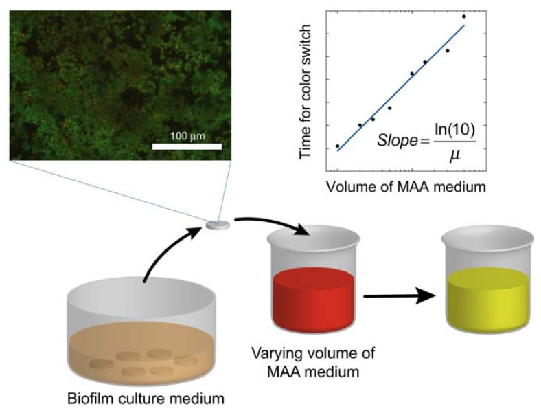
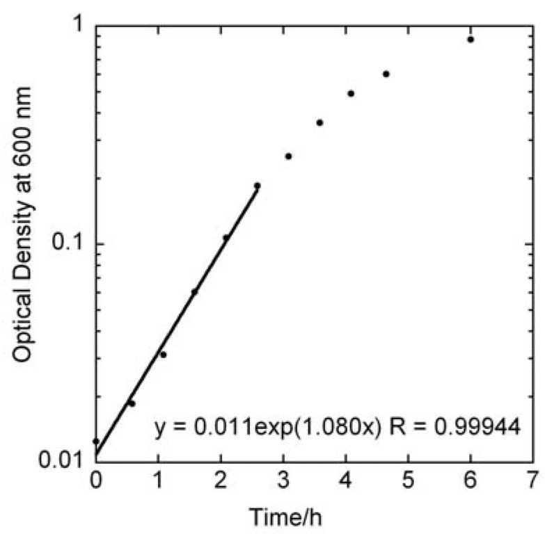
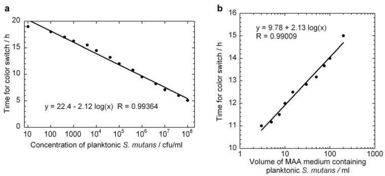
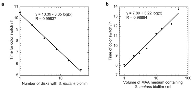
In this study we present a scheme for quantitative determination of biofilm viability offering significant improvement over existing methods with metabolic assays. Existing metabolic assays for quantifying viable bacteria in biofilms usually utilize calibration curves derived from planktonic bacteria, which can introduce large errors due to significant differences in the metabolic and/or growth rates of biofilm bacteria in the assay media compared to their planktonic counterparts. In the presented method we derive the specific growth rate of Streptococcus mutans bacteria biofilm from a series of metabolic assays using the pH indicator phenol red, and show that this information could be used to more accurately quantify the relative number of viable bacteria in a biofilm. We found that the specific growth rate of S. mutans in biofilm mode of growth was 0.70 h−1, compared to 1.09 h−1 in planktonic growth. This method should be applicable to other bacteria types, as well as other metabolic assays, and, for example, to quantify the effect of antibacterial treatments or the performance of bactericidal implant surfaces.
Overview
The claims and statistics of this paper haven’t been checked yet.
Run the check — free, no account needed. It reads the open-access text and checks the sources it cites.
Integrity signals
Registry
Cites
Statistics
Figures
Verification
Claims the paper makes
The claims and statistics of this paper haven’t been checked yet.
Run the check — free, no account needed. It reads the open-access text and checks the sources it cites.
Claims other papers make about this one
Each is checked against this paper’s own text: does it say what the citing paper says it does?
In the citing paper
“The culture-based technique was used to assess biofilm presence; however, the primary issue in accurately determining biofilm presence using the culture technique can be attributed to the inherent complexity and heterogeneity of biofilm structures.79”
From SEMTWIST Quantification of Biofilm Infection in Human Chronic Wound Using Scanning Electron Microscopy and Machine Learning (2025) · cites this paper as [79]
Self-check
Percentages, sums and reported differences re-computed from the paper’s own numbers.
The self-check needs the paper’s full text.
Citations
Cites
Oral multispecies biofilm development and the key role of cell–cell distance
Kolenbrander, P. E., Palmer, R. J., Periasamy, S., & Jakubovics, N. S. (2010) · Nature Reviews Microbiology · doi:10.1038/nrmicro2381 · Cited by 1,309
Research works printed but not linked: 27
- Davey, 2000, Microbial biofilms: From ecology to molecular genetics, Microbiol. Mol. Biol. Rev., 64, 847, 10.1128/MMBR.64.4.847-867.2000 · DOI
- Sutherland, 2001, The biofilm matrix—An immobilized but dynamic microbial environment, Trends Microbiol., 9, 222, 10.1016/S0966-842X(01)02012-1 · DOI
- Mah, 2001, Mechanisms of biofilm resistance to antimicrobial agents, Trends Microbiol., 9, 34, 10.1016/S0966-842X(00)01913-2 · DOI · Paper page
- Stewart, 2001, Antibiotic resistance of bacteria in biofilms, Lancet, 358, 135, 10.1016/S0140-6736(01)05321-1 · DOI
- Roberts, 2004, Modeling antibiotic tolerance in biofilms by accounting for nutrient limitation, Antimicrob. Agents Chemother., 48, 48, 10.1128/AAC.48.1.48-52.2004 · DOI
- Ito, 2009, Increased antibiotic resistance of escherichia coli in mature biofilms, Appl. Environ. Microbiol., 75, 4093, 10.1128/AEM.02949-08 · DOI
- Konig, 2001, Factors compromising antibiotic activity against biofilms of Staphylococcus epidermidis, Eur. J. Clin. Microbiol. Infect. Dis., 20, 20, 10.1007/PL00011232 · DOI
- Kuramitsu, 2006, Role of bacteriocin immunity proteins in the antimicrobial sensitivity of Streptococcus mutans, J. Bacteriol., 188, 8095, 10.1128/JB.00908-06 · DOI
- Ceri, 1999, The calgary biofilm device: New technology for rapid determination of antibiotic susceptibilities of bacterial biofilms, J. Clin. Microbiol., 37, 1771, 10.1128/JCM.37.6.1771-1776.1999 · DOI · Paper page
- Berney, 2007, Assessment and interpretation of bacterial viability by using the LIVE/DEAD baclight kit in combination with flow cytometry, Appl. Environ. Microbiol., 73, 3283, 10.1128/AEM.02750-06 · DOI
- Pognan, 2001, Investigation of the alamar blue (resazurin) fluorescent dye for the assessment of mammalian cell cytotoxicity, Toxicology, 164, 132
- Pettit, 2005, Microplate alamar blue assay for Staphylococcus epidermidis biofilm susceptibility testing, Antimicrob. Agents Chemother., 49, 2612, 10.1128/AAC.49.7.2612-2617.2005 · DOI
- Mariscal, 2009, Fluorescent assay based on resazurin for detection of activity of disinfectants against bacterial biofilm, Appl. Microbiol. Biotechnol., 82, 773, 10.1007/s00253-009-1879-x · DOI
- Honraet, 2005, Comparison of three assays for the quantification of candida biomass in suspension and CDC reactor grown biofilms, J. Microbiol. Methods, 63, 287, 10.1016/j.mimet.2005.03.014 · DOI
- Prieto, 2004, Biofilm quantification on stone surfaces: comparison of various methods, Sci. Total Environ., 333, 1, 10.1016/j.scitotenv.2004.05.003 · DOI
- Peeters, 2008, Comparison of multiple methods for quantification of microbial biofilms grown in microtiter plates, J. Microbiol. Meth., 72, 157, 10.1016/j.mimet.2007.11.010 · DOI
- Berlutti, 2003, Quantitative evaluation of bacteria adherent to polyelectrolyte HEMA-based hydrogels, J. Biomed. Mater. Res. A, 67A, 18, 10.1002/jbm.a.10026 · DOI
- Pantanella, 2008, Biotimer assay, a new method for counting Staphylococcus spp. in biofilm without sample manipulation applied to evaluate antibiotic susceptibility of biofilm, J. Microbiol. Methods, 75, 478, 10.1016/j.mimet.2008.07.027 · DOI
- Donlan, 2001, Biofilm formation: A clinically relevant microbiological process, Clin. Infect. Dis., 33, 1387, 10.1086/322972 · DOI · Paper page
- Donlan, 2002, Biofilms: Survival mechanisms of clinically relevant microorganisms, Clin. Microbiol. Rev., 15, 167, 10.1128/CMR.15.2.167-193.2002 · DOI · Paper page
- Sandberg, 2009, Pros and cons of using resazurin staining for quantification of viable Staphylococcus aureus biofilms in a screening assay, J. Microbiol. Methods, 78, 104, 10.1016/j.mimet.2009.04.014 · DOI
- Brohede, 2009, Multifunctional implant coatings providing possibilities for fast antibiotics loading with subsequent slow release, J. Mater. Sci. Mater. Med., 20, 1859, 10.1007/s10856-009-3749-6 · DOI
- Forsgren, 2009, Fast loading, slow release—A new strategy for incorporating antibiotics to hydroxyapatite, Key Eng. Mater., 396–398, 523
- Welch, 2010, Dental adhesives with bioactive and on-demand bactericidal properties, Dent. Mater., 26, 491, 10.1016/j.dental.2010.01.008 · DOI
- Banas, 2004, Virulence properties of Streptococcus mutans, Front. Biosci., 9, 1267, 10.2741/1305 · DOI
- Merritt, 2003, Mutation of luxS affects biofilm formation in Streptococcus mutans, Infect. Immun., 71, 1972, 10.1128/IAI.71.4.1972-1979.2003 · DOI
- Kaewsrichan, 2008, Ion-paired RP-HPLC for determining fermentation end products of Streptococcus mutans grown under different conditions, Scienceasia, 34, 193, 10.2306/scienceasia1513-1874.2008.34.193 · DOI
Cited by
Sorted by citations
Recent developments in the use of nanoparticles for treatment of biofilms
Han, C., Romero, N., Fischer, S., Dookran, J., Berger, A., & Doiron, A. L. (2017) · Nanotechnology Reviews · doi:10.1515/ntrev-2016-0054 · Cited by 114
Singh, S., Muniz De Oliveira, F., Wang, C., Kumar, M., Xuan, Y., DeMazumder, D., Sen, C. K., & Roy, S. (2025) · Advances in Wound Care · doi:10.1089/wound.2024.0291 · Cited by 6
Metadata
- Authors
- Welch, K., Cai, Y., & Strømme, M.
- Venue
- Journal of Functional Biomaterials
- Publisher
- MDPI AG
- Published
- 2012-06-01
- Volume
- 3
- Issue
- 2
- Pages
- 418-431
- ISSN
- 2079-4983
- PMC
- PMC4047939
- Cited by
- 96
- References
- 1
- Language
- en
About these checks
Registry, retraction, Cites and statistics results are computed, not judged, with one exception: a printed reference that no index lists is read once by a model before it counts as a citation error. Claims are extracted by a model and kept only when their quote appears verbatim in the text; each cited source is then checked, with a model’s help, for whether it says what the claim relies on. Those verdicts can be wrong — read the claim’s quote and the cited source before relying on one.
Statistics and arithmetic are recomputed, not judged.
Confidence bands are shown only for fields calibrated on our annotated test set. None is calibrated yet, so no band is shown.
What counts as a claim ›How we measure confidence ›How we check this ›
Are you an author, or do you see a problem with this page? Request a review
