

The DOI matches its Crossref record; 63 of the 65 references it cites were found in a registry or an index.
Enhanced Expression of p53 and Suppression of PI3K/Akt/mTOR by Three Red Sea Algal Extracts: Insights on Their Composition by LC-MS-Based Metabolic Profiling and Molecular Networking
Open accessAbstract
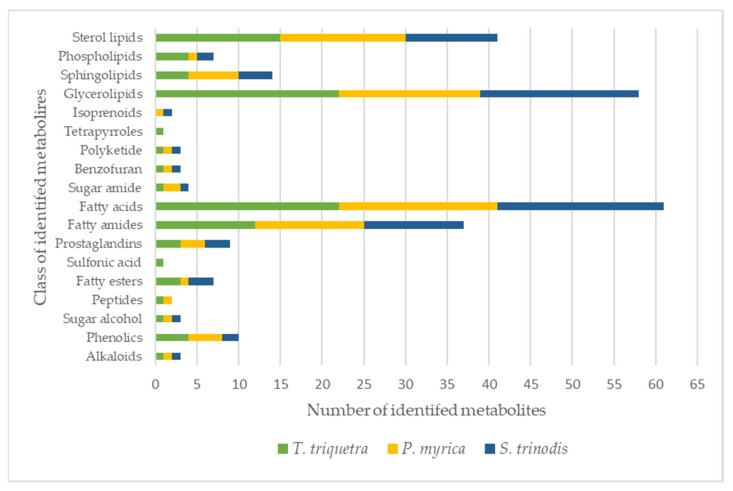
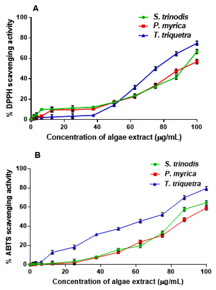
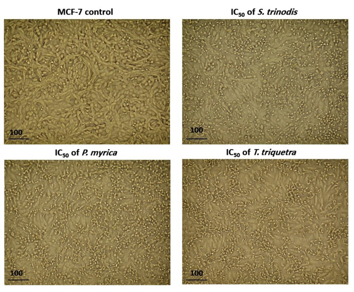
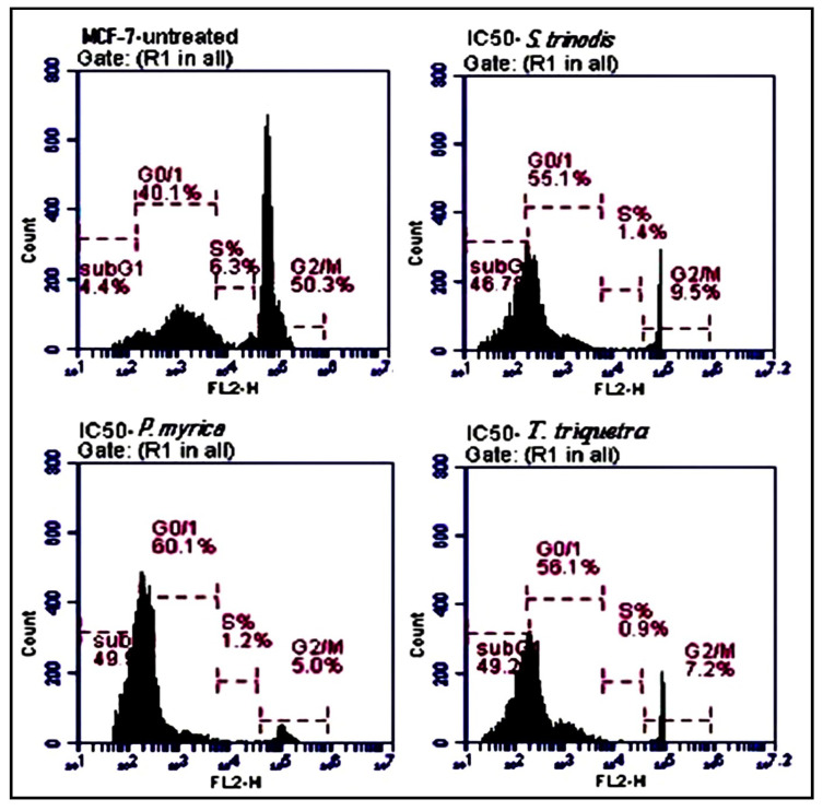
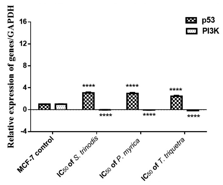
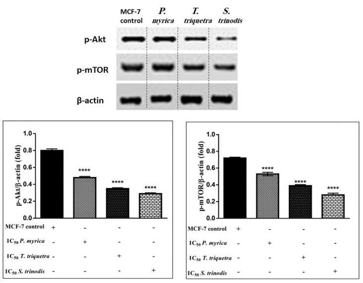
Brown algae comprise up to 2000 species with wide dissemination in temperate zones. A comprehensive untargeted metabolic profiling guided by molecular networking of three uninvestigated Red-Sea-derived brown algae, namely Sirophysalis trinodis, Polycladia myrica, and Turbinaria triquetra, led to the identification of over 115 metabolites categorized as glycerolipids, fatty acids, sterol lipids, sphingolipids, and phospholipids. The three algae exhibited low-to-moderate antioxidant capacity using DPPH and ABTS assays. Preliminary in vitro antiproliferative studies showed that the algal extracts displayed high cytotoxic activity against a panel of cancer cell lines. The most potent activity was recorded against MCF-7 with IC50 values of 51.37 ± 1.19, 63.44 ± 1.13, and 59.70 ± 1.22 µg/mL for S. trinodis, P. myrica, and T. triquetra, respectively. The cytotoxicity of the algae was selective to MCF-7 without showing notable effects on the proliferation of normal human WISH cells. Morphological studies revealed that the algae caused cell shrinkage, increased cellular debris, triggered detachment, cell rounding, and cytoplasmic condensation in MCF-7 cancer cells. Mechanistic investigations using flow cytometry, qPCR, and Western blot showed that the algae induced apoptosis, initiated cell cycle arrest in the sub-G0/G1 phase, and inhibited the proliferation of cancer cells via increasing mRNA and protein expression of p53, while reducing the expression of PI3K, Akt, and mTOR.
Overview
The claims and statistics of this paper haven’t been checked yet.
Run the check — free, no account needed. It reads the open-access text and checks the sources it cites.
Integrity signals
Registry
Cites
Statistics
Figures
Verification
Claims the paper makes
The claims and statistics of this paper haven’t been checked yet.
Run the check — free, no account needed. It reads the open-access text and checks the sources it cites.
Claims other papers make about this one
Each is checked against this paper’s own text: does it say what the citing paper says it does?
The source backs part of the claim, not all of it — or backs it for a different population, condition or comparison.
In the citing paper
“The presence of the same fatty acids was also confirmed in the methanolic extracts of the red seaweed species Turbinaria triquetra and Polycladia myrica [30].”
From Improvement of Alginate Extraction from Brown Seaweed (Laminaria digitata L.) and Valorization of Its Remaining Ethanolic Fraction (2024) · cites this paper as [30]
Experimental — accuracy not yet measuredThe cited source says what the claim attributes to it.
In the citing paper
“Similar to the previous study [30], sterol lipid (22), which had an [M − H]− ion at m/z 439.29, was also detected in the analyzed extract.”
From Improvement of Alginate Extraction from Brown Seaweed (Laminaria digitata L.) and Valorization of Its Remaining Ethanolic Fraction (2024) · cites this paper as [30]
Self-check
Percentages, sums and reported differences re-computed from the paper’s own numbers.
The self-check needs the paper’s full text.
Citations
Cites
Bradford, M. M. (1976) · Analytical Biochemistry · doi:10.1016/0003-2697(76)90527-3 · Cited by 255,302
Analysis of Relative Gene Expression Data Using Real-Time Quantitative PCR and the 2−ΔΔCT Method
Livak, K. J., & Schmittgen, T. D. (2001) · Methods · doi:10.1006/meth.2001.1262 · Cited by 180,860
Antioxidant activity applying an improved ABTS radical cation decolorization assay
Re, R., Pellegrini, N., Proteggente, A. R., Pannala, A. S., Yang, M. H., & Rice-Evans, C. A. (1999) · Free Radical Biology and Medicine · doi:10.1016/s0891-5849(98)00315-3 · Cited by 26,986
Mutation of the mouse klotho gene leads to a syndrome resembling ageing
Kuro-o, M., Matsumura, Y., Aizawa, H., Kawaguchi, H., Suga, T., Utsugi, T., Ohyama, Y., Kurabayashi, M., Kaname, T., Kume, E., et al. (1997) · Nature · doi:10.1038/36285 · Cited by 3,837
Antioxidant activity of Nigella sativa essential oil
Burits, M., & Bučar, F. (2000) · Phytotherapy Research · doi:10.1002/1099-1573(200008)14:5 · Cited by 1,859
Targeting PI3K/Akt/mTOR Signaling in Cancer
Porta, C., Paglino, C., & Mosca, A. (2014) · Frontiers in Oncology · doi:10.3389/fonc.2014.00064 · Cited by 1,547
Drug development from marine natural products
Molinski, T. F., Dalisay, D. S., Lievens, S. C., & Saludes, J. P. (2008) · Nature Reviews Drug Discovery · doi:10.1038/nrd2487 · Cited by 1,226
The global burden of cancer: priorities for prevention
Thun, M. J., DeLancey, J. O. L., Jemal, A., & Ward, E. (2009) · Carcinogenesis · doi:10.1093/carcin/bgp263 · Cited by 1,093
Targeting PI3K/mTOR Signaling in Cancer
Arcaro, A. (2014) · Frontiers in Oncology · doi:10.3389/fonc.2014.00084 · Cited by 1,027
An overview on the role of dietary phenolics for the treatment of cancers
Anantharaju, P. G., Prathima, C., Vimalambike, M. G., & Madhunapantula, S. V. (2016) · Nutrition Journal · doi:10.1186/s12937-016-0217-2 · Cited by 525
Research works printed but not linked: 3
- Kharkwal, 2012, Algae as future drugs, Asian J. Pharm. Clin. Res, 5, 1
- Lu, W.-Y., Li, H.-J., and Wu, Y.-C. (2023). Marine Biochemistry, CRC Press.
- Salem, 2014, Terpenes and sterols composition of marine brown algae Padina pavonica (Dictyotales) and Hormophysa triquetra (Fucales), Int. J. Pharmacogn. Phytochem., 6, 894
Cited by
Sorted by citations
Savić Gajić, I. M., Savić, I. M., Ivanovska, A. M., Vunduk, J. D., Mihalj, I. S., & Svirčev, Z. B. (2024) · Marine Drugs · doi:10.3390/md22060280 · Cited by 29
Noser, A. A., El-Barbary, A. A., Salem, M. M., Salam, H. A. A. E., & shahien, M. (2024) · Scientific Reports · doi:10.1038/s41598-024-53517-y · Cited by 27
Self-citationBoškov, I. A., Savic, I. M., Grozdanić, N., Kundaković, T., Selgrad, J. S. R., & Savić, I. (2024) · Polymers · doi:10.3390/polym16050688 · Cited by 20
El-Demerdash, F. M., Mhanna, A. B. A., El-Sayed, R. A., Mohamed, T. M., & Salem, M. M. (2024) · Tissue and Cell · doi:10.1016/j.tice.2024.102332 · Cited by 18
Self-citationExtraction and Characterization of Antioxidants and Cellulose from Green Walnut Husks
Savic, I. M., & Savić, I. (2025) · Foods · doi:10.3390/foods14030409 · Cited by 17
Metadata
- Authors
- Fahmy, N. M., El-Din, M. I. G., Salem, M. M., Rashedy, S. H., Lee, G. S., Jang, Y. S., Kim, K. H., Kim, C. S., El-Shazly, M., & Fayez, S.
- Affiliations
- Ain Shams University (EG)
- Department of Pharmacognosy, Faculty of Pharmacy, Ain-Shams University, Cairo 11566, Egypt
- Tanta University (EG)
- National Institute of Oceanography and Fisheries (EG)
- Sungkyunkwan University (KR)
- Corresponding author
- Kim, Chung Sub
- ORCID
- Venue
- Marine Drugs
- Publisher
- MDPI AG
- Published
- 2023-07-17
- Volume
- 21
- Issue
- 7
- Pages
- 404
- ISSN
- 1660-3397 (ISSN-L)
- PMC
- PMC10381385
- OpenAlex
- W4384636495
- Cited by
- 28
- References
- 65
- Citations by year
- 2026: 5 · 2025: 10 · 2024: 13
- FWCI
- 5.74
- Type
- journal article
- Language
- en
- Open access
- gold
- doi.org
- Licence
- Locations
- Marine Drugs (publishedVersion, cc-by)
- PubMed Central (publishedVersion, cc-by)
- PubMed (publishedVersion)
- DOAJ (DOAJ: Directory of Open Access Journals) (submittedVersion, cc-by-sa)
- Funders
- National Research Foundation (NRF) of Korea (2022R1A6A1A03054419)
- Topics
- Life Sciences › Agricultural and Biological Sciences › Aquatic Science › Seaweed-derived Bioactive Compounds
- Physical Sciences › Chemistry › Organic Chemistry › Phytochemical Studies and Bioactivities
- Physical Sciences › Energy › Renewable Energy, Sustainability and the Environment › Algal biology and biofuel production
- Keywords
- algal metabolites, untargeted metabolomics, molecular networking, glycerolipids, sphingolipids, phospholipids, antioxidant activity, antiproliferative activity, MCF-7 cells, apoptosis, cell cycle arrest, p53 expression, PI3K/AKT/mTOR pathway, cytotoxicity
- MeSH terms
- Chromatography, Liquid, Humans, Signal Transduction, Tumor Suppressor Protein p53, Apoptosis, Indian Ocean, Phosphatidylinositol 3-Kinases, Cell Line, Tumor, Cell Proliferation, Proto-Oncogene Proteins c-akt, Tandem Mass Spectrometry, TOR Serine-Threonine Kinases, MCF-7 Cells
About these checks
Registry, retraction, Cites and statistics results are computed, not judged, with one exception: a printed reference that no index lists is read once by a model before it counts as a citation error. Claims are extracted by a model and kept only when their quote appears verbatim in the text; each cited source is then checked, with a model’s help, for whether it says what the claim relies on. Those verdicts can be wrong — read the claim’s quote and the cited source before relying on one.
Statistics and arithmetic are recomputed, not judged.
Confidence bands are shown only for fields calibrated on our annotated test set. None is calibrated yet, so no band is shown.
What counts as a claim ›How we measure confidence ›How we check this ›
Are you an author, or do you see a problem with this page? Request a review
