

The DOI matches its Crossref record; 2 of the 2 references it cites were found in a registry or an index.
Scanning Electron Microscope: A New Potential Tool to Replace Gram Staining for Microbe Identification in Blood Cultures
Abstract
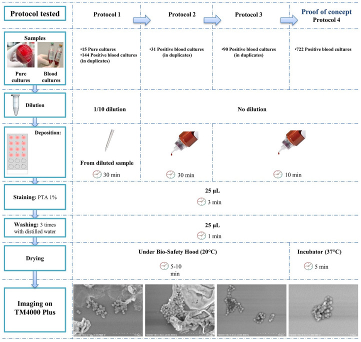
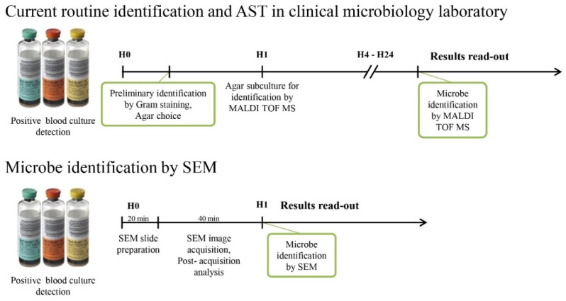
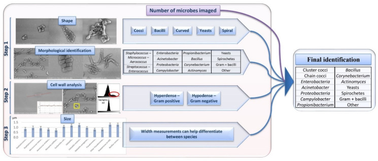
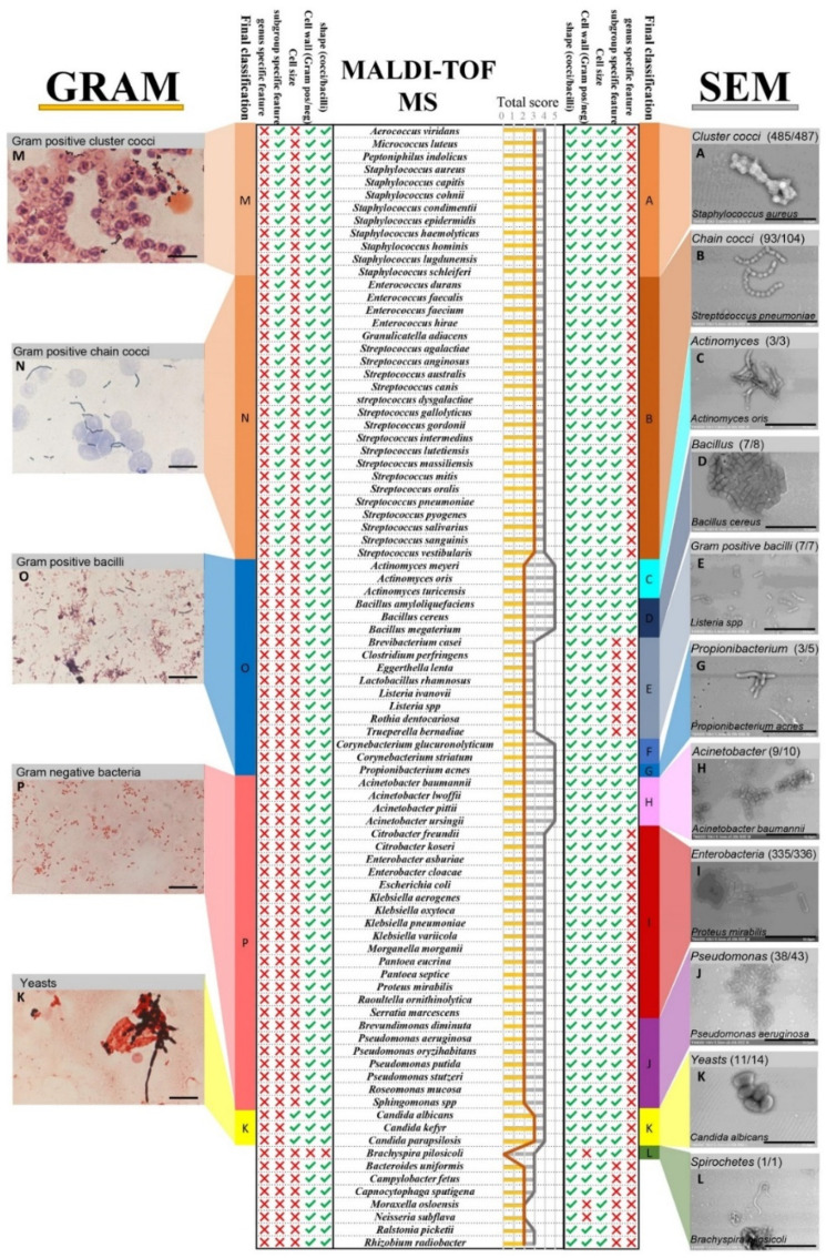
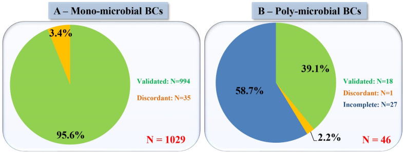
Blood culture is currently the most commonly used method for diagnosing sepsis and bloodstream infections. However, the long turn-around-time to achieve microbe identification remains a major concern for clinical microbiology laboratories. Gram staining for preliminary identification remains the gold standard. We developed a new rapid strategy using a tabletop scanning electron microscope (SEM) and compared its performance with Gram staining for the detection of micro-organisms and preliminary identification directly from blood cultures. We first optimised the sample preparation for twelve samples simultaneously, saving time on imaging. In this work, SEM proved its ability to identify bacteria and yeasts in morphotypes up to the genus level in some cases. We blindly tested 1075 blood cultures and compared our results to the Gram staining preliminary identification, with MALDI-TOF/MS as a reference. This method presents major advantages such as a fast microbe identification, within an hour of the blood culture being detected positive, low preparation costs, and data traceability. This SEM identification strategy can be developed into an automated assay from the sample preparation, micrograph acquisition, and identification process. This strategy could revolutionise urgent microbiological diagnosis of infectious diseases.
Overview
The claims and statistics of this paper haven’t been checked yet.
Run the check — free, no account needed. It reads the open-access text and checks the sources it cites.
Integrity signals
Registry
Cites
Statistics
Figures
Verification
Claims the paper makes
The claims and statistics of this paper haven’t been checked yet.
Run the check — free, no account needed. It reads the open-access text and checks the sources it cites.
Claims other papers make about this one
Each is checked against this paper’s own text: does it say what the citing paper says it does?
In the citing paper
“The high resolution of SEM allows detailed analysis of biofilm structures, allowing direct visualization by achieving a resolution of 50 to 100 nm.25”
From SEMTWIST Quantification of Biofilm Infection in Human Chronic Wound Using Scanning Electron Microscopy and Machine Learning (2025) · cites this paper as [25]
Self-check
Percentages, sums and reported differences re-computed from the paper’s own numbers.
The self-check needs the paper’s full text.
Citations
Cites
Seng, P., Drancourt, M., Gouriet, F., La Scola, B., Fournier, P., Rolain, J. M., & Raoult, D. (2009) · Clinical Infectious Diseases · doi:10.1086/600885 · Cited by 1,983 · cited 1×
“As a reference method, colonies from subcultures were also identified using MALDI TOF/MS (Microflex, Bruker Daltonics, Bremen, Germany) as previously described [27].”
MethodSelf-citationMutters, N. T., Hodiamont, C. J., Jong, M. D. D., Overmeijer, H. P. J., Boogaard, M. V. D., & E.Visser, C. (2014) · Annals of Laboratory Medicine · doi:10.3343/alm.2014.34.2.111 · Cited by 82
Research works printed but not linked: 32
- Mingorance, 2012, Identification and Susceptibility Testing of Microorganism by Direct Inoculation from Positive Blood Culture Bottles by Combining MALDI-TOF and Vitek-2 Compact Is Rapid and Effective, J. Infect., 65, 513, 10.1016/j.jinf.2012.08.013 · DOI
- Kok, J., Thomas, L.C., Olma, T., Chen, S.C.A., and Iredell, J.R. (2011). Identification of Bacteria in Blood Culture Broths Using Matrix-Assisted Laser Desorption-Ionization SepsityperTM and Time of Flight Mass Spectrometry. PLoS ONE, 6. · DOI
- Dubourg, 2016, Emerging Methodologies for Pathogen Identification in Positive Blood Culture Testing, Expert Rev. Mol. Diagn., 16, 97, 10.1586/14737159.2016.1112274 · DOI
- Dubourg, 2019, Emerging Methodologies for Pathogen Identification in Bloodstream Infections: An Update, Expert Rev. Mol. Diagn., 19, 161, 10.1080/14737159.2019.1568241 · DOI
- Verroken, A., Defourny, L., Waroux, O.P., de Belkhir, L., Laterre, P.-F., Delmée, M., and Glupczynski, Y. (2016). Clinical Impact of MALDI-TOF MS Identification and Rapid Susceptibility Testing on Adequate Antimicrobial Treatment in Sepsis with Positive Blood Cultures. PLoS ONE, 11. · DOI
- Wolk, 2019, Rapid Diagnostics for Blood Cultures: Supporting Decisions for Antimicrobial Therapy and Value-Based Care, J. Appl. Lab. Med., 3, 686, 10.1373/jalm.2018.028159 · DOI
- Aoki, 2010, Refinement of presumptive antimicrobial therapy based on initial microbiological information on positive blood culture, Rinsho Byori, 58, 498
- Boyanova, 2018, Direct Gram Staining and Its Various Benefits in the Diagnosis of Bacterial Infections, Postgrad. Med., 130, 105, 10.1080/00325481.2018.1398049 · DOI
- 1995, The Impact of the First Notification of Positive Blood Cultures on Antibiotic Therapy, APMIS, 103, 37, 10.1111/j.1699-0463.1995.tb01077.x · DOI
- Barenfanger, 2008, Decreased Mortality Associated With Prompt Gram Staining of Blood Cultures, Am. J. Clin. Pathol., 130, 870, 10.1309/AJCPVMDQU2ZJDPBL · DOI
- Nagata, 2010, Usefulness and limit of Gram staining smear examination, Rinsho Byori, 58, 490
- Beveridge, 1990, Mechanism of Gram Variability in Select Bacteria, J. Bacteriol., 172, 1609, 10.1128/jb.172.3.1609-1620.1990 · DOI
- Esteban, 1996, Failure of Gram Stain to Detect Propionibacterium Acnes in Specimens from Clinically Significant Infections, J. Clin. Microbiol., 34, 2051, 10.1128/jcm.34.8.2051-2051.1996 · DOI
- Pliakos, E.E., Andreatos, N., Shehadeh, F., Ziakas, P.D., and Mylonakis, E. (2018). The Cost-Effectiveness of Rapid Diagnostic Testing for the Diagnosis of Bloodstream Infections with or without Antimicrobial Stewardship. Clin. Microbiol. Rev., 31. · DOI
- Verroken, A., Despas, N., Rodriguez-Villalobos, H., and Laterre, P.-F. (2019). The Impact of a Rapid Molecular Identification Test on Positive Blood Cultures from Critically Ill with Bacteremia: A Pre-Post Intervention Study. PLoS ONE, 14. · DOI
- Hou, 2019, Current Status of MALDI-TOF Mass Spectrometry in Clinical Microbiology, J. Food Drug Anal., 27, 404, 10.1016/j.jfda.2019.01.001 · DOI
- MacVane, 2016, Benefits of Adding a Rapid PCR-Based Blood Culture Identification Panel to an Established Antimicrobial Stewardship Program, J. Clin. Microbiol., 54, 2455, 10.1128/JCM.00996-16 · DOI · Paper page
- Mancini, 2014, Potential Impact of a Microarray-Based Nucleic Acid Assay for Rapid Detection of Gram-Negative Bacteria and Resistance Markers in Positive Blood Cultures, J. Clin. Microbiol., 52, 1242, 10.1128/JCM.00142-14 · DOI
- Peker, 2018, Diagnosis of Bloodstream Infections from Positive Blood Cultures and Directly from Blood Samples: Recent Developments in Molecular Approaches, Clin. Microbiol. Infect., 24, 944, 10.1016/j.cmi.2018.05.007 · DOI
- Zrodlowski, 2017, Fluorescent in Situ Hybridization and Gram-stained Smears of Whole Blood as Complementary Screening Tools in the Diagnosis of Sepsis, Pol. Arch. Intern. Med., 127, 122, 10.20452/pamw.3949 · DOI
- Koh, 2016, Combinational Approach Using in Situ Hybridization Targeting 23S Ribosomal RNA Genes and Blood Cultures for Bacterial Identification in Patients with Neutropenia and Fever, J. Infect. Chemother., 22, 697, 10.1016/j.jiac.2016.08.001 · DOI
- Berild, 2006, Adjustment of Antibiotic Treatment According to the Results of Blood Cultures Leads to Decreased Antibiotic Use and Costs, J. Antimicrob. Chemother., 57, 326, 10.1093/jac/dki463 · DOI
- Bourbeau, 2013, Automation in Clinical Microbiology, J. Clin. Microbiol., 51, 1658, 10.1128/JCM.00301-13 · DOI
- Dauwalder, 2016, Does Bacteriology Laboratory Automation Reduce Time to Results and Increase Quality Management?, Clin. Microbiol. Infect., 22, 236, 10.1016/j.cmi.2015.10.037 · DOI
- Schindelin, 2012, Fiji: An Open-Source Platform for Biological-Image Analysis, Nat. Methods, 9, 676, 10.1038/nmeth.2019 · DOI · Paper page
- Sogaard, 2007, First Notification of Positive Blood Cultures and the High Accuracy of the Gram Stain Report, J. Clin. Microbiol., 45, 1113, 10.1128/JCM.02523-06 · DOI
- Fenollar, 2000, Comparison of a Commercial Disk Test with Vancomycin and Colimycin Susceptibility Testing for Identification of Bacteria with Abnormal Gram Staining Reactions, EJCMID, 19, 33, 10.1007/s100960050006 · DOI
- Haddad, 2020, Rapid Scanning Electron Microscopy Detection and Sequencing of Severe Acute Respiratory Syndrome Coronavirus 2 and Other Respiratory Viruses, Front. Microbiol., 11, 596180, 10.3389/fmicb.2020.596180 · DOI
- Haddad, G., Fontanini, A., Bellali, S., Takakura, T., Ominami, Y., Hisada, A., Hadjadj, L., Rolain, J.M., Raoult, D., and Bou Khalil, J. (2021). Rapid Detection of Imipenem Resistance in Gram-Negative Bacteria Using Tabletop Scanning Electron Microscopy: A Preliminary Evaluation. Front. Microbiol., 658322. · DOI
- Hannachi, N., Lepidi, H., Fontanini, A., Takakura, T., Bou-Khalil, J., Gouriet, F., Habib, G., Raoult, D., Camoin-Jau, L., and Baudoin, J.-P. (2020). A Novel Approach for Detecting Unique Variations among Infectious Bacterial Species in Endocarditic Cardiac Valve Vegetation. Cells, 9. · DOI
- Verroken, 2015, Reducing Time to Identification of Positive Blood Cultures with MALDI-TOF MS Analysis after a 5-h Subculture, Eur. J. Clin. Microbiol. Infect. Dis., 34, 405, 10.1007/s10096-014-2242-4 · DOI
- Gaibani, 2009, Blood Culture Systems: Rapid Detection—How and Why?, Int. J. Antimicrob. Agents, 34, S13, 10.1016/S0924-8579(09)70559-X · DOI
Cited by
Sorted by citations
Singh, S., Muniz De Oliveira, F., Wang, C., Kumar, M., Xuan, Y., DeMazumder, D., Sen, C. K., & Roy, S. (2025) · Advances in Wound Care · doi:10.1089/wound.2024.0291 · Cited by 6
Metadata
- Authors
- Haddad, G., Bellali, S., Takakura, T., Fontanini, A., Ominami, Y., Bou Khalil, J., & Raoult, D.
- Venue
- Microorganisms
- Publisher
- MDPI AG
- Published
- 2021-05-28
- Volume
- 9
- Issue
- 6
- Pages
- 1170
- ISSN
- 2076-2607
- PMC
- PMC8227564
- Cited by
- 20
- References
- 2
- Language
- en
- Funders
- Fondation Méditerranée Infection (ANR-10-IAHU-03)
About these checks
Registry, retraction, Cites and statistics results are computed, not judged, with one exception: a printed reference that no index lists is read once by a model before it counts as a citation error. Claims are extracted by a model and kept only when their quote appears verbatim in the text; each cited source is then checked, with a model’s help, for whether it says what the claim relies on. Those verdicts can be wrong — read the claim’s quote and the cited source before relying on one.
Statistics and arithmetic are recomputed, not judged.
Confidence bands are shown only for fields calibrated on our annotated test set. None is calibrated yet, so no band is shown.
What counts as a claim ›How we measure confidence ›How we check this ›
Are you an author, or do you see a problem with this page? Request a review
How to cite
Cite CoChat for the checks on this page, and the paper for its own findings.
- This page
- CoChat. (2026). Citation check: Scanning Electron Microscope: A New Potential Tool to Replace Gram Staining for Microbe Identification in Blood Cultures. https://app.cochat.ai/papers/10.3390/microorganisms9061170
- The paper
- Haddad, G., Bellali, S., Takakura, T., Fontanini, A., Ominami, Y., Bou Khalil, J., & Raoult, D. (2021). Scanning Electron Microscope: A New Potential Tool to Replace Gram Staining for Microbe Identification in Blood Cultures. Microorganisms. https://doi.org/10.3390/microorganisms9061170
