

The DOI matches its Crossref record.
Applications of Magnetotactic Bacteria, Magnetosomes and Magnetosome Crystals in Biotechnology and Nanotechnology: Mini-Review
Open accessAbstract
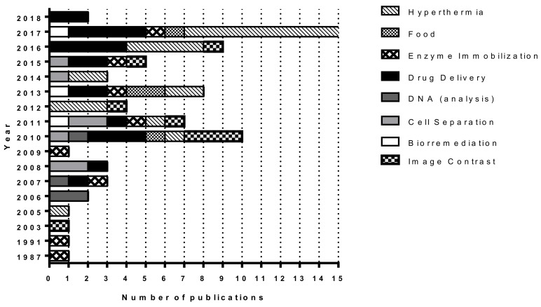
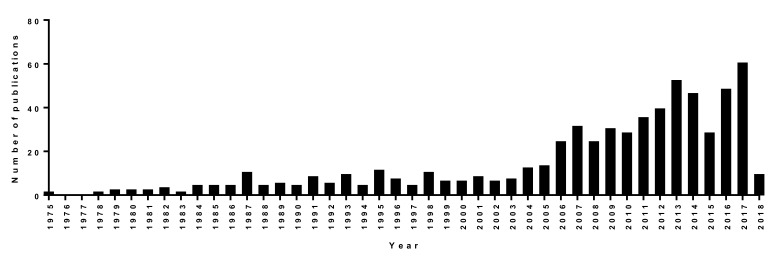
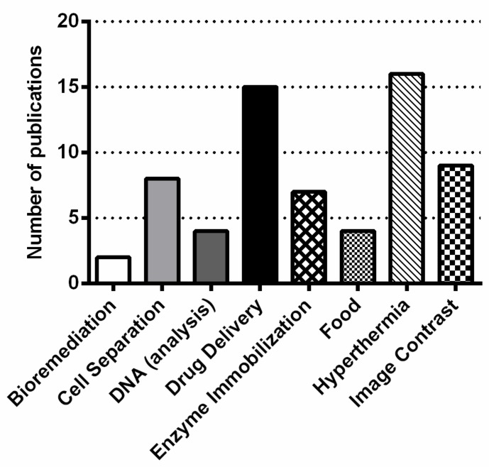
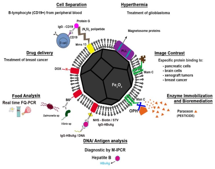
Magnetotactic bacteria (MTB) biomineralize magnetosomes, which are defined as intracellular nanocrystals of the magnetic minerals magnetite (Fe₃O₄) or greigite (Fe₃S₄) enveloped by a phospholipid bilayer membrane. The synthesis of magnetosomes is controlled by a specific set of genes that encode proteins, some of which are exclusively found in the magnetosome membrane in the cell. Over the past several decades, interest in nanoscale technology (nanotechnology) and biotechnology has increased significantly due to the development and establishment of new commercial, medical and scientific processes and applications that utilize nanomaterials, some of which are biologically derived. One excellent example of a biological nanomaterial that is showing great promise for use in a large number of commercial and medical applications are bacterial magnetite magnetosomes. Unlike chemically-synthesized magnetite nanoparticles, magnetosome magnetite crystals are stable single-magnetic domains and are thus permanently magnetic at ambient temperature, are of high chemical purity, and display a narrow size range and consistent crystal morphology. These physical/chemical features are important in their use in biotechnological and other applications. Applications utilizing magnetite-producing MTB, magnetite magnetosomes and/or magnetosome magnetite crystals include and/or involve bioremediation, cell separation, DNA/antigen recovery or detection, drug delivery, enzyme immobilization, magnetic hyperthermia and contrast enhancement of magnetic resonance imaging. Metric analysis using Scopus and Web of Science databases from 2003 to 2018 showed that applied research involving magnetite from MTB in some form has been focused mainly in biomedical applications, particularly in magnetic hyperthermia and drug delivery.
Overview
The claims and statistics of this paper haven’t been checked yet.
Run the check — free, no account needed. It reads the open-access text and checks the sources it cites.
Integrity signals
Registry
Cites
Statistics
Figures
Verification
Claims the paper makes
The claims and statistics of this paper haven’t been checked yet.
Run the check — free, no account needed. It reads the open-access text and checks the sources it cites.
Claims other papers make about this one
Each is checked against this paper’s own text: does it say what the citing paper says it does?
The cited source says what the claim attributes to it.
In the citing paper
“Finally, in a pathway of iron biomineralization approximating asbestos body formation, several bacteria incorporate iron from their environment to synthesize intracellular nanoparticles of magnetite or greigite (Fe3S4) in organelles called magnetosomes [137,138].”
From Asbestos and Iron (2023) · cites this paper as [137]
Self-check
Percentages, sums and reported differences re-computed from the paper’s own numbers.
The self-check needs the paper’s full text.
Citations
Cites
No references could be resolved yet.
Research works printed but not linked: 131
- Bazylinski, 2004, Magnetosome formation in prokaryotes, Nat. Rev. Microbiol., 2, 217, 10.1038/nrmicro842 · DOI
- Frankel, 2006, How magnetotactic bacteria make magnetosomes queue up, Trends Microbiol., 14, 329, 10.1016/j.tim.2006.06.004 · DOI
- Bellini, 2009, Ulteriori Studi Sui “batteri Magnetosensibili”. Universita di Pavia, Pavia, Italy, 1963, apud Frankel, R.B. The discovery of magnetotactic/magnetosensitive bactéria, Chin. J. Oceanol. Limnol., 27, 1
- Blakemore, 1975, Magnetotactic bacteria, Science, 190, 377, 10.1126/science.170679 · DOI
- Lower, 2013, The bacterial magnetosome: A unique prokaryotic organelle, J. Mol. Microbiol. Biotechnol., 23, 63
- Lefevre, 2013, Ecology, diversity, and evolution of magnetotactic bacteria, Microbiol. Mol. Biol. Rev., 77, 497, 10.1128/MMBR.00021-13 · DOI
- Scopus (2018). Elsevier′s scopus, the largest abstract and citation database of peer-reviewed literature. Elsevier′s Scopus, Elsevier. Available online: https://www.scopus.com/.
- Yan, 2017, Bacterial magnetosome and its potential application, Microbiol. Res., 203, 19, 10.1016/j.micres.2017.06.005 · DOI
- Jacob, 2016, Magnetotactic bacteria and magnetosomes - scope and challenges, Mater. Sci. Eng. C Mater. Biol. Appl., 68, 919, 10.1016/j.msec.2016.07.049 · DOI
- Matsunaga, 1987, Use of magnetic particles isolated from magnetotactic bacteria for enzyme immobilization, Appl. Microbiol. Biotechnol., 26, 328, 10.1007/BF00256663 · DOI
- Gorby, 1988, Characterization of the bacterial magnetosome membrane, J. Bacteriol., 170, 834, 10.1128/jb.170.2.834-841.1988 · DOI
- Faivre, 2008, Magnetotactic bacteria and magnetosomes, Chem. Rev., 108, 4875, 10.1021/cr078258w · DOI
- Murat, 2010, Cell biology of prokaryotic organelles, Cold Spring Harb. Perspect. Biol., 2, a000422, 10.1101/cshperspect.a000422 · DOI
- Abreu, 2013, Cell adhesion, multicellular morphology, and magnetosome distribution in the multicellular magnetotactic prokaryote Candidatus Magnetoglobus multicellularis, Microsc. Microanal., 19, 535, 10.1017/S1431927613000329 · DOI
- Komeili, 2012, Molecular mechanisms of compartmentalization and biomineralization in magnetotactic bacteria, FEMS Microbiol. Rev., 36, 232, 10.1111/j.1574-6976.2011.00315.x · DOI
- Kundu, 2009, On the change in bacterial size and magnetosome features for Magnetospirillum magnetotacticum (MS-1) under high concentrations of zinc and nickel, Biomaterials, 30, 4211, 10.1016/j.biomaterials.2009.04.039 · DOI
- Otto, 2004, Biochemical and proteomic analysis of the magnetosome membrane in Magnetospirillum gryphiswaldense, Appl. Environ. Microbiol., 70, 1040, 10.1128/AEM.70.2.1040-1050.2004 · DOI
- Amemiya, 2007, Controlled formation of magnetite crystal by partial oxidation of ferrous hydroxide in the presence of recombinant magnetotactic bacterial protein Mms6, Biomaterials, 28, 5381, 10.1016/j.biomaterials.2007.07.051 · DOI
- Arakaki, 2003, A novel protein tightly bound to bacterial magnetic particles in Magnetospirillum magneticum strain AMB-1, J. Biol. Chem., 278, 8745, 10.1074/jbc.M211729200 · DOI
- Murat, 2010, Comprehensive genetic dissection of the magnetosome gene island reveals the step-wise assembly of a prokaryotic organelle, Proc. Natl. Acad. Sci. USA, 107, 5593, 10.1073/pnas.0914439107 · DOI
- Quinlan, 2011, The htra/degp family protease mame is a bifunctional protein with roles in magnetosome protein localization and magnetite biomineralization, Mol. Microbiol., 80, 1075, 10.1111/j.1365-2958.2011.07631.x · DOI
- Scheffel, 2008, The major magnetosome proteins mamgfdc are not essential for magnetite biomineralization in Magnetospirillum gryphiswaldense but regulate the size of magnetosome crystals, J. Bacteriol., 190, 377, 10.1128/JB.01371-07 · DOI
- Tanaka, 2006, Origin of magnetosome membrane: Proteomic analysis of magnetosome membrane and comparison with cytoplasmic membrane, Proteomics, 6, 5234, 10.1002/pmic.200500887 · DOI
- Faivre, 2015, From bacteria to mollusks: The principles underlying the biomineralization of iron oxide materials, Angew. Chem. Int. Ed., 54, 4728, 10.1002/anie.201408900 · DOI
- DeLong, E.F., Lory, S., Stackebrandt, E., and Thompson, F. (2013). Magnetotactic Bacteria. The Prokaryotes, Springer.
- Baumgartner, 2013, Magnetotactic bacteria form magnetite from a phosphate-rich ferric hydroxide via nanometric ferric (oxyhydr) oxide intermediates, Proc. Natl. Acad. Sci. USA, 110, 883, 10.1073/pnas.1307119110 · DOI
- Buseck, 1998, Iron sulfides from magnetotactic bacteria: Structure, composition, and phase transitions, Am. Mineral., 83, 1469, 10.2138/am-1998-11-1235 · DOI
- Li, 2010, Magnetite nanoparticles with high heating efficiencies for application in the hyperthermia of cancer, Mater. Sci Eng., 30, 990, 10.1016/j.msec.2010.04.016 · DOI
- Sun, 2002, Size-Controlled Synthesis of Magnetite Nanoparticles, J. Am. Chem. Soc., 124, 8204, 10.1021/ja026501x · DOI
- Schwab, 2004, Polymeric precursors to refractory metal borides, J. Mat. Sci., 39, 6051, 10.1023/B:JMSC.0000041701.01103.41 · DOI
- Qi, 2011, A non-alkoxide sol-gel method for the preparation of magnetite (Fe3O4) nanoparticles, Curr. Nanosci., 7, 381, 10.2174/157341311795542426 · DOI
- Wang, 2010, Shape-control and characterization of magnetite prepared via a one-step solvothermal route, Cryst. Growth Des., 10, 2863, 10.1021/cg900472d · DOI
- Peigneux, 2016, Learning from magnetotactic bacteria: A review on the synthesis of biomimetic nanoparticles mediated by magnetosome-associated proteins, J. Struct. Biol., 196, 75, 10.1016/j.jsb.2016.06.026 · DOI · Paper page
- 2014, Applications of magnetosomes synthesized by magnetotactic bacteria in medicine, Front. Bioen. Biotechnol., 2, 5
- Bazylinski, 1995, Controlled biomineralization of magnetite Fe3O4 and greigite Fe3O4 in a magnetotactic bacterium, Appl. Environ. Microbiol., 61, 3232, 10.1128/aem.61.9.3232-3239.1995 · DOI
- Bazylinski, 1994, Electron microscopic studies of magnetosomes in magnetotactic bacteria, Microsc. Res. Tech., 27, 389, 10.1002/jemt.1070270505 · DOI
- Balkwill, 1980, Ultra-structure of a magnetotactic spirillum, J. Bacteriol., 141, 1399, 10.1128/jb.141.3.1399-1408.1980 · DOI
- Mann, 1984, Structure, morphology and crystal growth of bacterial magnetite, Nature, 310, 405, 10.1038/310405a0 · DOI
- Mann, 1984, A high resolution electron microscopic investigation of bacterial magnetite. Implications for crystal growth, Proc. R. Soc. Lond. B., 310, 385
- Kiseleva, 2015, Magnet-facilitated selection of electrogenic bacteria from marine sediment, Biomed. Res. Int., 2015, 582471, 10.1155/2015/582471 · DOI
- Frankel, 2011, Culture-independent characterization of a novel, uncultivated magnetotactic member of the nitrospirae phylum, Environ. Microbiol., 13, 538, 10.1111/j.1462-2920.2010.02361.x · DOI
- Li, 2015, Crystal growth of bullet-shaped magnetite in magnetotactic bacteria of the nitrospirae phylum, J. R Soc. Interface, 12, 20141288, 10.1098/rsif.2014.1288 · DOI
- Mann, 1988, Ultrastructure, morphology and organization of biogenic magnetite from sockeye salmon, oncorhynchus nerka: Implications for magnetoreception, J. Exp. Biol., 140, 35, 10.1242/jeb.140.1.35 · DOI
- Lovely, D.R. (2000). Biologically controlled mineralization of magnetic iron minerals by MTB. Environmental Microbe-Mineral Interactions, ASM Press. · DOI
- Baeuerlein, E. (2000). Magnetic iron oxide and iron sulfide minerals within organisms. Biomineralization: From Biology to Biotechnology and Medical Application, Wiley-VCH.
- Lins, 2005, Habits of magnetosome crystals in coccoid magnetotactic bacteria, Appl. Environ. Microbiol., 71, 4902, 10.1128/AEM.71.8.4902-4905.2005 · DOI
- Spring, 1998, Phylogenetic affiliation and ultrastructure of uncultured magnetic bacteria with unusually large magnetosomes, Arch. Microbiol., 169, 136, 10.1007/s002030050553 · DOI
- Albrecht, 2005, Scanning force microspy study of biogenic nanoparticles for medical applications, J. Magn. Magn. Mater., 290–291, 269, 10.1016/j.jmmm.2004.11.206 · DOI
- Butler, 1975, Theoretical single-domain grain size range in magnetite and titanomagnetite, J. Geophysics, 80, 4049, 10.1029/JB080i029p04049 · DOI
- Frankel, 1980, Navigational compass in magnetic bacteria, J. Magn. Magn. Mater, 1562, 15
- Moskowitz, 1995, Biomineralization of magnetic minerals, Rev. Geophys., 33, 123, 10.1029/95RG00443 · DOI
- Feng, 2013, Bioinspired Greigite Magnetic Nanocrystals: Chemical Synthesis and Biomedicine Applications, Sci. Rep., 3, 2994, 10.1038/srep02994 · DOI
- Xie, 2009, Production, modification and bio-applications of magnetic nanoparticles gestated by magnetotactic bacteria, Nano Res., 2, 261, 10.1007/s12274-009-9025-8 · DOI
- Yan, 2012, Magnetotactic bacteria, magnetosomes and their application, Microbiol. Res., 167, 507, 10.1016/j.micres.2012.04.002 · DOI
- Heyen, 2003, Growth and magnetosome formation by microaerophilic Magnetospirillum strains in an oxygen-controlled fermentor, Appl. Microbiol. Biotechnol., 61, 536, 10.1007/s00253-002-1219-x · DOI
- Xu, 2014, Bio and nanomaterials based on Fe3O4, Molecules, 19, 21506, 10.3390/molecules191221506 · DOI
- Liu, 2010, Large-scale production of magnetosomes by chemostat culture of Magnetospirillum gryphiswaldense at high cell density, Microb. Cell. Fact., 9, 99, 10.1186/1475-2859-9-99 · DOI
- Zhang, 2011, Semicontinuous culture of Magnetospirillum gryphiswaldense MSR-1 cells in an autofermentor by nutrient-balanced and isosmotic feeding strategies, Appl. Environ. Microbiol., 77, 5851, 10.1128/AEM.05962-11 · DOI
- Sun, 2008, High-yield growth and magnetosome formation by Magnetospirillum gryphiswaldense MSR-1 in an oxygen-controlled fermentor supplied solely with air, Appl. Microbiol. Biotechnol., 79, 389, 10.1007/s00253-008-1453-y · DOI
- Araujo, 2015, Magnetotactic bacteria as potential sources of bioproducts, Mar. Drugs, 13, 389, 10.3390/md13010389 · DOI
- Xiang, 2007, Purified and sterilized magnetosomes from Magnetospirillum gryphiswaldense MSR-1 were not toxic to mouse fibroblasts in vitro, Lett. Appl. Microbiol., 45, 75, 10.1111/j.1472-765X.2007.02143.x · DOI
- Sun, 2008, Preparation and anti-tumor efficiency evaluation of doxorubicin-loaded bacterial magnetosomes: Magnetic nanoparticles as drug carriers isolated from Magnetospirillum gryphiswaldense, Biotechnol. Bioeng., 101, 1313, 10.1002/bit.22011 · DOI
- Amor, 2015, Chemical signature of magnetotactic bacteria, Proc. Natl. Acad. Sci. USA, 112, 1699, 10.1073/pnas.1414112112 · DOI
- Sun, 2011, Bacterial magnetosome: A novel biogenetic magnetic targeted drug carrier with potential multifunctions, J. Nanomater, 2011, 469031, 10.1155/2011/469031 · DOI
- Yoshino, 2005, Development of efficient expression system for protein display on bacterial magnetic particles, Biochem. Biophys. Res. Commun., 338, 1678, 10.1016/j.bbrc.2005.10.148 · DOI
- Kuhara, 2004, Magnetic cell separation using antibody binding with protein a expressed on bacterial magnetic particles, Anal. Chem., 76, 6207, 10.1021/ac0493727 · DOI
- Tanaka, 2000, Fully automated chemiluminescence immunoassay of insulin using antibody-Protein A–Bacterial magnetic particle complexes, Anal. Chem., 72, 3518, 10.1021/ac9912505 · DOI
- Schultheiss, 2003, Development of a genetic system for Magnetospirillum gryphiswaldense, Arch. Microbiol., 179, 89, 10.1007/s00203-002-0498-z · DOI
- Mickoleit, 2018, In vivo coating of bacterial magnetic nanoparticles by magnetosome expression of spider silk-inspired peptides, Biomacromolecules, 19, 962, 10.1021/acs.biomac.7b01749 · DOI
- Ceyhan, 2006, Semisynthetic biogenic magnetosome nanoparticles for the detection of proteins and nucleic acids, Small, 2, 1251, 10.1002/smll.200600282 · DOI
- Wacker, 2007, Magneto immuno-PCR: A novel immunoassay based on biogenic magnetosome nanoparticles, Biochem. Biophys. Res. Commun., 357, 391, 10.1016/j.bbrc.2007.03.156 · DOI
- Liu, 2015, Preparation and in vitro antitumor effects of cytosine arabinoside-loaded genipin-poly-l-glutamic acid-modified bacterial magnetosomes, Int. J. Nanomed., 10, 1387, 10.2147/IJN.S76123 · DOI
- Cheng, 2016, Co-delivery of doxorubicin and recombinant plasmid phsp70-plk1-shrna by bacterial magnetosomes for osteosarcoma therapy, Int. J. Nanomed., 11, 5277, 10.2147/IJN.S115364 · DOI
- Guan, 2015, Ganglioside-magnetosome complex formation enhances uptake of gangliosides by cells, Int. J. Nanomed., 10, 6919
- Li, 2010, Rapid separation and immunoassay for low levels of Salmonella in foods using magnetosome-antibody complex and real-time fluorescence quantitative pcr, J. Sep. Sci., 33, 3437, 10.1002/jssc.201000441 · DOI
- Wu, 2013, A novel electrochemical immunosensor based on magnetosomes for detection of Staphylococcal enterotoxin B in milk, Talanta, 106, 360, 10.1016/j.talanta.2012.12.053 · DOI
- Felfoul, 2016, Magneto-aerotactic bacteria deliver drug-containing nanoliposomes to tumour hypoxic regions, Nat. Nanotechnol., 11, 941, 10.1038/nnano.2016.137 · DOI
- Shimoshige, H., Nakajima, Y., Kobayashi, H., Yanagisawa, K., Nagaoka, Y., Shimamura, S., Mizuki, T., Inoue, A., and Maekawa, T. (2017). Formation of core-shell nanoparticles composed of magnetite and samarium oxide in Magnetospirillum magneticum strain RSS-1. PLoS ONE, 12. · DOI
- Zhou, 2017, Genetic improvement of Magnetospirillum gryphiswaldense for enhanced biological removal of phosphate, Biotechnol. Lett., 39, 1509, 10.1007/s10529-017-2383-5 · DOI
- Tanaka, 2012, Highest levels of Cu, Mn and Co doped into nanomagnetic magnetosomes through optimized biomineralisation, J. Mater. Chem., 22, 11919, 10.1039/c2jm31520c · DOI
- Prozorov, 2007, Cobalt ferrite nanocrystals: Out-performing magnetotactic bacteria, ACS Nano, 1, 228, 10.1021/nn700194h · DOI
- Staniland, 2008, Controlled cobalt doping of magnetosomes in vivo, Nat. Nanotechnol., 3, 158, 10.1038/nnano.2008.35 · DOI
- Tanaka, 2010, Simultaneously discrete biomineralization of magnetite and tellurium nanocrystals in magnetotactic bacteria, Appl. Environ. Microbiol., 76, 5526, 10.1128/AEM.00589-10 · DOI
- Tanaka, 2016, Biomagnetic recovery and bioaccumulation of selenium granules in magnetotactic bacteria, Appl. Environ. Microbiol., 82, 3886, 10.1128/AEM.00508-16 · DOI
- Ali, 2018, An overview of heavy metal removal from wastewater using magnetotactic bacteria, J. Chem. Technol. Biotechnol., 93, 2817, 10.1002/jctb.5648 · DOI
- Arakaki, 2002, Cadmium recovery by a sulfate-reducing magnetotactic bacterium, Desulfovibrio magneticus RS-1, using magnetic separation, Appl. Biochem. Biotechnol., 98–100, 833, 10.1385/ABAB:98-100:1-9:833 · DOI
- Smit, 2018, Magnetotactic bacteria used to generate electricity based on faraday′s law of electromagnetic induction, Lett. Appl. Microbiol., 66, 362, 10.1111/lam.12862 · DOI
- Long, R., Liu, Y., Dai, Q., Wang, S., Deng, Q., and Zhou, X. (2016). A natural bacterium-produced membrane-bound nanocarrier for drug combination therapy. Materials, 9. · DOI
- Wang, 2018, An enhanced anti-tumor effect of apoptin-cecropin b on human hepatoma cells by using bacterial magnetic particle gene delivery system, Biochem. Biophys. Res. Commun., 496, 719, 10.1016/j.bbrc.2018.01.108 · DOI
- Yoshino, 2008, Magnetic cell separation using nano-sized bacterial magnetic particles with reconstructed magnetosome membrane, Biotechnol. Bioeng., 101, 470, 10.1002/bit.21912 · DOI
- Takahashi, 2010, Surface modification of magnetic nanoparticles using asparagines-serine polypeptide designed to control interactions with cell surfaces, Biomaterials, 31, 4952, 10.1016/j.biomaterials.2010.02.048 · DOI
- Grouzdev, D.S., Dziuba, M.V., Kurek, D.V., Ovchinnikov, A.I., Zhigalova, N.A., Kuznetsov, B.B., and Skryabin, K.G. (2014). Optimized method for preparation of igg-binding bacterial magnetic nanoparticles. PLoS ONE, 9. · DOI
- Xu, 2014, Surface expression of protein a on magnetosomes and capture of pathogenic bacteria by magnetosome/antibody complexes, Front. Microbiol., 5, 136, 10.3389/fmicb.2014.00136 · DOI
- Lee, 2011, Magnetosome-like ferrimagnetic iron oxide nanocubes for highly sensitive MRI of single cells and transplanted pancreatic islets, Proc. Natl. Acad. Sci. USA, 108, 2662, 10.1073/pnas.1016409108 · DOI
- Orlando, 2016, Characterization of magnetic nanoparticles from Magnetospirillum gryphiswaldense as potential theranostics tools, Contrast Media Mol Imaging, 11, 139, 10.1002/cmmi.1673 · DOI
- Boucher, 2017, Genetically tailored magnetosomes used as MRI probe for molecular imaging of brain tumor, Biomaterials, 121, 167, 10.1016/j.biomaterials.2016.12.013 · DOI
- Goldhawk, D.E., Neil Gelman, T.R.T., and Prato, F.S. (2017). Forming magnetosome-like nanoparticles in mammalian cells for molecular MRI. Design and Applications of Nanoparticles in Biomedical Imaging, Springer International Publishing. · DOI
- Xiang, 2017, Tumor detection using magnetosome nanoparticles functionalized with a newly screened egfr/her2 targeting peptide, Biomaterials, 115, 53, 10.1016/j.biomaterials.2016.11.022 · DOI
- Idbaih, 2017, Development of non-pyrogenic magnetosome minerals coated with poly-l-lysine leading to full disappearance of intracranial U87-luc glioblastoma in 100% of treated mice using magnetic hyperthermia, Biomaterials, 141, 210, 10.1016/j.biomaterials.2017.06.026 · DOI
- Faure, 2011, Chains of magnetosomes extracted from AMB-1 magnetotactic bacteria for application in alternative magnetic field cancer therapy, ACS Nano, 5, 6279, 10.1021/nn201290k · DOI
- Mannucci, 2018, Magnetosomes extracted from Magnetospirillum gryphiswaldense as theranostic agents in an experimental model of glioblastoma, Contrast Media Mol. Imaging, 2018, 2198703, 10.1155/2018/2198703 · DOI
- Ginet, N., Pardoux, R., Adryanczyk, G., Garcia, D., Brutesco, C., and Pignol, D. (2011). Single-step production of a recyclable nanobiocatalyst for organophosphate pesticides biodegradation using functionalized bacterial magnetosomes. PLoS ONE, 6. · DOI
- Honda, 2015, Stoichiometrically controlled immobilization of multiple enzymes on magnetic nanoparticles by the magnetosome display system for efficient cellulose hydrolysis, Biomacromolecules, 16, 3863, 10.1021/acs.biomac.5b01174 · DOI
- Martel, 2017, Targeting active cancer cells with smart bullets, Ther. Deliv., 8, 301, 10.4155/tde-2016-0088 · DOI
- Takeuchi, 1999, Differential roles of tlr2 and TLR4 in recognition of gram-negative and gram-positive bacterial cell wall components, Immunity, 11, 443, 10.1016/S1074-7613(00)80119-3 · DOI
- Weinbaum, 1971, Isolation and chemical and immunological characterization of bacterial lipopolysaccharides, Microbial Toxins, Volume 4, 145
- Wang, 2010, Endotoxins: Lipopolysaccharides of gram-negative bacteria, Subcell. Biochem., 53, 3, 10.1007/978-90-481-9078-2_1 · DOI
- Tanaka, 2008, Development of a cell surface display system in a magnetotactic bacterium, “Magnetospirillum magneticum” AMB-1, Appl. Environ. Microbiol., 74, 3342, 10.1128/AEM.02276-07 · DOI
- Blondeau, 2018, Magnetic-field induced rotation of magnetosome chains in silicified magnetotactic bacteria, Sci. Rep., 8, 7699, 10.1038/s41598-018-25972-x · DOI
- Pierce, 2017, Tuning bacterial hydrodynamics with magnetic fields, Phys. Rev. E, 95, 062612, 10.1103/PhysRevE.95.062612 · DOI
- Sun, 2007, In vitro and in vivo antitumor effects of doxorubicin loaded with bacterial magnetosomes (DBMs) on H22 cells: The magnetic bio-nanoparticles as drug carriers, Cancer Lett., 258, 109, 10.1016/j.canlet.2007.08.018 · DOI
- Liu, 2015, The preparation and characterization of micelles from poly (γ-glutamic acid)-graft-poly(l-lactide) and the cellular uptake thereof, J. Mater. Sci. Mater. Med., 26, 187, 10.1007/s10856-015-5519-y · DOI
- Matsunaga, T., and Kamiya, S. (1987, January 27–30). Indroduction of bacterial magnetic particles into red blood cells with cell fusion. Proceedings of the 6th International Conference on Biomagnetisms, Toyko, Japan.
- Matsunaga, 1989, Phagocytosis of bacterial magnetite by leucocytes, Appl. Microbiol. Biotechnol., 31, 401, 10.1007/BF00257612 · DOI
- Herborn, 2003, Magnetosomes as biological model for iron binding: Relaxivity determination with MRI, RoFo, 175, 830
- Boucher, 2015, Magnetosomes, biogenic magnetic nanomaterials for brain molecular imaging with 17.2 t MRI scanner, Adv. Healthc. Mater., 4, 1076, 10.1002/adhm.201400756 · DOI
- Kraupner, 2017, Bacterial magnetosomes - nature’s powerful contribution to mpi tracer research, Nanoscale, 9, 5788, 10.1039/C7NR01530E · DOI
- Gilchrist, 1957, Selective inductive heating of lymph nodes, Ann. Surg., 146, 596, 10.1097/00000658-195710000-00007 · DOI
- Moroz, 2002, Magnetically mediated hyperthermia: Current status and future directions, Int. J. Hyperthermia, 18, 267, 10.1080/02656730110108785 · DOI
- Idbaih, 2017, Chains of magnetosomes with controlled endotoxin release and partial tumor occupation induce full destruction of intracranial U87-luc glioma in mice under the application of an alternating magnetic field, J. Control. Release, 262, 259, 10.1016/j.jconrel.2017.07.020 · DOI
- Lee, 2015, Natural magnetic nanoparticle containing droplet for smart drug delivery and heat treatment, Conf. Proc. IEEE Eng. Med. Biol. Soc., 2015, 3541
- Mathuriya, 2016, Magnetotactic bacteria: Nanodrivers of the future, Crit. Rev. Biotechnol., 36, 788, 10.3109/07388551.2015.1046810 · DOI
- Chebbi, 2013, Use of bacterial magnetosomes in the magnetic hyperthermia treatment of tumours: A review, Int. J. Hyperthermia, 29, 801, 10.3109/02656736.2013.821527 · DOI
- Guyot, 2012, Preparation of chains of magnetosomes, isolated from Magnetospirillum magneticum strain AMB-1 magnetotactic bacteria, yielding efficient treatment of tumors using magnetic hyperthermia, Int. J. Pharm., 434, 444, 10.1016/j.ijpharm.2012.06.015 · DOI
- Yoshino, 2018, Biosynthesis of thermoresponsive magnetic nanoparticles by magnetosome display system, Bioconjug. Chem., 29, 1756, 10.1021/acs.bioconjchem.8b00195 · DOI
- Hergt, 2005, Magnetic properties of bacterial magnetosomes as potential diagnostic and therapeutic tools, J. Magn. Magn. Mater., 293, 80, 10.1016/j.jmmm.2005.01.047 · DOI
- Alphandery, 2018, Fluorescent magnetosomes for controlled and repetitive drug release under the application of an alternating magnetic field under conditions of limited temperature increase (<2.5 °C), Nanoscale, 10, 10918, 10.1039/C8NR02164C · DOI
- Preveral, 2018, Targeted thermal therapy with genetically engineered magnetite magnetosomes@RGD: Photothermia is far more efficient than magnetic hyperthermia, J. Control. Release, 279, 271, 10.1016/j.jconrel.2018.04.036 · DOI
- Johnson, 2011, Enzyme nanoparticle fabrication: Magnetic nanoparticle synthesis and enzyme immobilization, Methods Mol. Biol., 679, 183, 10.1007/978-1-60761-895-9_15 · DOI
- Kolinko, 2014, Biosynthesis of magnetic nanostructures in a foreign organism by transfer of bacterial magnetosome gene clusters, Nat. Nanotechnol., 9, 193, 10.1038/nnano.2014.13 · DOI
- Ke, 2017, In situ synthesis of fluorescent magnetosomes using an organic membrane as a soft template, Nanoscale, 9, 5408, 10.1039/C7NR00542C · DOI
Cited by
Sorted by citations
Magnetic Iron Nanoparticles: Synthesis, Surface Enhancements, and Biological Challenges
Vargas-Ortiz, J. R., Gonzalez, C., & Esquivel, K. (2022) · Processes · doi:10.3390/pr10112282 · Cited by 62
Recent Advances in the Development of Magnetic Nanoparticles for Biomedical Applications
Monteserín, M., Larumbe, S., Martínez, A. V., Burgui, S., & Francisco Martín, L. (2021) · Journal of Nanoscience and Nanotechnology · doi:10.1166/jnn.2021.19062 · Cited by 27
Ghio, A. J., Stewart, M., Sangani, R. G., Pavlisko, E. N., & Roggli, V. L. (2023) · International Journal of Molecular Sciences · doi:10.3390/ijms241512390 · Cited by 15
“Finally, in a pathway of iron biomineralization approximating asbestos body formation, several bacteria incorporate iron from their environment to synthesize intracellular nanoparticles of magnetite or greigite (Fe3S4) in organelles called magnetosomes [137,138].”
Background
Metadata
- Authors
- Vargas, G., Cypriano, J., Correa, T., Leão, P., Bazylinski, D. A., & Abreu, F.
- Affiliations
- Universidade Federal do Rio de Janeiro (BR)
- Instituto de Microbiologia Paulo de Góes, Universidade Federal do Rio de Janeiro, Avenida Carlos Chagas Filho, 373, CCS, UFRJ, Rio de Janeiro, RJ 21941-902, Brazil
- University of Nevada, Las Vegas (US)
- Corresponding author
- Abreu, Fernanda
- Venue
- Molecules
- Publisher
- MDPI AG
- Published
- 2018-09-24
- Volume
- 23
- Issue
- 10
- Pages
- 2438
- ISSN
- 1420-3049 (ISSN-L)
- PMC
- PMC6222368
- OpenAlex
- W2890606087
- Cited by
- 180
- References
- 0
- Citations by year
- 2026: 16 · 2025: 21 · 2024: 15 · 2023: 26 · 2022: 29 · 2021: 35 · 2020: 32 · 2019: 5 · 2018: 1
- FWCI
- 1.79
- Type
- journal article
- Language
- en
- Open access
- gold
- dx.doi.org
- Licence
- Locations
- Molecules (publishedVersion, cc-by)
- Europe PMC (PubMed Central) (publishedVersion, other-oa)
- PubMed Central (publishedVersion, cc-by)
- PubMed (publishedVersion)
- Digital Scholarship - UNLV (University of Nevada Reno) (submittedVersion, cc-by)
- DOAJ (DOAJ: Directory of Open Access Journals) (submittedVersion, cc-by-sa)
- Funders
- National Science Foundation (EAR-1423939)
- Fundação Carlos Chagas Filho de Amparo à Pesquisa do Estado do Rio de Janeiro (233179, E_26/200.019/2017)
- Coordenação de Aperfeiçoamento de Pessoal de Nível Superior (E_26/200.019/2017)
- Topics
- Physical Sciences › Earth and Planetary Sciences › Geophysics › Geomagnetism and Paleomagnetism Studies
- Physical Sciences › Materials Science › Materials Chemistry › Magnetic Properties and Synthesis of Ferrites
- Physical Sciences › Engineering › Biomedical Engineering › Characterization and Applications of Magnetic Nanoparticles
- Keywords
- magnetotactic bacteria, magnetosomes, magnetite, greigite, biomineralization, magnetic nanoparticles, magnetic hyperthermia, drug delivery, bioremediation, cell isolation, DNA recovery, enzyme immobilization, MRI contrast enhancement
- MeSH terms
- Bacteria, Bacterial Proteins, Biotechnology, Iron, Sulfides, Ferrosoferric Oxide, Nanoparticles, Magnetosomes
About these checks
Registry, retraction, Cites and statistics results are computed, not judged, with one exception: a printed reference that no index lists is read once by a model before it counts as a citation error. Claims are extracted by a model and kept only when their quote appears verbatim in the text; each cited source is then checked, with a model’s help, for whether it says what the claim relies on. Those verdicts can be wrong — read the claim’s quote and the cited source before relying on one.
Statistics and arithmetic are recomputed, not judged.
Confidence bands are shown only for fields calibrated on our annotated test set. None is calibrated yet, so no band is shown.
What counts as a claim ›How we measure confidence ›How we check this ›
Are you an author, or do you see a problem with this page? Request a review
