

The DOI matches its Crossref record; 152 of the 152 references it cites were found in a registry or an index; 48 of the 159 sources its claims cite were checked for support.
Asbestos and Iron
Open accessAbstract
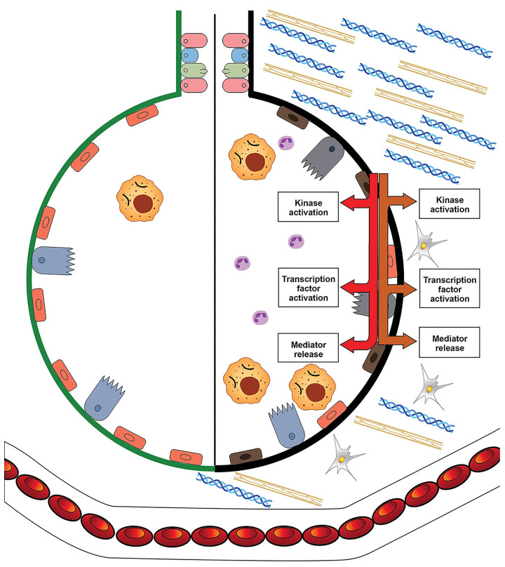
Theories of disease pathogenesis following asbestos exposure have focused on the participation of iron. After exposure, an open network of negatively charged functional groups on the fiber surface complexes host metals with a preference for iron. Competition for iron between the host and the asbestos results in a functional metal deficiency. The homeostasis of iron in the host is modified by the cell response, including increased import to correct the loss of the metal to the fiber surface. The biological effects of asbestos develop in response to and are associated with the disruption of iron homeostasis. Cell iron deficiency in the host following fiber exposure activates kinases and transcription factors, which are associated with the release of mediators coordinating both inflammatory and fibrotic responses. Relative to serpentine chrysotile, the clearance of amphiboles is incomplete, resulting in translocation to the mesothelial surface of the pleura. Since the biological effect of asbestos is dependent on retention of the fiber, the sequestration of iron by the surface, and functional iron deficiency in the cell, the greater clearance (i.e., decreased persistence) of chrysotile results in its diminished impact. An inability to clear asbestos from the lower respiratory tract initiates a host process of iron biomineralization (i.e., asbestos body formation). Host cells attempt to mobilize the metal sequestered by the fiber surface by producing superoxide at the phagosome membrane. The subsequent ferrous cation is oxidized and undergoes hydrolysis, creating poorly crystalline iron oxyhydroxide (i.e., ferrihydrite) included in the coat of the asbestos body.
Overview
CoChat Summary
TL;DR
This review argues that asbestos-related disease pathogenesis depends on disruption of host iron homeostasis after surface complexation of iron by the fiber, rather than on structural iron or oxidant chemistry alone. It proposes that amphiboles are more biopersistent and therefore have greater biological effects than chrysotile, and that asbestos body formation reflects a host iron biomineralization response.
Key findings
- Iron content in standard asbestos reference samples was greater in amosite (28%) and crocidolite (27%) than in chrysotile (0–2%), but the paper concludes current evidence does not support structural iron in the crystal lattice as the main driver of biological effect.
- Asbestos surfaces have negatively charged silanol groups that complex Fe3+ with high affinity; crystalline silicates show critical stability constants up to 1 × 10^17, while transferrin binds iron with a Ksc of approximately 10^20 M−1 at pH 7.4, and the labile iron pool is less than 1–5 µM.
- Functional iron deficiency after asbestos exposure increases DMT1 and ferritin expression, activates MAP kinases and transcription factors, and increases inflammatory mediators such as IL-6 and IL-8; iron pretreatment diminishes these responses.
- Asbestos bodies form on biopersistent fibers: uncoated fibers account for 95–97% of total inhaled asbestos, fibers less than 20 µm rarely become coated while virtually all fibers 80 µm or greater are coated, chrysotile asbestos bodies are usually less than 1–3% (mean 0.14%), and the coat can contain iron up to 10% w/w and calcium up to 1% w/w, with ferrihydrite as the major iron oxyhydroxide and more crystalline forms absent or below 5%.
Methods
- Narrative review and conceptual synthesis of literature on asbestos and iron; no primary experimental design is reported.
- Evidence cited includes animal models, human lung tissue and autopsy studies, in vitro macrophage and respiratory cell exposures, mineralogical/spectroscopic analyses, and epidemiologic/pathology reports.
- Main analysis integrates asbestos crystal lattice chemistry, surface complexation, cell iron homeostasis, inflammatory/fibrotic signaling, and ferruginous body formation.
Limitations
- The paper states no formal limitations section and describes no systematic search or meta-analytic method.
- The text notes that the precise composition of the asbestos body coat is not known, that there are no data supporting the fiber as the source of coat iron, and that dissolution has not been delineated during asbestos body formation.
- Evident from the text: the mechanistic hypothesis is largely inferential and synthesized from heterogeneous cited studies rather than tested by new primary data.
Model-written summary. Check it against the paper before relying on it.
Integrity signals
Registry
Cites
Statistics
Figures



Verification
Claims the paper makes
37 Valid · 6 Not valid · 42 Not checked · 58 Own results and interpretations
At least one cited source doesn’t support this claim or points to a different paper, and none confirmed it — see the verdicts below.
In the paper
“Iron in the coat of asbestos bodies does not appear to directly participate in the generation of reactive oxygen species [100].”
§ Asbestos and Iron > 5. The Formation of Ferruginous Bodies and Iron
What the sources say
- Disputedcites [100] Bardelli, 2021, Chemo-physical properties of asbestos bodies in human lung tissues studied at the nano-scale by non-invasive, label free X-ray imaging and spectroscopic techniques, Toxicol. Lett. sourceExperimental — accuracy not yet measured
The Introduction (page not provided) says that experiments found asbestos bodies induce free-radical generation and that iron in their coating is catalytically active. That is contrary to the claim that coating iron does not appear to participate directly in reactive-oxygen-species generation. (LLM, conf=0.85)
Checked by a reasoning model — may be wrong.
hedged- Asserted: passes — The authors put it forward as true.
- Checkable: passes — It can be checked against evidence.
- Load-bearing: unclear — It is unclear whether the paper’s conclusions depend on it.
- Based: passes — The paper gives a reason why it holds.
- Disputed
The sources with a verdict support this claim; the rest weren’t checked yet, have no full text we could read, or their verdicts aren’t rated yet.
In the paper
“There is support for metals’ involvement in the biological effect of asbestos, with iron chelation therapy and phlebotomy demonstrating possible preventive effects against asbestos-induced carcinogenesis in animal models [5,6].”
§ Asbestos and Iron > 1. Introduction
What the sources say
- Not checked — no full text of the sourcecites [5] Nagai, 2013, Deferasirox induces mesenchymal-epithelial transition in crocidolite-induced mesothelial carcinogenesis in rats, Cancer Prev. Res. source
- Supportedcites [6] Ohara, 2018, Phlebotomy as a preventive measure for crocidolite-induced mesothelioma in male rats, Cancer Sci. source
The Introduction reports preventive effects of iron chelators against asbestos-induced mesothelioma in rats. The Discussion reports that phlebotomy reduced tumor burden and improved survival in male rats, while cautiously describing it as a possible preventive measure. (LLM, conf=0.93)
hedgedstates a possible cause- Asserted: passes — The authors put it forward as true.
- Checkable: passes — It can be checked against evidence.
- Load-bearing: passes — The paper’s conclusions depend on it.
- Based: passes — The paper gives a reason why it holds.
The cited sources support only part of this claim — read each source’s verdict below.
In the paper
“An inability to clear asbestos from the lower respiratory tract initiates a host process of iron biomineralization (a process through which a living organism produces a mineral including an oxide) [97,98].”
§ Asbestos and Iron > 5. The Formation of Ferruginous Bodies and Iron
What the sources say
- Partly supportedcites [97] Pascolo, 2011, Synchrotron soft X-ray imaging and fluorescence microscopy reveal novel features of asbestos body morphology and composition in human lung tissues, Part. Fibre Toxicol. sourceExperimental — accuracy not yet measured
The Abstract Background describes asbestos bodies in lung tissue as products of biomineralization that deposits endogenous iron around inhaled fibres. The Background also describes their formation as a host protective mechanism and mentions fibre retention in the distal lung. It does not establish that an inability to clear asbestos initiates that process; the formation mechanism is described as not well defined. Page numbers are unavailable in our copy. (LLM, conf=0.82)
- Supportedcites [98] Bardelli, 2017, New insights on the biomineralisation process developing in human lungs around inhaled asbestos fibres, Sci. Rep. source
The Abstract states that asbestos in the lungs induces biomineralisation, forming an iron-rich coating around the fibres. The Discussion describes the fibres’ high persistence in tissue and iron deposition during their prolonged residence in the lungs, supporting the claim’s description of a process following failure to clear them. (LLM, conf=0.89)
plainstates a cause- Asserted: passes — The authors put it forward as true.
- Checkable: passes — It can be checked against evidence.
- Load-bearing: passes — The paper’s conclusions depend on it.
- Based: passes — The paper gives a reason why it holds.
- Partly supported
The cited sources support only part of this claim — read each source’s verdict below.
In the paper
“Following human inhalation, the greatest deposition of particles/fibers is in the 16th to 19th generations of airways, which includes the respiratory bronchioles [85,86,87,88].”
§ Asbestos and Iron > 4. Asbestos, Cell Iron Homeostasis, and Their Biological Effects
What the sources say
- Partly supportedcites [85] Lippmann, 1980, Deposition, retention, and clearance of inhaled particles, Br. J. Ind. Med. sourceExperimental — accuracy not yet measured
The Deposition Mechanisms section (page unavailable) says inhaled particles can deposit in bronchioles and alveolar spaces, supporting deposition in small airways generally. It does not identify the 16th–19th airway generations, establish that they include respiratory bronchioles, or say that they receive the greatest deposition of particles or fibers. (LLM, conf=0.78)
- Not checked — no full text of the sourcecites [86] Hiller, 1984, Deposition of sidestream cigarette smoke in the human respiratory tract, Prev. Med. source
- Not checked — no full text of the sourcecites [87] Choi, 2007, Mathematical analysis of particle deposition in human lungs: An improved single path transport model, Inhal. Toxicol. source
- Could not verifycites [88] Kim, 2018, Active and passive smoking impacts on asthma with quantitative and temporal relations: A Korean Community Health Survey, Sci. Rep. sourceExperimental — accuracy not yet measured
The Discussion passages address tobacco smoke, airway inflammation, and asthma, but not where inhaled particles or fibers deposit. Searches for deposition in the 16th–19th airway generations and for respiratory bronchioles found no passage addressing the claim; page numbers are unavailable in our copy. (LLM, conf=0.74)
plainstates a cause- Asserted: passes — The authors put it forward as true.
- Checkable: passes — It can be checked against evidence.
- Load-bearing: unclear — It is unclear whether the paper’s conclusions depend on it.
- Based: passes — The paper gives a reason why it holds.
- Partly supported
The cited sources support only part of this claim — read each source’s verdict below.
In the paper
“Particle/fiber deposition can often be greatest in the lower lung fields [89].”
§ Asbestos and Iron > 4. Asbestos, Cell Iron Homeostasis, and Their Biological Effects
What the sources say
- Partly supportedcites [89] Lambert, 2011, Regional deposition of particles in an image-based airway model: Large-eddy simulation and left-right lung ventilation asymmetry, Aerosol. Sci. Technol. sourceExperimental — accuracy not yet measured
The Discussion states that particle deposition was greatest in the lower lung lobes and reports a similar finding in a prior study. This supports the claim for particles, but our copy does not establish the same pattern for fibers. (LLM, conf=0.83)
plain- Asserted: passes — The authors put it forward as true.
- Checkable: passes — It can be checked against evidence.
- Load-bearing: unclear — It is unclear whether the paper’s conclusions depend on it.
- Based: passes — The paper gives a reason why it holds.
- Partly supported
91 more claims · 22 with their cited sources checked
Create a free account to see all 143 claims and to verify the 5 references still open.
- It may state two separate claims as one. 41
- Probably not a substantial claim. 7
Claims other papers make about this one
Each is checked against this paper’s own text: does it say what the citing paper says it does?
The cited source says what the claim attributes to it.
In the citing paper
“Increasing the bioavailability of iron, however, can overcome this process [84].”
From Environmental Contaminants, Iron Deficiency, and Iron‐Deficiency Anemia: A Review of the Literature (2025) · cites this paper as [84]
Citations
Cites
Oxyl radicals, redox-sensitive signalling cascades and antioxidants
Genestra, M. (2007) · Cellular Signalling · doi:10.1016/j.cellsig.2007.04.009 · Cited by 4,930
Production of Reactive Oxygen Species by Mitochondria
Chen, Q., Vazquez, E. J., Moghaddas, S., Hoppel, C. L., & Lesnefsky, E. J. (2003) · Journal of Biological Chemistry · doi:10.1074/jbc.m304854200 · Cited by 1,616 · cited 1×
“The electron transport chain in the mitochondria can be a major source of superoxide production [46,47].”
BackgroundMesoscale Iron Enrichment Experiments 1993-2005: Synthesis and Future Directions
Boyd, P. W., Jickells, T., Law, C. S., Blain, S., Boyle, E. A., Buesseler, K. O., Coale, K. H., Cullen, J. J., de Baar, H. J. W., Follows, M., et al. (2007) · Science · doi:10.1126/science.1131669 · Cited by 1,542 · cited 1×
“Iron is essential for almost every organism, and its lack of availability can restrict life in environments ranging in size from the ecosystem of the Pacific Ocean to a bacterium [33,34].”
BackgroundKakhlon, O., & Cabantchik, Z. I. (2002) · Free Radical Biology and Medicine · doi:10.1016/s0891-5849(02)01006-7 · Cited by 839 · cited 1×
“Intracellular iron levels available for complexation by the fiber surface can approach the concentration of the labile iron pool (less than 1–5 μM) [39,40].”
BackgroundMitogen-activated protein kinase cascades and regulation of gene expression
Su, B., & Karin, M. (1996) · Current Opinion in Immunology · doi:10.1016/s0952-7915(96)80131-2 · Cited by 747
Mortality from Lung Cancer in Asbestos Workers
Doll, R. (1955) · Occupational and Environmental Medicine · doi:10.1136/oem.12.2.81 · Cited by 717 · cited 1×
“In 1955, epidemiologic studies initially established an association between lung cancer and asbestos exposure [2,3].”
BackgroundHansel, C. M., Benner, S. G., Neiss, J., Dohnalkova, A., Kukkadapu, R. K., & Fendorf, S. (2003) · Geochimica et Cosmochimica Acta · doi:10.1016/s0016-7037(03)00276-x · Cited by 634 · cited 1×
“Subsequently, ferrihydrite in the coat can further react through hydrolysis– precipitation processes to produce goethite (Fe(OH)O), lepidocrocite (γ-FeO(OH)), magnetite, and hematite (Fe2O3) [113].”
BackgroundCompeting Fe (II)-induced mineralization pathways of ferrihydrite
Hansel, C. M., Benner, S. G., & Fendorf, S. (2005) · Environmental Science & Technology · doi:10.1021/es050666z · Cited by 619
Heyder, J. (2004) · Proceedings of the American Thoracic Society · doi:10.1513/pats.200409-046ta · Cited by 601
Deposition, retention, and clearance of inhaled particles.
Lippmann, M., Yeates, D. B., & Albert, R. E. (1980) · Occupational and Environmental Medicine · doi:10.1136/oem.37.4.337 · Cited by 557
Cited by
Exploring microbial diversity and interactions for asbestos modifying properties
Wong, L. C., Rodenburg, U., Leite, R. R., Korthals, G. W., Pover, J., Koerten, H. K., Kuramae, E. E., & Bodelier, P. L. E. (2024) · The Science of The Total Environment · doi:10.1016/j.scitotenv.2024.175577 · Cited by 7
Environmental Contaminants, Iron Deficiency, and Iron‐Deficiency Anemia: A Review of the Literature
Lichtler, R., & Cowley, M. (2025) · Scientifica · doi:10.1155/sci5/5007983 · Cited by 6
“Increasing the bioavailability of iron, however, can overcome this process [84].”
Zahiu, T., Mihu, C. M., Boşca, A. B., Mărginean, M. V., Mocan, L. P., Lupean, R.-A., Suflețel, R. T., Mihu, C., & Melincovici, C. S. (2025) · Diagnostics · doi:10.3390/diagnostics15111323 · Cited by 6
“As-bestos exposure lowers intracellular iron levels by binding the metal with fiber surfaces, disrupting essential cellular functions and causing cell death or apoptosis [23,24].”
Cellular defense mechanisms against asbestos fibers
Barlow, C. A., & Mossman, B. T. (2025) · Frontiers in Public Health · doi:10.3389/fpubh.2025.1566473 · Cited by 5
“Since amosite and crocidolite asbestos are high iron-containing and the most potent in the induction of human mesotheliomas, most studies have focused on their mechanisms of oxidant stress and defense (40, 48, 49).”
InfluentialGualtieri, A. F., Ferrari, É., Rigamonti, L., Ruozi, B., Mirata, S., Almonti, V., Passalacqua, M., Vernazza, S., Valerio, S. D., Tossetta, G., et al. (2024) · Current Research in Toxicology · doi:10.1016/j.crtox.2024.100202 · Cited by 3
Metadata
- Authors
- Ghio, A. J., Stewart, M., Sangani, R. G., Pavlisko, E. N., & Roggli, V. L.
- Affiliations
- Environmental Protection Agency (US)
- Johns Hopkins University (US)
- West Virginia University (US)
- Duke Medical Center (US)
- Corresponding author
- Ghio, Andrew J.
- Venue
- International Journal of Molecular Sciences
- Publisher
- MDPI AG
- Published
- 2023-08-03
- Volume
- 24
- Issue
- 15
- Pages
- 12390
- ISSN
- 1422-0067 (ISSN-L)
- PMC
- PMC10419076
- OpenAlex
- W4385507347
- Semantic Scholar
- 3adf5b7d7da4ae390667d7b3a13b9a9b7649da01
- Cited by
- 15
- References
- 152
- Citations by year
- 2026: 5 · 2025: 6 · 2024: 4
- FWCI
- 0.69
- Type
- journal article
- Language
- en
- Open access
- gold
- doi.org
- Licence
- Locations
- International Journal of Molecular Sciences (publishedVersion, cc-by)
- PubMed Central (publishedVersion, cc-by)
- PubMed (publishedVersion)
- Topics
- Health Sciences › Medicine › Pulmonary and Respiratory Medicine › Occupational and environmental lung diseases
- Health Sciences › Medicine › Hematology › Iron Metabolism and Disorders
- Health Sciences › Medicine › Pulmonary and Respiratory Medicine › Interstitial Lung Diseases and Idiopathic Pulmonary Fibrosis
- Keywords
- asbestos, iron, iron homeostasis, chrysotile, amphibole fibers, serpentine asbestos, metal sequestration, iron deficiency, pleural mesothelium, ferrihydrite, iron biomineralization, oxidative stress, inflammation, fibrosis, pathogenesis
- MeSH terms
- Animals, Asbestos, Asbestosis, Homeostasis, Humans, Iron, Asbestos, Serpentine
About these checks
Registry, retraction, Cites and statistics results are computed, not judged, with one exception: a printed reference that no index lists is read once by a model before it counts as a citation error. Claims are extracted by a model and kept only when their quote appears verbatim in the text; each cited source is then checked, with a model’s help, for whether it says what the claim relies on. Those verdicts can be wrong — read the claim’s quote and the cited source before relying on one.
The claim verdicts this report counts are experimental: their accuracy is not yet measured.
A language model drew the claims from the paper on 2026-10-10, under version 3 of the claim definition (model deepseek-v4.1-flash, prompt extract-v3).
A second model labelled each claim on 2026-10-10 (model jev-1.13-20260917, definition revision gold-guideline-v0.2).
Cited sources are checked with a language model’s help (gpt-6-sol).
Statistics and arithmetic are recomputed, not judged.
Confidence bands are shown only for fields calibrated on our annotated test set. None is calibrated yet, so no band is shown.
What counts as a claim ›How we measure confidence ›How we check this ›
Are you an author, or do you see a problem with this page? Request a review
How to cite
Cite CoChat for the checks on this page, and the paper for its own findings.
- This page
- CoChat. (2026). Citation check: Asbestos and Iron. https://app.cochat.ai/papers/10.3390/ijms241512390
- The paper
- Ghio, A. J., Stewart, M., Sangani, R. G., Pavlisko, E. N., & Roggli, V. L. (2023). Asbestos and Iron. International Journal of Molecular Sciences. https://doi.org/10.3390/ijms241512390
Data sources
Bibliographic data: Semantic Scholar, OpenAlex, Crossref.
Contains information from the Semantic Scholar Academic Graph Datasets, made available under the ODC Attribution License (ODC-BY 1.0).
Semantic Scholar’s data is described in: Kinney, R., et al. (2023). The Semantic Scholar Open Data Platform. arXiv:2301.10140.
