

The DOI matches its Crossref record; 103 of the 103 references it cites were found in a registry or an index.
Functionalized Optical Microcavities for Sensing Applications
Open accessAbstract
Functionalized optical microcavities constitute an emerging highly sensitive and highly selective sensing technology. By combining optical microcavities with novel materials, microcavity sensors offer exceptional precision, unlocking considerable potential for medical diagnostics, physical and chemical analyses, and environmental monitoring. The high capabilities of functionalized microcavities enable subwavelength light detection and manipulation, facilitating the precise detection of analytes. Furthermore, recent advancements in miniaturization have paved the way for their integration into portable platforms. For leveraging the potential of microcavity sensors, it is crucial to address challenges related to the need for increasing cost-effectiveness, enhancing selectivity and sensitivity, enabling real-time measurements, and improving fabrication techniques. New strategies include the use of advanced materials, the optimization of signal processing, hybrid design approaches, and the employment of artificial intelligence. This review outlines the key strategies toward enhancing the performance of optical microcavities, highlights their broad applicability across various fields, and discusses the challenges that should be overcome to unlock their full potential.
Overview
The claims and statistics of this paper haven’t been checked yet.
Run the check — free, no account needed. It reads the open-access text and checks the sources it cites.
Integrity signals
Registry
Cites
Statistics
Figures
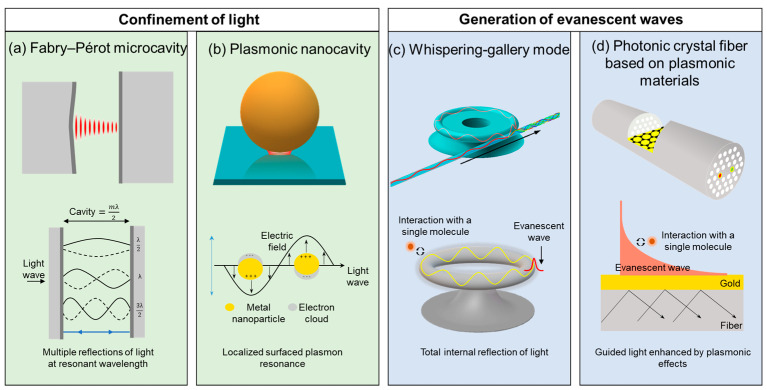
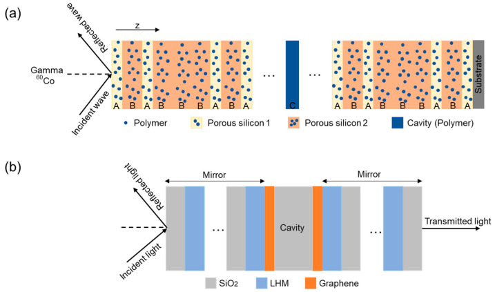
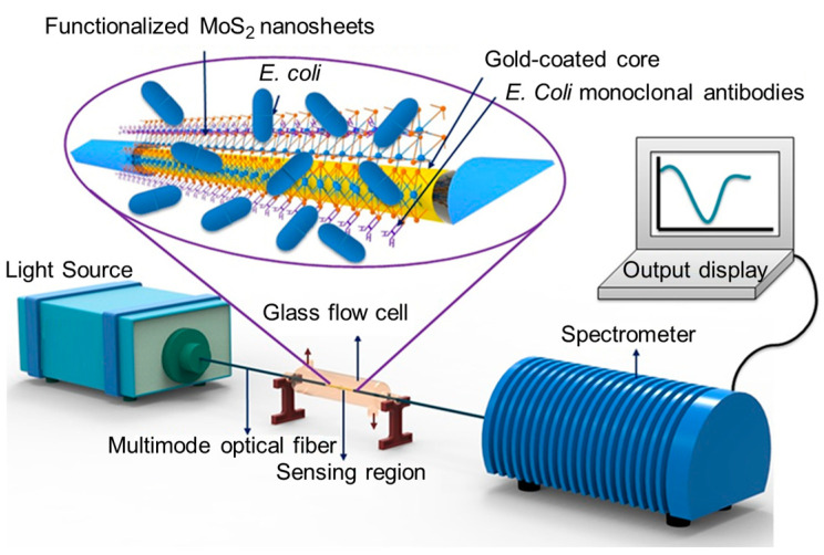
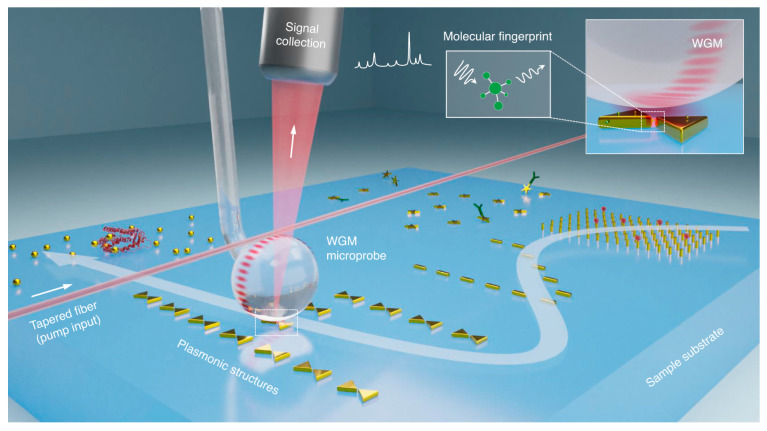
Verification
Claims the paper makes
The claims and statistics of this paper haven’t been checked yet.
Run the check — free, no account needed. It reads the open-access text and checks the sources it cites.
Claims other papers make about this one
Each is checked against this paper’s own text: does it say what the citing paper says it does?
The cited source says what the claim attributes to it.
In the citing paper
“The resonance phenomenon occurs when the distance between the mirror surfaces (i.e., optical path length) is a multiple of λ c /2, permitting light to be confined within the cavity layer [262].”
From Metal Oxide Thin Films for Advanced Photonic Applications (2026) · cites this paper as [262]
Self-check
Percentages, sums and reported differences re-computed from the paper’s own numbers.
The self-check needs the paper’s full text.
Citations
Cites
Graphene Thickness Determination Using Reflection and Contrast Spectroscopy
Ni, Z., Wang, H., Kasim, J., Fan, H., Yu, T., Wu, Y., Feng, Y. P., & Shen, Z. (2007) · Nano Letters · doi:10.1021/nl071254m · Cited by 1,190
Kaliteevski, M. A., Iorsh, I. V., Brand, S., et al. (2007) · Physical Review B · doi:10.1103/physrevb.76.165415 · Cited by 930
Plasmon light scattering in biology and medicine: new sensing approaches, visions and perspectives
Aslan, K., Lakowicz, J. R., & Geddes, C. D. (2005) · Current Opinion in Chemical Biology · doi:10.1016/j.cbpa.2005.08.021 · Cited by 303
Chaudhary, V. S., Kumar, D., & Kumar, S. (2021) · IEEE Sensors Journal · doi:10.1109/jsen.2021.3085829 · Cited by 231
Rasheed, S., Kanwal, T., Ahmad, N., Fatima, B., Haq, M. N. U., & Hussain, D. (2024) · TrAC Trends in Analytical Chemistry · doi:10.1016/j.trac.2024.117640 · Cited by 228
Chaudhary, V. S., Kumar, D., Pandey, B. P., et al. (2022) · IEEE Sensors Journal · doi:10.1109/jsen.2022.3222969 · Cited by 186
Kaushik, S., Tiwari, U., Pal, S. S., & Sinha, R. (2019) · Biosensors & bioelectronics · doi:10.1016/j.bios.2018.11.006 · Cited by 179
Ramanujam, N. R., Amiri, I. S., Taya, S. A., et al. (2018) · Microsystem Technologies · doi:10.1007/s00542-018-3947-6 · Cited by 172
Recent advances in quantum dots-based biosensors for antibiotics detection
Ding, R., Chen, Y., Wang, Q., Wu, Z., Zhang, X., Li, B., & Lin, L. (2021) · Journal of Pharmaceutical Analysis · doi:10.1016/j.jpha.2021.08.002 · Cited by 159
Tan, T., Yuan, Z., Zhang, H., Yan, G., Zhou, S., An, N., Peng, B., Soavi, G., Rao, Y., & Yao, B. (2021) · Nature Communications · doi:10.1038/s41467-021-26740-8 · Cited by 149
Recent advances and applications in QDs-based sensors
Ma, Q., & Su, X. (2011) · The Analyst · doi:10.1039/c1an15741h · Cited by 131
Li, W., Zhao, W., Cheng, S., et al. (2024) · Optics and Lasers in Engineering · doi:10.1016/j.optlaseng.2024.108368 · Cited by 128
Liu, C., Lü, J., Liu, W., et al. (2021) · Chinese Optics Letters · doi:10.3788/col202119.102202 · Cited by 127
Lv, J., Wang, J., Yang, L., et al. (2024) · Sensors & Diagnostics · doi:10.1039/d4sd00045e · Cited by 124
Resonance Surface Plasmon Spectroscopy: Low Molecular Weight Substrate Binding to Cytochrome P450
Zhao, J., Das, A., Zhang, X., Schatz, G. C., Sligar, S. G., & Duyne, R. P. V. (2006) · Journal of the American Chemical Society · doi:10.1021/ja0636082 · Cited by 110
Graphene-based 1D defective photonic crystal biosensor for real-time detection of cancer cells
Panda, A., Pukhrambam, P. D., Wu, F., & Belhadj, W. (2021) · The European Physical Journal Plus · doi:10.1140/epjp/s13360-021-01796-z · Cited by 99
Advances in Miniaturized Computational Spectrometers
Xue, Q., Yang, Y., Ma, W., et al. (2024) · Advanced Science · doi:10.1002/advs.202404448 · Cited by 96
Whispering Gallery Mode Optical Microresonators: Structures and Sensing Applications
Cai, L., Pan, J., Zhao, Y., Wang, J., & Xiao, S. (2020) · physica status solidi (a) · doi:10.1002/pssa.201900825 · Cited by 81
Xia, F., Song, H., Zhao, Y., et al. (2020) · Measurement · doi:10.1016/j.measurement.2020.108083 · Cited by 80
Yadav, A., Kumar, S., Kumar, A., & Sharan, P. (2023) · Frontiers in Materials · doi:10.3389/fmats.2022.1106251 · Cited by 76
Fiber Bragg grating assisted surface plasmon resonance sensor with graphene oxide sensing layer
Arasu, P. T., Noor, A. S. M., Shabaneh, A. A. A., et al. (2016) · Optics Communications · doi:10.1016/j.optcom.2016.05.081 · Cited by 70
Yang, J., Chen, Y., Rao, Z., Zheng, Z., Song, C., Chen, Y., Xiong, K., Chen, P.-X., Zhang, C., Wu, W., et al. (2024) · Light Science & Applications · doi:10.1038/s41377-024-01384-7 · Cited by 70
Chao, C.-T. C., Chen, S., Huang, H. J., et al. (2023) · Crystals · doi:10.3390/cryst13050813 · Cited by 65
A whispering-gallery scanning microprobe for Raman spectroscopy and imaging
Mao, W., Li, Y., Jiang, X., Liu, Z., & Yang, L. (2023) · Light Science & Applications · doi:10.1038/s41377-023-01276-2 · Cited by 56
Wu, X., Wang, Y., Chen, Q., et al. (2018) · Photonics Research · doi:10.1364/prj.7.000050 · Cited by 55
Ultra-high-Q free-space coupling to microtoroid resonators
Suebka, S., McLeod, E. J., & Su, J. (2024) · Light Science & Applications · doi:10.1038/s41377-024-01418-0 · Cited by 51
Optimization of Novel 2D Material Based SPR Biosensor Using Machine Learning
Patel, S. K., Surve, J., Baz, A., et al. (2024) · IEEE Transactions on NanoBioscience · doi:10.1109/tnb.2024.3354810 · Cited by 49
Wekalao, J., Mandela, N., Apochi, O., Lefu, C., & Topisia, T. (2024) · Plasmonics · doi:10.1007/s11468-024-02489-w · Cited by 48
Review of different coupling methods with whispering gallery mode resonator cavities for sensing
Zhang, Y., Song, Q., Zhao, D., Tang, X., Zhang, Y. X., Liu, Z., & Yuan, L. (2022) · Optics & Laser Technology · doi:10.1016/j.optlastec.2022.108955 · Cited by 47
Basha, A. J., Maheshwari, R. U., Pandey, B. K., et al. (2024) · Plasmonics · doi:10.1007/s11468-024-02584-y · Cited by 45
Rabi-like splitting and refractive index sensing with hybrid Tamm plasmon-cavity modes
Jena, S., Tokas, R. B., Thakur, S., et al. (2022) · Journal of Physics D Applied Physics · doi:10.1088/1361-6463/ac49b3 · Cited by 44
Functionalizing nanophotonic structures with 2D van der Waals materials
Meng, Y., Zhong, H., Xu, Z., He, T., Kim, J. S., Han, S., Kim, S., Park, S., Shen, Y., Gong, M., et al. (2023) · Nanoscale Horizons · doi:10.1039/d3nh00246b · Cited by 44
Awawdeh, K., Buttkewitz, M. A., Bahnemann, J., & Segal, E. (2024) · Microsystems & Nanoengineering · doi:10.1038/s41378-024-00738-w · Cited by 43
Gowda, R. B., Saara, K., & Sharan, P. (2021) · Optik · doi:10.1016/j.ijleo.2021.167599 · Cited by 42
Printed quantum dot photodetectors for applications from the high-energy to the infrared region
Zou, T., Choi, T., Liu, A., Zhu, H., & Noh, Y. (2024) · Nano Energy · doi:10.1016/j.nanoen.2024.109539 · Cited by 42
Sun, J., Shi, Z., Wang, L., Zhang, X., Luo, C., Hua, J., Feng, M., Chen, Z., Wang, M., & Xu, C. (2023) · Talanta · doi:10.1016/j.talanta.2023.124677 · Cited by 40
Zhan, K., Jiang, Y., Qin, P., Chen, Y., & Heinke, L. (2023) · Advanced Materials Interfaces · doi:10.1002/admi.202300329 · Cited by 39
Label-Free Biochemical Sensing Using Processed Optical Fiber Interferometry: A Review
Jha, R., Gorai, P., Shrivastav, A. M., & Pathak, A. P. (2024) · ACS Omega · doi:10.1021/acsomega.3c03970 · Cited by 39
Lu, M., Zhu, H., Lin, M., Wang, F., Hong, L., Masson, J., & Peng, W. B. (2020) · Sensors and Actuators B Chemical · doi:10.1016/j.snb.2020.129094 · Cited by 37
Micro/nanoporous polymer chips as templates for highly sensitive SERS sensors
Akil, S., Jradi, S., Plain, J., et al. (2012) · RSC Advances · doi:10.1039/c2ra21186f · Cited by 36
Deep-learning powered whispering gallery mode sensor based on multiplexed imaging at fixed frequency
Saetchnikov, A. V., Tcherniavskaia, E. A., Saetchnikov, V. A., et al. (2020) · Opto-Electronic Advances · doi:10.29026/oea.2020.200048 · Cited by 36
Paul, A. K., Mollah, M. A., Hassan, M. Z., et al. (2021) · Photonics · doi:10.3390/photonics8050155 · Cited by 36
Machine learning enabled 2D photonic crystal biosensor for early cancer detection
Balaji, V. R., A, I. J. M., Sridarshini, T., et al. (2023) · Measurement · doi:10.1016/j.measurement.2023.113858 · Cited by 36
Zhou, S., Li, J., Zhang, Q., Tong, Y., Qi, X., Duan, Y., Zhang, X., Luo, Z., & Li, Y. (2024) · Optics & Laser Technology · doi:10.1016/j.optlastec.2024.110783 · Cited by 36
Qiu, H., Yao, Y., Dong, Y., et al. (2024) · Biosensors and Bioelectronics · doi:10.1016/j.bios.2024.116265 · Cited by 35
Patel, S. K., & Alsalman, O. (2024) · Optical and Quantum Electronics · doi:10.1007/s11082-024-07068-1 · Cited by 35
2D-material-integrated whispering-gallery-mode microcavity
Wang, L., Zhou, X., Yang, S., Huang, G., & Mei, Y. (2019) · Photonics Research · doi:10.1364/prj.7.000905 · Cited by 34
Kumar, N., Suthar, B., Nayak, C., & Bhargava, A. (2023) · Physica Scripta · doi:10.1088/1402-4896/accfca · Cited by 34
Photothermal gas detection using a miniaturized fiber Fabry-Perot cavity
Krzempek, K., Jaworski, P., Tenbrake, L., et al. (2023) · Sensors and Actuators B Chemical · doi:10.1016/j.snb.2023.135040 · Cited by 34
Vatani, S., Biney, J., Faramarzi, V., Jabbour, G. E., & Park, J. (2024) · ChemistrySelect · doi:10.1002/slct.202401265 · Cited by 34
Zaky, Z. A., Al-Dossari, M., Hendy, A. S., et al. (2024) · International Journal of Modern Physics B · doi:10.1142/s0217979224504095 · Cited by 33
Li, X., Singh, R., Zhang, B., et al. (2023) · IEEE Sensors Journal · doi:10.1109/jsen.2023.3346180 · Cited by 32
Suthar, B., Kumar, N., & Taya, S. A. (2022) · The European Physical Journal Plus · doi:10.1140/epjp/s13360-022-03533-6 · Cited by 31
Advancements in optical biosensing techniques: From fundamentals to future prospects
Kaur, B., Kumar, S., Nedoma, J., Martínek, R., & Marques, C. (2024) · APL Photonics · doi:10.1063/5.0216621 · Cited by 31
Polymer Waveguide Coupled Surface Plasmon Refractive Index Sensor: A Theoretical Study
Ji, L., Yang, S., Shi, R., et al. (2020) · Photonic Sensors · doi:10.1007/s13320-020-0589-y · Cited by 30
A plasmonic refractive index sensor with an ultrabroad dynamic sensing range
Cheng, Y., Chang, Y.-J., Chuang, Y.-C., et al. (2019) · Scientific Reports · doi:10.1038/s41598-019-41353-4 · Cited by 29
Yang, J., Zhang, H., Wang, T., et al. (2022) · Physical Review Applied · doi:10.1103/physrevapplied.18.014056 · Cited by 29
Yang, X., Hou, S., Xie, C., et al. (2023) · Plasmonics · doi:10.1007/s11468-023-02017-2 · Cited by 28
Microcavity-based SERS chip for ultrasensitive immune detection of cardiac biomarkers
Wang, J., Xu, C., Lei, M., Ma, Y., Wang, X., Wang, R., Sun, J., & Wang, R. (2021) · Microchemical Journal · doi:10.1016/j.microc.2021.106875 · Cited by 27
Kausaite‐Minkstimiene, A., Popov, A., & Ramanavičienė, A. (2023) · TrAC Trends in Analytical Chemistry · doi:10.1016/j.trac.2023.117468 · Cited by 25
Two-Dimensional Materials-Based Plasmonic Sensors for Health Monitoring Systems—A Review
Gupta, V. K., Choudhary, K., & Kumar, S. (2023) · IEEE Sensors Journal · doi:10.1109/jsen.2023.3268175 · Cited by 24
Visual/quantitative SERS biosensing chip based on Au-decorated polystyrene sphere microcavity arrays
Sun, J., Wang, R., Wang, L., et al. (2023) · Sensors and Actuators B Chemical · doi:10.1016/j.snb.2023.133869 · Cited by 24
Multimode sensing based on optical microcavities
Wu, Y., Duan, B., Li, C., & Yang, D. (2023) · Frontiers of Optoelectronics · doi:10.1007/s12200-023-00084-1 · Cited by 24
Plasmonic Nanoarchitectures for Single‐Molecule Explorations: An Overview
Koya, A. N. (2021) · Advanced Photonics Research · doi:10.1002/adpr.202100325 · Cited by 23
Sarychev, A. K., Sukhanova, A. V., Иванов, А. В., et al. (2022) · Biosensors · doi:10.3390/bios12050300 · Cited by 23
Emerging nanophotonic biosensor technologies for virus detection
Nanjunda, S. B., Seshadri, V. N., Krishnan, C., et al. (2022) · Nanophotonics · doi:10.1515/nanoph-2022-0571 · Cited by 23
Chaity, A. C. (2023) · Plasmonics · doi:10.1007/s11468-023-02128-w · Cited by 22
Ankita, A., Suthar, B., Bissa, S., & Bhargava, A. (2024) · Optical and Quantum Electronics · doi:10.1007/s11082-024-07065-4 · Cited by 22
Abbaszadeh, A., & Ahmadi, S. R. (2024) · Optical and Quantum Electronics · doi:10.1007/s11082-024-06853-2 · Cited by 21
Panda, A., Nguyên, C. V., Pukhrambam, P. D., et al. (2023) · Optical and Quantum Electronics · doi:10.1007/s11082-023-04748-2 · Cited by 20
Liu, S., Zhang, X., Wang, Q., et al. (2023) · IEEE Sensors Journal · doi:10.1109/jsen.2023.3273905 · Cited by 17
Rho, D., & Kim, S. (2020) · Biosensors · doi:10.3390/bios11010004 · Cited by 16
Highly sensitive, modification-free, and dynamic real-time stereo-optical immuno-sensor
Zhao, X., Guo, Z., Zhou, Y., Guo, J., Liu, Z., Luo, M., Li, Y., Wang, Q., Zhang, M., Yang, X., et al. (2023) · Biosensors and Bioelectronics · doi:10.1016/j.bios.2023.115477 · Cited by 16
Gu, C., Singh, R., Li, G., et al. (2024) · Journal of Lightwave Technology · doi:10.1109/jlt.2024.3379290 · Cited by 16
FM-Level Detection of Glucose Using a Grating Based Sensor Enhanced With Graphene Oxide
Chen, X., Lin, W., Xu, P., et al. (2022) · Journal of Lightwave Technology · doi:10.1109/jlt.2022.3174039 · Cited by 15
Optimizing an Optical Cavity-Based Biosensor for Enhanced Sensitivity
Sypabekova, M., Hagemann, A., Kleiss, J., et al. (2023) · IEEE Sensors Journal · doi:10.1109/jsen.2023.3317678 · Cited by 15
Zhang, Z., Chen, Y., Tian, Y., Li, H., Lu, Z., & Yang, L. (2024) · ACS Applied Nano Materials · doi:10.1021/acsanm.3c06060 · Cited by 15
Highly sensitive photonic crystal fiber optic sensor for cancer cell detection
Bing, P., Zhao, J., Zhang, X., et al. (2024) · The European Physical Journal Plus · doi:10.1140/epjp/s13360-024-05360-3 · Cited by 15
Puspita, I., Irawati, N., Madurani, K. A., Kurniawan, F., Koentjoro, S., & Hatta, A. M. (2022) · Photonic Sensors · doi:10.1007/s13320-022-0652-y · Cited by 14
Silicon Microring Resonator Biosensor for Detection of Nucleocapsid Protein of SARS-CoV-2
Uchida, Y., Arakawa, T., Higo, A., et al. (2024) · Sensors · doi:10.3390/s24103250 · Cited by 14
Microfluidics and Nanofluidics in Strong Light–Matter Coupling Systems
Granizo, E., Kriukova, I. S., Escudero‐Villa, P., Samokhvalov, P. S., & Nabiev, I. R. (2024) · Nanomaterials · doi:10.3390/nano14181520 · Cited by 14
Self-citationA versatile tunable microcavity for investigation of light–matter interaction
Mochalov, K. E., Vaskan, I. S., Dovzhenko, D. S., Ракович, Ю. П., & Nabiev, I. R. (2018) · Review of Scientific Instruments · doi:10.1063/1.5021055 · Cited by 12
Self-citationLiao, J., & Yang, L. (2024) · IEEE Transactions on Instrumentation and Measurement · doi:10.1109/tim.2024.3352712 · Cited by 11
Fluorescence enhanced biomolecule detection using direct laser written micro-ring resonators
Makela, M., Lin, Z., Coté, G. L., et al. (2024) · Optics & Laser Technology · doi:10.1016/j.optlastec.2024.110629 · Cited by 11
Hajiani, T., Shirkani, H., & Sadeghi, Z. (2024) · Optics Communications · doi:10.1016/j.optcom.2024.130801 · Cited by 11
Tao, Y., Ye, H., Ding, Y., et al. (2022) · Photonic Sensors · doi:10.1007/s13320-022-0641-1 · Cited by 10
Firouzi, F., Vahedi, A., & Haghipour, S. (2024) · Physica B Condensed Matter · doi:10.1016/j.physb.2024.416766 · Cited by 10
Femtosecond laser fabricated polymer microring resonator for sensing applications
Zhang, D., Men, L. Q., & Chen, Q.-Y. (2018) · Electronics Letters · doi:10.1049/el.2017.4585 · Cited by 9
WS2 Monolayer in Fabry–Perot Cavity Support for Plasmonic Fano Resonance
Chen, F., Li, Y., Yang, W., et al. (2023) · Plasmonics · doi:10.1007/s11468-023-01865-2 · Cited by 9
Tkach, A. P., Miropoltsev, M. A., Kundelev, E. V., et al. (2024) · Optics & Laser Technology · doi:10.1016/j.optlastec.2024.111359 · Cited by 9
Kim, H. S., Jun, S. W., & Ahn, Y. H. (2023) · Sensors · doi:10.3390/s23135797 · Cited by 8
Tran, K. N., Tran, H. N. Q., Lim, S. Y., et al. (2024) · ACS Applied Materials & Interfaces · doi:10.1021/acsami.4c03804 · Cited by 7
High Sensitivity Refractive Index Sensor Based on TiO2-Ag Double-Layer Coated Photonic Crystal Fiber
Liu, Q., Zhao, X., Zhang, Q., et al. (2024) · Plasmonics · doi:10.1007/s11468-024-02459-2 · Cited by 7
Bianki, M.-A., Guertin, R., Lemieux‐Leduc, C., & Peter, Y.-A. (2024) · Sensors and Actuators B Chemical · doi:10.1016/j.snb.2024.136460 · Cited by 7
High-Q Fabry-Pérot Cavity Based on Micro-Lens Array for Refractive Index Sensing
Wang, Q., Zhao, X., Luo, M., et al. (2024) · Photonic Sensors · doi:10.1007/s13320-024-0716-2 · Cited by 6
Enabling new frontiers of nanophotonics with metamaterials, photonic crystals, and plasmonics
Badloe, T., & Rho, J. (2024) · Nanophotonics · doi:10.1515/nanoph-2024-0100 · Cited by 5
Li, Y., Wang, P., Wang, H., et al. (2024) · Talanta · doi:10.1016/j.talanta.2024.126627 · Cited by 5
Elsayed, A. M., Ahmed, A. M., Aly, A. H., et al. (2024) · RSC Advances · doi:10.1039/d4ra05116e · Cited by 5
Wu, Y., Chen, K., Fahad, A. K., et al. (2024) · Spectrochimica Acta Part A Molecular and Biomolecular Spectroscopy · doi:10.1016/j.saa.2024.125293 · Cited by 5
Enhanced Biosensor Based on Assembled Porous Silicon Microcavities Using CdSe/ZnS Quantum Dots
Sun, M., Zhang, S., Wang, J., et al. (2021) · IEEE photonics journal · doi:10.1109/jphot.2021.3101656 · Cited by 4
High performance SERS boosting by Fabry- Pérot cavities of silica-gold-silicon multilayers
Zhang, M., Zhou, Y., Li, P., et al. (2024) · Optics Express · doi:10.1364/oe.542131 · Cited by 4
Elblbeisi, M. H., Taya, S. A., Almawgani, A. H. M., et al. (2023) · Plasmonics · doi:10.1007/s11468-023-02084-5 · Cited by 2
Nifontova, G., Gerasimovich, E., Fleury, F., et al. (2023) · EPJ Web of Conferences · doi:10.1051/epjconf/202328703007 · Cited by 1
Cited by
Sorted by citations
Metal Oxide Thin Films for Advanced Photonic Applications
Stîngescu, M.-L., Șopronyi, M.-A., Antohe, Ș., & Scărișoreanu, N.-D. (2026) · Crystals · doi:10.3390/cryst16030164 · Cited by 2
“The resonance phenomenon occurs when the distance between the mirror surfaces (i.e., optical path length) is a multiple of λ c /2, permitting light to be confined within the cavity layer [262].”
Metadata
- Authors
- Granizo, E., Samokhvalov, P., & Nabiev, I.
- Affiliations
- National Research Nuclear University MEPhI (RU)
- Sechenov University (RU)
- Corresponding author
- Nabiev, Igor
- Venue
- Nanomaterials
- Publisher
- MDPI AG
- Published
- 2025-01-27
- Volume
- 15
- Issue
- 3
- Pages
- 206
- ISSN
- 2079-4991 (ISSN-L)
- PMC
- PMC11820503
- OpenAlex
- W4406911399
- Cited by
- 12
- References
- 103
- Citations by year
- 2026: 5 · 2025: 7
- FWCI
- 0.60
- Type
- journal article
- Language
- en
- Open access
- gold
- doi.org
- Licence
- Locations
- Nanomaterials (publishedVersion, cc-by)
- Preprints.org (acceptedVersion, cc-by)
- PubMed Central (publishedVersion, cc-by)
- PubMed (publishedVersion)
- HAL (submittedVersion, cc-by)
- DOAJ (DOAJ: Directory of Open Access Journals) (submittedVersion, cc-by-sa)
- Funders
- Russian Science Foundation (23-75-30016)
- Topics
- Physical Sciences › Engineering › Electrical and Electronic Engineering › Photonic and Optical Devices
- Physical Sciences › Physics and Astronomy › Atomic and Molecular Physics, and Optics › Strong Light-Matter Interactions
- Physical Sciences › Physics and Astronomy › Atomic and Molecular Physics, and Optics › Mechanical and Optical Resonators
- Keywords
- optical microcavities, optical sensing, subwavelength light manipulation, sensor sensitivity and selectivity, portable sensing platform, signal processing, medical diagnostics, environmental monitoring, fabrication techniques
About these checks
Registry, retraction, Cites and statistics results are computed, not judged, with one exception: a printed reference that no index lists is read once by a model before it counts as a citation error. Claims are extracted by a model and kept only when their quote appears verbatim in the text; each cited source is then checked, with a model’s help, for whether it says what the claim relies on. Those verdicts can be wrong — read the claim’s quote and the cited source before relying on one.
Statistics and arithmetic are recomputed, not judged.
Confidence bands are shown only for fields calibrated on our annotated test set. None is calibrated yet, so no band is shown.
What counts as a claim ›How we measure confidence ›How we check this ›
Are you an author, or do you see a problem with this page? Request a review
