Cited by 9

The DOI matches its Crossref record; 1 of the 1 references it cites were found in a registry or an index.

Upregulation of miR‑128 inhibits neuronal cell apoptosis following spinal cord injury via FasL downregulation by repressing ULK1

Liu, Ruixuan, Peng, Zhibin, Zhang, Yubo, Li, Rui, Wang, Yansong·2021·Molecular Medicine Reports

Overview

The claims and statistics of this paper haven’t been checked yet.

Run the check — free, no account needed. It reads the open-access text and checks the sources it cites.

A few free checks a day · papers already verified open instantly · results are public

Integrity signals

Registry

Crossref · 10.3892/mmr.2021.12306

Cites

1 of 1 references verified0 citation errors · 0 not checkable

Statistics

Figures

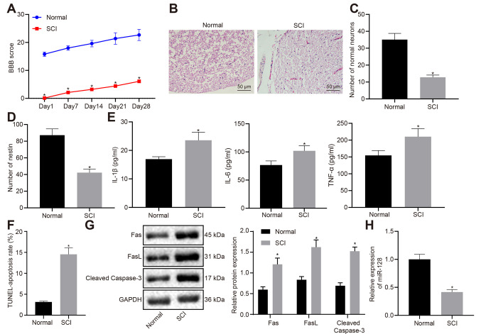
Figure 1. Rats with SCI exhibit increased neuronal apoptosis, intense inflammatory response and lowly expressed miR-128. (A) Locomotor functions of rats evaluated by BBB score. (B) Hematoxylin and eosin staining of morphological changes of neuronal cells (magnification, ×200). (C) The survival of neurons in spinal cord of rats determined by Nissl staining. (D) The number of nestin-positive cells detected using immunofluorescence. (E) The expression of inflammatory factors (IL-1β, IL-6 and TNF-α) in rat serum determined by ELISA. (F) Apoptosis of spinal cord cells detected using TUNEL. (G) Protein expression of apoptosis-related factors measured by western blot analysis. (H) The expression of miR-128 assessed by reverse transcription-quantitative PCR. An unpaired t-test was performed for comparisons of data between two groups. BBB score was used for comparison of data at different time points. Repeated-measures analysis of ANOVA was performed, followed by Bonferroni post-hoc test. *P<0.05 vs. normal. SCI, spinal cord injury; miR, microRNA; BBB, Basso, Beattie and Bresnahan; ANOVA, analysis of variance.
Figure 1. Rats with SCI exhibit increased neuronal apoptosis, intense inflammatory response and lowly expressed miR-128. (A) Locomotor functions of rats evaluated by BBB score. (B) Hematoxylin and eosin staining of morphological changes of neuronal cells (magnification, ×200). (C) The survival of neurons in spinal cord of rats determined by Nissl staining. (D) The number of nestin-positive cells detected using immunofluorescence. (E) The expression of inflammatory factors (IL-1β, IL-6 and TNF-α) in rat serum determined by ELISA. (F) Apoptosis of spinal cord cells detected using TUNEL. (G) Protein expression of apoptosis-related factors measured by western blot analysis. (H) The expression of miR-128 assessed by reverse transcription-quantitative PCR. An unpaired t-test was performed for comparisons of data between two groups. BBB score was used for comparison of data at different time points. Repeated-measures analysis of ANOVA was performed, followed by Bonferroni post-hoc test. *P<0.05 vs. normal. SCI, spinal cord injury; miR, microRNA; BBB, Basso, Beattie and Bresnahan; ANOVA, analysis of variance.CC BY-NC-ND · PubMed Central
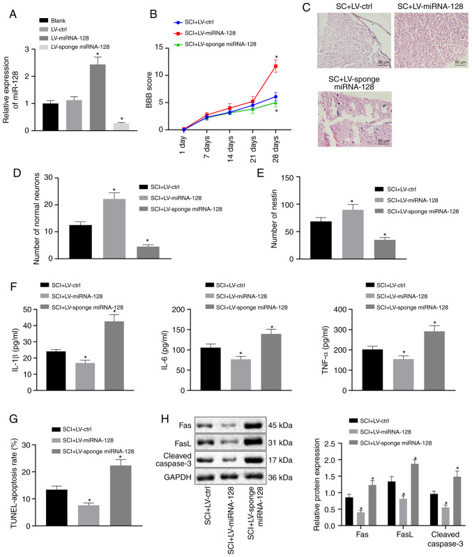
Figure 2. Upregulation of miR-128 can alleviate SCI in rats. Vectors containing LV-control, LV-miR-128, or LV-sponge miR-128 were injected into rats with SCI (n=10/group). (A) miR-128 expression was assessed by reverse transcription-quantitative PCR. *P<0.05 vs. SCI rats injected with LV-control. (B) Locomotor functions of rats indicated by BBB score. (C) Hematoxylin and eosin staining of morphological changes of neuronal cells (magnification, ×200). (D) The survival of neuronal cells in rat spinal cord determined by Nissl staining. (E) The number of nestin-positive cells evaluated by immunofluorescence. (F) The levels of inflammatory factors in rat serum detected by ELISA. (G) Cell apoptosis was assessed by TUNEL staining. (H) The expression of apoptosis-related factor protein in rat spinal cord determined by western blot analysis. An unpaired t-test was performed for comparisons of data between two groups. ANOVA was performed for comparisons of data among multiple groups, followed by Tukey's post-hoc test. BBB score was used as the comparisons of different time points using repeated-measures analysis of ANOVA, followed by Bonferroni post-hoc test. *P<0.05 vs. SCI rats infected with LV-control. miR, microRNA; SCI, spinal cord injury; LV, Lentiviral vector; ctrl, control; BBB, Basso, Beattie and Bresnahan; ANOVA, analysis of variance.
Figure 2. Upregulation of miR-128 can alleviate SCI in rats. Vectors containing LV-control, LV-miR-128, or LV-sponge miR-128 were injected into rats with SCI (n=10/group). (A) miR-128 expression was assessed by reverse transcription-quantitative PCR. *P<0.05 vs. SCI rats injected with LV-control. (B) Locomotor functions of rats indicated by BBB score. (C) Hematoxylin and eosin staining of morphological changes of neuronal cells (magnification, ×200). (D) The survival of neuronal cells in rat spinal cord determined by Nissl staining. (E) The number of nestin-positive cells evaluated by immunofluorescence. (F) The levels of inflammatory factors in rat serum detected by ELISA. (G) Cell apoptosis was assessed by TUNEL staining. (H) The expression of apoptosis-related factor protein in rat spinal cord determined by western blot analysis. An unpaired t-test was performed for comparisons of data between two groups. ANOVA was performed for comparisons of data among multiple groups, followed by Tukey's post-hoc test. BBB score was used as the comparisons of different time points using repeated-measures analysis of ANOVA, followed by Bonferroni post-hoc test. *P<0.05 vs. SCI rats infected with LV-control. miR, microRNA; SCI, spinal cord injury; LV, Lentiviral vector; ctrl, control; BBB, Basso, Beattie and Bresnahan; ANOVA, analysis of variance.CC BY-NC-ND · PubMed Central
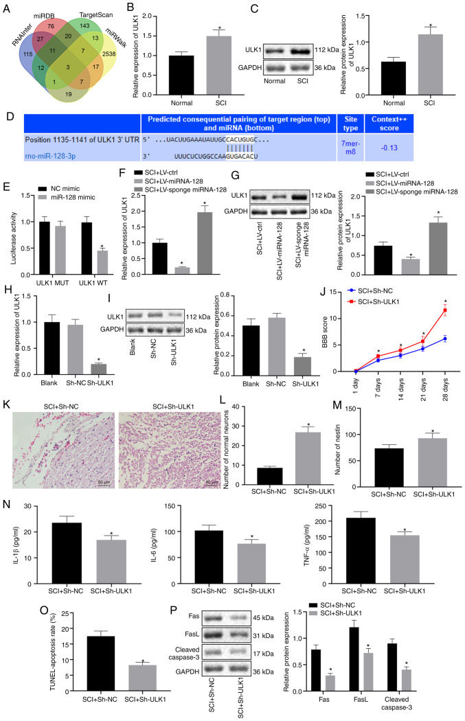
Figure 3. miR-128 inhibits the expression of ULK1 and knockdown of ULK1 attenuates SCI in rats. Vectors containing LV-control, LV-miR-128, or LV-sponge miR-128 were constructed and injected into rats with SCI in A-G, while vectors containing sh-NC or sh-ULK1 were injected into rats with SCI in H-P (n=10/group). (A) Venn diagram of the target genes of miR-128 predicted using RNAInter, miRDB, TargetScan and miRWalk. (B) The mRNA expression of ULK1 in normal rats and rats with SCI detected using RT-qPCR. *P<0.05 vs. normal rats. (C) Protein expression of ULK1 detected by western blot analysis. *P<0.05 vs. normal rats. (D) The binding site of miR-128 and ULK1 predicted using TargetScanHuman 7.2. (E) The targeting relationship between ULK1 and miR-128 measured by dual-luciferase report assay. *P<0.05 vs. NC mimic. (F) The mRNA expression of ULK1 examined by RT-qPCR. *P<0.05 vs. SCI rats injected with LV-control. (G) The protein expression of ULK1 measured using western blot analysis. *P<0.05 vs. SCI rats infected with LV-control. (H) The mRNA expression of ULK1 measured using RT-qPCR. *P<0.05 vs. Blank or sh-NC. (I) The protein expression of ULK1 expressing sh-NC and sh-ULK1 evaluated by western blot analysis. *P<0.05 vs. Blank or sh-NC. (J) Locomotor functions of rats evaluated by BBB score. (K) H&E staining of morphological changes of neurons (magnification, ×200). (L) Survival of neuronal cells detected by Nissl staining. (M) The number of nestin-positive cells detected by immunofluorescence. (N) The content of inflammatory factors in rat serum determined by ELISA. (O) Apoptosis of rat spinal cord cells determined using TUNEL. (P) The protein expression of apoptosis-related factor in rats with SCI evaluated by western blot analysis. J-P, *P<0.05 vs. SCI rats transfected with sh-NC. An unpaired t-test was performed for comparisons of data between two groups. ANOVA was performed for comparisons of data among multiple groups, followed by Tukey's post-hoc test. BBB score was used as comparisons of data at different time points using repeated-measures analysis of ANOVA, followed by Bonferroni post-hoc test. miR, microRNA; ULK1, Unc-51 like autophagy activating kinase 1; SCI, spinal cord injury; RT-qPCR, reverse transcription-quantitative PCR; NC, negative control; LV, Lentiviral vector; ctrl, control; sh, short hairpin; ANOVA, analysis of variance.
Figure 3. miR-128 inhibits the expression of ULK1 and knockdown of ULK1 attenuates SCI in rats. Vectors containing LV-control, LV-miR-128, or LV-sponge miR-128 were constructed and injected into rats with SCI in A-G, while vectors containing sh-NC or sh-ULK1 were injected into rats with SCI in H-P (n=10/group). (A) Venn diagram of the target genes of miR-128 predicted using RNAInter, miRDB, TargetScan and miRWalk. (B) The mRNA expression of ULK1 in normal rats and rats with SCI detected using RT-qPCR. *P<0.05 vs. normal rats. (C) Protein expression of ULK1 detected by western blot analysis. *P<0.05 vs. normal rats. (D) The binding site of miR-128 and ULK1 predicted using TargetScanHuman 7.2. (E) The targeting relationship between ULK1 and miR-128 measured by dual-luciferase report assay. *P<0.05 vs. NC mimic. (F) The mRNA expression of ULK1 examined by RT-qPCR. *P<0.05 vs. SCI rats injected with LV-control. (G) The protein expression of ULK1 measured using western blot analysis. *P<0.05 vs. SCI rats infected with LV-control. (H) The mRNA expression of ULK1 measured using RT-qPCR. *P<0.05 vs. Blank or sh-NC. (I) The protein expression of ULK1 expressing sh-NC and sh-ULK1 evaluated by western blot analysis. *P<0.05 vs. Blank or sh-NC. (J) Locomotor functions of rats evaluated by BBB score. (K) H&E staining of morphological changes of neurons (magnification, ×200). (L) Survival of neuronal cells detected by Nissl staining. (M) The number of nestin-positive cells detected by immunofluorescence. (N) The content of inflammatory factors in rat serum determined by ELISA. (O) Apoptosis of rat spinal cord cells determined using TUNEL. (P) The protein expression of apoptosis-related factor in rats with SCI evaluated by western blot analysis. J-P, *P<0.05 vs. SCI rats transfected with sh-NC. An unpaired t-test was performed for comparisons of data between two groups. ANOVA was performed for comparisons of data among multiple groups, followed by Tukey's post-hoc test. BBB score was used as comparisons of data at different time points using repeated-measures analysis of ANOVA, followed by Bonferroni post-hoc test. miR, microRNA; ULK1, Unc-51 like autophagy activating kinase 1; SCI, spinal cord injury; RT-qPCR, reverse transcription-quantitative PCR; NC, negative control; LV, Lentiviral vector; ctrl, control; sh, short hairpin; ANOVA, analysis of variance.CC BY-NC-ND · PubMed Central
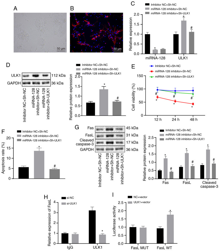
Figure 4. Upregulation of miR-128 repressed ULK1 expression and thereby attenuating SCI via the downregulation of FasL. The neuronal cells were transfected with inhibitor NC and sh-NC, miR-128 inhibitor and sh-NC or miR-128 inhibitor and sh-ULK1. (A) The morphology of spinal cord neurons observed under an inverted microscope (magnification, ×200). (B) Neuronal cell nestin-positive expression determined by immunofluorescence (magnification, ×200). (C) The expression of miR-128 and ULK1 detected using reverse transcription-quantitative PCR. (D) The protein expression of ULK1 measured by western blot analysis. (E) Cell proliferation assessed by MTT assay. (F) Cell apoptosis determined by flow cytometry. (G) The protein expression of apoptosis-related factors determined by western blot analysis. (H) The enrichment of ULK1 and FasL determined by ChIP assay. (I) The regulatory relationship of ULK1 on FasL assessed using dual-luciferase reporter assay. *P<0.05 vs. cells co-transfected with inhibitor NC and sh-NC or #P<0.05 vs. cells co-transfected with miR-128 inhibitor and sh-NC, or cells co-transfected with si-NC. An unpaired t-test was performed for comparisons of data between two groups. ANOVA was conducted for comparison of data among multiple groups, followed by Tukey's post-hoc test. miR, microRNA; ULK1, Unc-51 like autophagy activating kinase 1; SCI, spinal cord injury; FasL, Fas ligand; NC, negative control; sh, short hairpin; WT, wild type; MUT, mutated; ChIP, Chromatin immunoprecipitation; IgG, immunoglobulin G.
Figure 4. Upregulation of miR-128 repressed ULK1 expression and thereby attenuating SCI via the downregulation of FasL. The neuronal cells were transfected with inhibitor NC and sh-NC, miR-128 inhibitor and sh-NC or miR-128 inhibitor and sh-ULK1. (A) The morphology of spinal cord neurons observed under an inverted microscope (magnification, ×200). (B) Neuronal cell nestin-positive expression determined by immunofluorescence (magnification, ×200). (C) The expression of miR-128 and ULK1 detected using reverse transcription-quantitative PCR. (D) The protein expression of ULK1 measured by western blot analysis. (E) Cell proliferation assessed by MTT assay. (F) Cell apoptosis determined by flow cytometry. (G) The protein expression of apoptosis-related factors determined by western blot analysis. (H) The enrichment of ULK1 and FasL determined by ChIP assay. (I) The regulatory relationship of ULK1 on FasL assessed using dual-luciferase reporter assay. *P<0.05 vs. cells co-transfected with inhibitor NC and sh-NC or #P<0.05 vs. cells co-transfected with miR-128 inhibitor and sh-NC, or cells co-transfected with si-NC. An unpaired t-test was performed for comparisons of data between two groups. ANOVA was conducted for comparison of data among multiple groups, followed by Tukey's post-hoc test. miR, microRNA; ULK1, Unc-51 like autophagy activating kinase 1; SCI, spinal cord injury; FasL, Fas ligand; NC, negative control; sh, short hairpin; WT, wild type; MUT, mutated; ChIP, Chromatin immunoprecipitation; IgG, immunoglobulin G.CC BY-NC-ND · PubMed Central
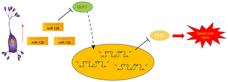
Figure 5. Molecular mechanism of miR-128/ULK1/FasL axis in protecting spinal cord injury. The upregulation of miR-128 could suppress ULK1 to inhibit the apoptosis of neuronal cells, thereby alleviating SCI via the downregulation of FasL. miR, microRNA; ULK1, Unc-51 like autophagy activating kinase 1; FasL, Fas ligand.
Figure 5. Molecular mechanism of miR-128/ULK1/FasL axis in protecting spinal cord injury. The upregulation of miR-128 could suppress ULK1 to inhibit the apoptosis of neuronal cells, thereby alleviating SCI via the downregulation of FasL. miR, microRNA; ULK1, Unc-51 like autophagy activating kinase 1; FasL, Fas ligand.CC BY-NC-ND · PubMed Central

Verification

Claims the paper makes

The claims and statistics of this paper haven’t been checked yet.

Run the check — free, no account needed. It reads the open-access text and checks the sources it cites.

A few free checks a day · papers already verified open instantly · results are public

Claims other papers make about this one

Each is checked against this paper’s own text: does it say what the citing paper says it does?

Self-check

Percentages, sums and reported differences re-computed from the paper’s own numbers.

The self-check needs the paper’s full text.

Citations

Cites

Research works printed but not linked: 31

  1. Saxena, 2015, Nanocarrier-mediated inhibition of macrophage migration inhibitory factor attenuates secondary injury after spinal cord injury, ACS Nano, 9, 1492, 10.1021/nn505980z · DOI
  2. Ramer, 2014, Restoring function after spinal cord injury: Towards clinical translation of experimental strategies, Lancet Neurol, 13, 1241, 10.1016/S1474-4422(14)70144-9 · DOI
  3. Assinck, 2017, Cell transplantation therapy for spinal cord injury, Nat Neurosci, 20, 637, 10.1038/nn.4541 · DOI
  4. Zhang, 2018, Tanshinone IIA promotes the differentiation of bone marrow mesenchymal stem cells into neuronal-like cells in a spinal cord injury model, J Transl Med, 16, 193, 10.1186/s12967-018-1571-y · DOI
  5. Ning, 2014, microRNAs in spinal cord injury: Potential roles and therapeutic implications, Int J Biol Sci, 10, 997, 10.7150/ijbs.9058 · DOI
  6. Yang, 2017, Down-regulation of miRNA-128 contributes to neuropathic pain following spinal cord injury via activation of P38, Med Sci Monit, 23, 405, 10.12659/MSM.898788 · DOI
  7. Wei, 2018, Deficiency of unc-51 like kinase 1 (Ulk1) protects against mice traumatic brain injury (TBI) by suppression of p38 and JNK pathway, Biochem Biophys Res Commun, 503, 467, 10.1016/j.bbrc.2018.04.154 · DOI
  8. Lu, 2018, Overexpression of ULK1 represents a potential diagnostic marker for clear cell renal carcinoma and the antitumor effects of SBI-0206965, EBioMedicine, 34, 85, 10.1016/j.ebiom.2018.07.034 · DOI
  9. Pang, 2019, ULK1 affects cell viability of goat Sertoli cell by modulating both autophagy and apoptosis, In Vitro Cell Dev Biol Anim, 55, 604, 10.1007/s11626-019-00371-2 · DOI
  10. Krzyzowska, 2014, Role of Fas/FasL signaling in regulation of anti-viral response during HSV-2 vaginal infection in mice, Immunobiology, 219, 932, 10.1016/j.imbio.2014.07.021 · DOI
  11. Zhao, 2019, MiR-543-5p inhibits inflammation and promotes nerve regeneration through inactivation of the NF-kB in rats after spinal cord injury, Eur Rev Med Pharmacol Sci, 23, S39
  12. von Leden, 2017, Age exacerbates microglial activation, oxidative stress, inflammatory and NOX2 gene expression, and delays functional recovery in a middle-aged rodent model of spinal cord injury, J Neuroinflammation, 14, 161, 10.1186/s12974-017-0933-3 · DOI
  13. Takenaga, 2006, Lecithinized superoxide dismutase (PC-SOD) improved spinal cord injury-induced motor dysfunction through suppression of oxidative stress and enhancement of neurotrophic factor production, J Control Release, 110, 283, 10.1016/j.jconrel.2005.10.022 · DOI
  14. Livak, 2001, Analysis of relative gene expression data using real-time quantitative PCR and the 2(-Delta Delta C(T)) method, Methods, 25, 402, 10.1006/meth.2001.1262 · DOI · Paper page
  15. Li, 2016, A hydrogel bridge incorporating immobilized growth factors and neural stem/progenitor cells to treat spinal cord injury, Adv Healthc Mater, 5, 802, 10.1002/adhm.201500810 · DOI
  16. Li, 2018, A collagen microchannel scaffold carrying paclitaxel-liposomes induces neuronal differentiation of neural stem cells through Wnt/β-catenin signaling for spinal cord injury repair, Biomaterials, 183, 114, 10.1016/j.biomaterials.2018.08.037 · DOI
  17. He, 2017, Molecular Mechanism of MiR-136-5p targeting NF-kB/A20 in the IL-17-mediated inflammatory response after spinal cord injury, Cell Physiol Biochem, 44, 1224, 10.1159/000485452 · DOI
  18. Farrell, 2011, Developmentally regulated expression of Sox9 and microRNAs 124, 128 and 23 in neuroepithelial stem cells in the developing spinal cord, Int J Dev Neurosci, 29, 31, 10.1016/j.ijdevneu.2010.10.001 · DOI
  19. Shao, 2018, Silencing of PHLPP1 promotes neuronal apoptosis and inhibits functional recovery after spinal cord injury in mice, Life Sci, 209, 291, 10.1016/j.lfs.2018.08.030 · DOI
  20. Zheng, 2019, Carbon monoxide releasing molecule-3 alleviates neuron death after spinal cord injury via inflammasome regulation, EBioMedicine, 40, 643, 10.1016/j.ebiom.2018.12.059 · DOI
  21. Wang, 2018, Effects of dexamethasone on autophagy and apoptosis in acute spinal cord injury, Neuroreport, 29, 1084, 10.1097/WNR.0000000000001076 · DOI
  22. Zhong, 2019, Inhibition of MSK1 promotes inflammation and apoptosis and inhibits functional recovery after spinal cord injury, J Mol Neurosci, 68, 191, 10.1007/s12031-019-01298-9 · DOI
  23. Wang, 2018, The role of the Fas/FasL signaling pathway in environmental toxicant-induced testicular cell apoptosis: An update, Syst Biol Reprod Med, 64, 93, 10.1080/19396368.2017.1422046 · DOI
  24. Li, 2018, Restraint-induced corticotrophin-releasing hormone elevation triggers apoptosis of ovarian cells and impairs oocyte competence via activation of the Fas/FasL system, Biol Reprod, 99, 828, 10.1093/biolre/ioy091 · DOI
  25. Liu, 2018, MiR-21 regulates the apoptosis of keloid fibroblasts by caspase-8 and the mitochondria-mediated apoptotic signaling pathway via targeting FasL, Biochem Cell Biol, 96, 548, 10.1139/bcb-2017-0306 · DOI
  26. Lu, 2012, Functional recovery in rat spinal cord injury induced by hyperbaric oxygen preconditioning, Neurol Res, 34, 944, 10.1179/1743132812Y.0000000096 · DOI
  27. Chen, 2017, Downregulation of ULK1 by microRNA-372 inhibits the survival of human pancreatic adenocarcinoma cells, Cancer Sci, 108, 1811, 10.1111/cas.13315 · DOI
  28. Zheng, 2018, MiR-26a-5p regulates cardiac fibroblasts collagen expression by targeting ULK1, Sci Rep, 8, 2104, 10.1038/s41598-018-20561-4 · DOI
  29. Li, 2015, UNC-51-like kinase 1 blocks S6k1 phosphorylation contributes to neurodegeneration in Parkinson's disease model in vitro, Biochem Biophys Res Commun, 459, 196, 10.1016/j.bbrc.2015.02.008 · DOI
  30. Zhang, 2012, miR-21 represses FasL in microglia and protects against microglia-mediated neuronal cell death following hypoxia/ischemia, Glia, 60, 1888, 10.1002/glia.22404 · DOI · Paper page
  31. Gong, 2018, miR-411 suppresses acute spinal cord injury via downregulation of Fas ligand in rats, Biochem Biophys Res Commun, 501, 501, 10.1016/j.bbrc.2018.05.022 · DOI

Cited by

Sorted by citations

See all 9

Metadata

Authors
Liu, R., Peng, Z., Zhang, Y., Li, R., & Wang, Y.
Affiliations
  • Department of Orthopaedics, The First Affiliated Hospital of Harbin Medical University, Harbin, Heilongjiang 150001, P.R. China
  • Department of Orthopaedics, Binzhou Medical University Hospital, Binzhou, Shandong 256603, P.R. China
Venue
Molecular Medicine Reports
Published
2021-07-20
Volume
24
Issue
3
ISSN
  • 1791-2997
  • 1791-3004
Cited by
9
References
1

About these checks

Registry, retraction, Cites and statistics results are computed, not judged, with one exception: a printed reference that no index lists is read once by a model before it counts as a citation error. Claims are extracted by a model and kept only when their quote appears verbatim in the text; each cited source is then checked, with a model’s help, for whether it says what the claim relies on. Those verdicts can be wrong — read the claim’s quote and the cited source before relying on one.

Statistics and arithmetic are recomputed, not judged.

Confidence bands are shown only for fields calibrated on our annotated test set. None is calibrated yet, so no band is shown.

What counts as a claim ›How we measure confidence ›How we check this ›

Are you an author, or do you see a problem with this page? Request a review