

The DOI matches its Crossref record; 1 of the 1 references it cites were found in a registry or an index.
Prognostic significance of the expression of HER family members in primary osteosarcoma
Open accessAbstract
The prognosis of patients with metastatic osteosarcoma is poor and has shown no significant improvement in nearly 20 years. The human epidermal growth factor (EGF) receptor (HER) family is frequently overexpressed in the majority of human carcinomas, and is involved in promoting the proliferation and survival of cancer cells. However, the role of EGFR and HER‑2 expression in osteosarcoma survival remains controversial and no previous study has simultaneously investigated the association of the expression of all the four HER family members with the prognostic significance of osteosarcoma. Therefore, the present study investigated the expression levels of the complete members of the HER family in osteosarcoma specimens, as well as their associations with the clinicopathological parameters, progression‑free survival (PFS) and overall survival (OS) time of patients with osteosarcoma. The expression of HER family members was detected in osteosarcoma tumor specimens from 60 patients using immunohistochemistry. The association of the expression of HER receptors in osteosarcoma with clinicopathological parameters was analyzed using χ2 test and Fishers exact test. Survival analyses were evaluated by Kaplan‑Meier method and Cox proportional hazards regression model. Overall, 18 (30%), 13 (22%), 23 (38%) and 19 (32%) patients presented with high expression of EGFR, HER‑2, HER‑3 and HER‑4, respectively, and the co‑expression of 2, 3 and all 4 members of the HER family was observed. High expression of EGFR and HER‑4 was associated with distant metastasis. High HER‑3 expression was significantly associated with an advanced Enneking stage and distant metastasis. Multivariate analysis demonstrated that the expression of EGFR, HER‑3, HER‑4, EGFR/HER‑3, EGFR/HER‑4 and HER‑3/HER‑4 was an independent predictor of poor PFS and OS time in osteosarcoma patients with stage I‑IIB disease. In patients with stage IIB osteosarcoma, the expression of HER‑4 and EGFR/HER‑4 demonstrated a more significant effect on PFS and OS time. In conclusion, therapies targeting EGFR, HER‑3 and HER‑4 may provide promising strategies for primary osteosarcoma.
Overview
The claims and statistics of this paper haven’t been checked yet.
Run the check — free, no account needed. It reads the open-access text and checks the sources it cites.
Integrity signals
Registry
Cites
Statistics
Figures
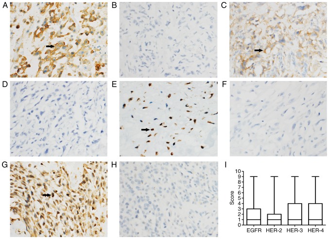
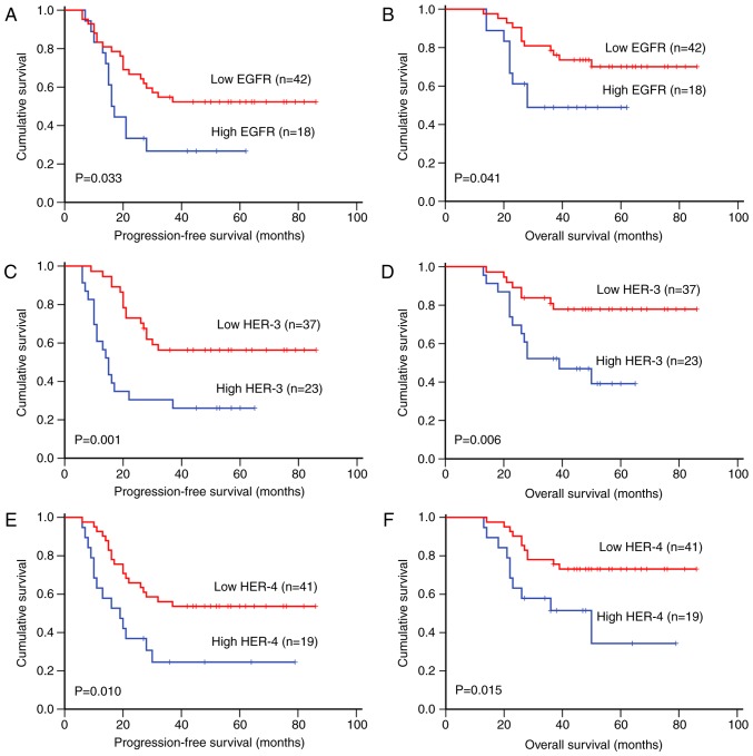
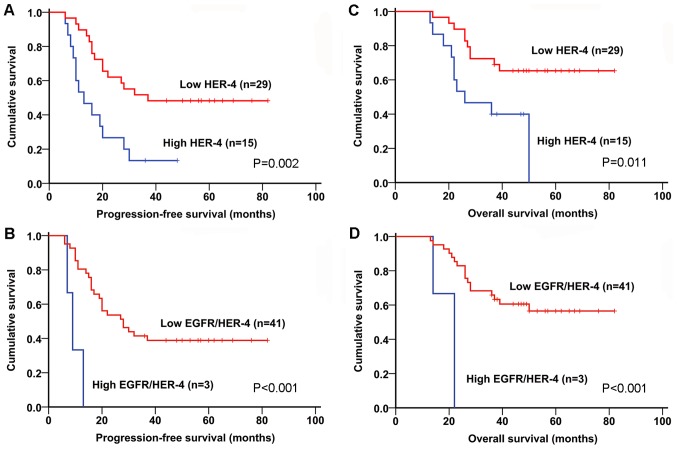
Verification
Claims the paper makes
The claims and statistics of this paper haven’t been checked yet.
Run the check — free, no account needed. It reads the open-access text and checks the sources it cites.
Claims other papers make about this one
Each is checked against this paper’s own text: does it say what the citing paper says it does?
The cited source doesn’t support the claim as written.
In the citing paper
“and is associated with adverse clinical outcomes [103]”
From Advances on immunotherapy for osteosarcoma (2024) · cites this paper as [103]
Checked by a reasoning model — may be wrong.
Experimental — accuracy not yet measured
Self-check
Percentages, sums and reported differences re-computed from the paper’s own numbers.
The self-check needs the paper’s full text.
Citations
Cites
Mirabello, L., Troisi, R. J., & Savage, S. A. (2009) · Cancer · doi:10.1002/cncr.24121 · Cited by 2,199
Research works printed but not linked: 41
- Zhou, 2014, Advances in targeted therapy for osteosarcoma, Discov Med, 17, 301
- De Luca, 2008, The role of the EGFR signaling in tumor microenvironment, J Cell Physiol, 214, 559, 10.1002/jcp.21260 · DOI
- Mendelsohn, 2006, Epidermal growth factor receptor targeting in cancer, Semin Oncol, 33, 369, 10.1053/j.seminoncol.2006.04.003 · DOI
- Renoir, 2013, Estrogen receptor signaling as a target for novel breast cancer therapeutics, Biochem Pharmacol, 85, 449, 10.1016/j.bcp.2012.10.018 · DOI
- Siu, 2014, Loss of EGFR signaling-regulated miR-203 promotes prostate cancer bone metastasis and tyrosine kinase inhibitors resistance, Oncotarget, 5, 3770, 10.18632/oncotarget.1994 · DOI
- Xu, 2016, Dacomitinib, a new pan-EGFR inhibitor, is effective in killing ovarian cancer cells, Discov Med, 22, 297
- Do, 2009, The expression of epidermal growth factor receptor and its downstream signaling molecules in osteosarcoma, Int J Oncol, 34, 797
- Lee, 2012, Epidermal growth factor receptor: Is it a feasible target for the treatment of osteosarcoma?, Cancer Res Treat, 44, 202, 10.4143/crt.2012.44.3.202 · DOI
- Kersting, 2007, Epidermal growth factor receptor expression in high-grade osteosarcomas is associated with a good clinical outcome, Clin Cancer Res, 13, 2998, 10.1158/1078-0432.CCR-06-2432 · DOI
- Baxevanis, 2004, Immunobiology of HER-2/neu oncoprotein and its potential application in cancer immunotherapy, Cancer Immunol Immunother, 53, 166, 10.1007/s00262-003-0475-7 · DOI
- Abdou, 2016, The prognostic role of Ezrin and HER2/neu expression in osteosarcoma, Appl Immunohistochem Mol Morphol: AIMM, 24, 355, 10.1097/PAI.0000000000000197 · DOI
- Yalcin, 2008, C-erbB-2 expression and prognostic significance in osteosarcoma, Pediatric Blood Cancer, 51, 222, 10.1002/pbc.21576 · DOI
- Zhang, 2016, HER-2 expression in biopsy and surgical specimen on prognosis of osteosarcoma: A systematic review and meta-analysis of 16 studies, Medicine, 95, e3661, 10.1097/MD.0000000000003661 · DOI
- Akatsuka, 2002, ErbB2 expression is correlated with increased survival of patients with osteosarcoma, Cancer, 94, 1397, 10.1002/cncr.10360 · DOI
- Geller, 2010, HER-2 targeted treatment of osteosarcoma: The challenges of developing targeted therapy and prognostic factors for rare malignancies, Exp Opin Pharmacother, 11, 51, 10.1517/14656560903419614 · DOI
- Ma, 2014, Targeting of erbB3 receptor to overcome resistance in cancer treatment, Mol Cancer, 13, 105, 10.1186/1476-4598-13-105 · DOI
- Hassan, 2012, Cell surface receptor expression patterns in osteosarcoma, Cancer, 118, 740, 10.1002/cncr.26339 · DOI
- Hughes, 2004, Cell surface expression of epidermal growth factor receptor and Her-2 with nuclear expression of Her-4 in primary osteosarcoma, Cancer Res, 64, 2047, 10.1158/0008-5472.CAN-03-3096 · DOI
- Hua, 2011, Abstract 197: Multi-cellular tumor spheroids in vitro model reveals a critical role of ERBB4 in survival of osteosarcoma, Cancer Res, 71, 197, 10.1158/1538-7445.AM2011-197 · DOI
- Enneking, 1980, A system for the surgical staging of musculoskeletal sarcoma, Clin Orthop Relat Res, 106, 10.1097/00003086-198011000-00013 · DOI
- Srirajaskanthan, 2010, Expression of the HER-1–4 family of receptor tyrosine kinases in neuroendocrine tumours, Oncol Rep, 23, 909, 10.3892/or_00000714 · DOI
- Liu, 2017, Survival time and prognostic factors of patients with initial noncurative colorectal liver metastases, Medicine (Baltimore), 96, e8831, 10.1097/MD.0000000000008831 · DOI
- Wu, 2007, Statistical analysis and correlation discovery of tumor respiratory motion, Phys Med Biol, 52, 4761, 10.1088/0031-9155/52/16/004 · DOI
- Nishikawa, 2013, Prognostic factors in patients with hepatitis B virus-related hepatocellular carcinoma undergoing nucleoside analog antiviral therapy, Oncol Lett, 6, 1213, 10.3892/ol.2013.1578 · DOI
- Pahl, 2012, Anti-EGFR antibody cetuximab enhances the cytolytic activity of natural killer cells toward osteosarcoma, Clin Cancer Res, 18, 432, 10.1158/1078-0432.CCR-11-2277 · DOI
- Huang, 2013, Dual targeting of EGFR and HER3 with MEHD7945A overcomes acquired resistance to EGFR inhibitors and radiation, Cancer Res, 73, 824, 10.1158/0008-5472.CAN-12-1611 · DOI
- Huang, 2012, Stretch-induced fetal type II cell differentiation is mediated via ErbB1-ErbB4 interactions, J Biol Chem, 287, 18091, 10.1074/jbc.M111.313163 · DOI
- Silva, 2014, Cooverexpression of ERBB1 and ERBB4 receptors predicts poor clinical outcome in pN+ oral squamous cell carcinoma with extranodal spread, Clin Exp Metastasis, 31, 307, 10.1007/s10585-013-9629-y · DOI
- Anninga, 2004, Overexpression of the HER-2 oncogene does not play a role in high-grade osteosarcomas, Eur J Cancer, 40, 963, 10.1016/j.ejca.2003.10.025 · DOI
- Maitra, 2001, Amplification of the HER-2/neu oncogene is uncommon in pediatric osteosarcomas, Cancer, 92, 677, 10.1002/1097-0142(20010801)92:3<677::AID-CNCR1370>3.0.CO;2-V · DOI
- Kilpatrick, 2001, Clinicopathologic analysis of HER-2/neu immunoexpression among various histologic subtypes and grades of osteosarcoma, Mod Pathol, 14, 1277, 10.1038/modpathol.3880474 · DOI
- Scotlandi, 2005, Prognostic and therapeutic relevance of HER2 expression in osteosarcoma and Ewing's sarcoma, Eur J Cancer, 41, 1349, 10.1016/j.ejca.2005.03.015 · DOI
- Yoshida, 2012, Mechanism of resistance to trastuzumab and molecular sensitization via ADCC activation by exogenous expression of HER2-extracellular domain in human cancer cells, Cancer Immunol Immunother, 61, 1905, 10.1007/s00262-012-1249-x · DOI
- Long, 2014, Lapatinib alters the malignant phenotype of osteosarcoma cells via downregulation of the activity of the HER2-PI3K/AKT-FASN axis in vitro, Oncol Rep, 31, 328, 10.3892/or.2013.2825 · DOI
- Jullien, 2012, Downregulation of ErbB3 by Wnt3a contributes to wnt-induced osteoblast differentiation in mesenchymal cells, J Cell Biochem, 113, 2047, 10.1002/jcb.24076 · DOI
- Cai, 2010, Inactive Wnt/beta-catenin pathway in conventional high-grade osteosarcoma, J Pathol, 220, 24, 10.1002/path.2628 · DOI
- Xu, 2014, Down-regulation of miR-3928 promoted osteosarcoma growth, Cell Physiol Biochem, 33, 1547, 10.1159/000358718 · DOI
- Bianchi, 2006, ErbB-receptors expression and survival in breast carcinoma: A 15-year follow-up study, J Cell Physiol, 206, 702, 10.1002/jcp.20535 · DOI
- Karamouzis, 2007, Breast cancer: The upgraded role of HER-3 and HER-4, Int J Biochem Cell Biol, 39, 851, 10.1016/j.biocel.2006.11.017 · DOI
- Citri, 2006, EGF-ERBB signalling: Towards the systems level, Nat Rev Mol Cell Biol, 7, 505, 10.1038/nrm1962 · DOI
- Canfield, 2015, Receptor tyrosine kinase ERBB4 mediates acquired resistance to ERBB2 inhibitors in breast cancer cells, Cell Cycle, 14, 648, 10.4161/15384101.2014.994966 · DOI
Cited by
Sorted by citations
Advances on immunotherapy for osteosarcoma
Yu, S., & Yao, X. (2024) · Molecular Cancer · doi:10.1186/s12943-024-02105-9 · Cited by 270
“HER2 expression has been reported in 60-70% of primary osteosarcoma [102] and is associated with adverse clinical outcomes [103].”
Background
Metadata
- Authors
- Wang, S., Zhong, G., Wang, X., Yu, F., Weng, D., Wang, X., & Lin, J.
- Affiliations
- Department of Orthopedics, The First Affiliated Hospital of Fujian Medical University, Fuzhou, Fujian 350005, P.R. China
- First Affiliated Hospital of Fujian Medical University (CN)
- ORCID
- Zhong, Guang‑Xian
- Venue
- Oncology Letters
- Publisher
- Spandidos Publications
- Published
- 2018-06-08
- Volume
- 16
- Issue
- 2
- Pages
- 2185-2194
- ISSN
- 1792-1074 (ISSN-L)
- 1792-1082
- PMC
- PMC6036504
- OpenAlex
- W2807420926
- Cited by
- 20
- References
- 1
- Citations by year
- 2024: 2 · 2023: 7 · 2021: 7 · 2020: 2 · 2019: 1 · 2018: 1
- FWCI
- 2.07
- Type
- journal article
- Language
- en
- Open access
- diamond
- doi.org
- Licence
- cc-by-nc-nd
- Locations
- Oncology Letters (publishedVersion, cc-by-nc-nd)
- Europe PMC (PubMed Central) (publishedVersion, cc-by-nc-nd)
- PubMed Central (publishedVersion, cc-by-nc-nd)
- PubMed (publishedVersion)
- Funders
- National Natural Science Foundation of China (31571292)
- Fujian Medical University
- Topics
- Health Sciences › Medicine › Oncology › Sarcoma Diagnosis and Treatment
- Health Sciences › Medicine › Oncology › HER2/EGFR in Cancer Research
- Health Sciences › Medicine › Orthopedics and Sports Medicine › Bone Tumor Diagnosis and Treatments
- Keywords
- primary osteosarcoma, ErbB receptors, EGFR, HER2, HER3, ErbB4, prognostic factors, progression-free survival, overall survival, distant metastasis, Enneking stage, targeted therapy
About these checks
Registry, retraction, Cites and statistics results are computed, not judged, with one exception: a printed reference that no index lists is read once by a model before it counts as a citation error. Claims are extracted by a model and kept only when their quote appears verbatim in the text; each cited source is then checked, with a model’s help, for whether it says what the claim relies on. Those verdicts can be wrong — read the claim’s quote and the cited source before relying on one.
Statistics and arithmetic are recomputed, not judged.
Confidence bands are shown only for fields calibrated on our annotated test set. None is calibrated yet, so no band is shown.
What counts as a claim ›How we measure confidence ›How we check this ›
Are you an author, or do you see a problem with this page? Request a review
