

The DOI matches its Crossref record; 44 of the 44 references it cites were found in a registry or an index.
HBV upregulates AP‑1 complex subunit mu‑1 expression via the JNK pathway to promote proliferation of liver cancer cells
Abstract
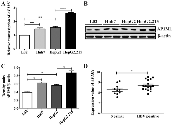
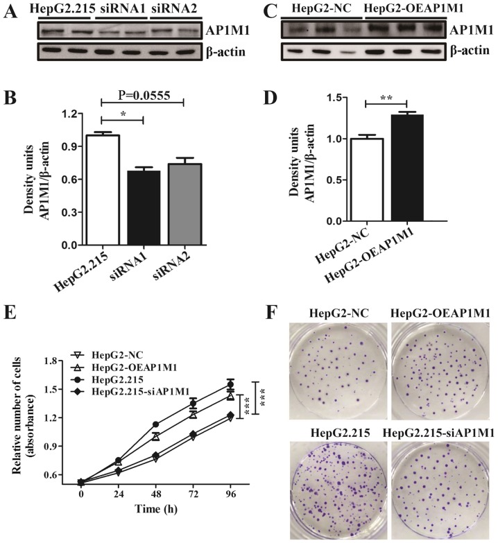
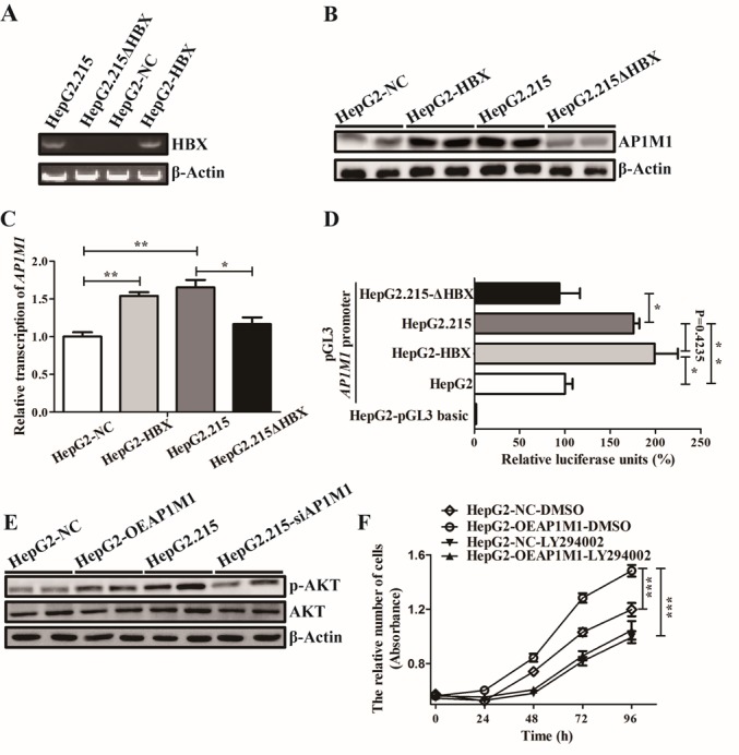
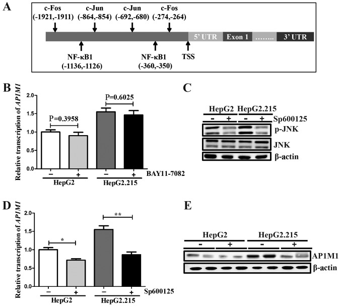
Although hepatitis B virus (HBV) infection is responsible for liver cancer, the exact mechanism of its action remains unclear. μ1 adaptin is an intrinsic part of the clathrin adaptor AP‑1 complex. In addition to its canonical biological function that involves cargo sorting and vesicular transport, recent studies have demonstrated that μ1 adaptin participates in cell growth and proliferation. The aim of the present study was to investigate the effects of the clathrin adaptor AP‑1 complex subunit mu‑1 (AP1M1) on liver cancer cell proliferation. The present study reports for the first time that AP1M1 is upregulated in the HBV‑transfected HepG2.215 liver cancer cells. Silencing of AP1M1 in HepG2.215 cells suppressed their proliferation, while the overexpression of AP1M1 in HepG2 cells promoted cell proliferation. The data suggested that AP1M1 is one of the crucial factors involved in the progression of liver cancer caused by HBV infection. In addition, it was demonstrated that HBV facilitated AP1M1 expression in a JNK‑dependent manner. The increased expression levels of AP1M1 enhanced phosphorylation of protein kinase B and accelerated cell proliferation. Unraveling the effects of AP1M1 on liver cancer cell proliferation and the mechanism of AP1M1 transcriptional regulation may provide new therapeutic targets for HBV‑positive liver cancer.
Overview
The claims and statistics of this paper haven’t been checked yet.
Run the check — free, no account needed. It reads the open-access text and checks the sources it cites.
Integrity signals
Registry
Cites
Statistics
Figures
Verification
Claims the paper makes
The claims and statistics of this paper haven’t been checked yet.
Run the check — free, no account needed. It reads the open-access text and checks the sources it cites.
Claims other papers make about this one
Each is checked against this paper’s own text: does it say what the citing paper says it does?
In the citing paper
“and of μ1A-adaptin (AP1M1) in hepatitis B virus-transfected HepG2 cells [39].”
From Clathrin adapters AP-1 and GGA2 support expression of epidermal growth factor receptor for cell growth (2021) · cites this paper as [39]
Self-check
Percentages, sums and reported differences re-computed from the paper’s own numbers.
The self-check needs the paper’s full text.
Citations
Cites
1 retracted
Bakhtiari, E., Hosseini, A., Boroushaki, M. T., & Mousavi, S. H. (2015) · Journal of Chemotherapy · doi:10.1179/1973947815y.0000000055 · Cited by 16
RetractedAnalysis of Relative Gene Expression Data Using Real-Time Quantitative PCR and the 2−ΔΔCT Method
Livak, K. J., & Schmittgen, T. D. (2001) · Methods · doi:10.1006/meth.2001.1262 · Cited by 180,860
Mutation of the mouse klotho gene leads to a syndrome resembling ageing
Kuro-o, M., Matsumura, Y., Aizawa, H., Kawaguchi, H., Suga, T., Utsugi, T., Ohyama, Y., Kurabayashi, M., Kaname, T., Kume, E., et al. (1997) · Nature · doi:10.1038/36285 · Cited by 3,837
Epidemiology of Viral Hepatitis and Hepatocellular Carcinoma
El-Serag, H. B. (2012) · Gastroenterology · doi:10.1053/j.gastro.2011.12.061 · Cited by 3,380
Interaction of Tyrosine-Based Sorting Signals with Clathrin-Associated Proteins
Ohno, H., Stewart, J., Fournier, M.-C., Bosshart, H., Rhee, I., Miyatake, S., Saito, T., Gallusser, A., Kirchhausen, T., & Bonifacino, J. S. (1995) · Science · doi:10.1126/science.7569928 · Cited by 969
CLATHRIN-COATED VESICLE FORMATION AND PROTEIN SORTING: An Integrated Process
Schmid, S. L. (1997) · Annual Review of Biochemistry · doi:10.1146/annurev.biochem.66.1.511 · Cited by 799
Forty Years of Clathrin‐coated Vesicles
Robinson, M. S. (2015) · Traffic · doi:10.1111/tra.12335 · Cited by 356
Doria, M., Klein, N. P., Lucito, R., & Schneider, R. J. (1995) · The EMBO Journal · doi:10.1002/j.1460-2075.1995.tb00156.x · Cited by 313
Benn, J., Su, F., Doria, M., & Schneider, R. J. (1996) · Journal of Virology · doi:10.1128/jvi.70.8.4978-4985.1996 · Cited by 281
μ1B, a novel adaptor medium chain expressed in polarized epithelial cells1
Ohno, H., Tomemori, T., Nakatsu, F., Okazaki, Y., Aguilar, R. C., Foelsch, H., Mellman, I., Saito, T., Shirasawa, T., & Bonifacino, J. S. (1999) · FEBS Letters · doi:10.1016/s0014-5793(99)00432-9 · Cited by 231
Cited by
Uemura, T., Suzuki, T., Dohmae, N., & Waguri, S. (2021) · Oncogenesis · doi:10.1038/s41389-021-00367-2 · Cited by 19
Metadata
- Authors
- Kou, Y., Yan, X., Liu, Q., Wei, X., Zhang, B., Li, X., Pan, W., Kong, F., Wang, Y., Zheng, K., & Tang, R.
- Affiliations
- Jiangsu Key Laboratory of Immunity and Metabolism, Department of Pathogenic Biology and Immunology, Xuzhou Medical University, Xuzhou, Jiangsu 221004, P.R. China
- Institute of Emergency and Rescue Medicine, Laboratory of Emergency Medicine, Xuzhou Medical University, Xuzhou, Jiangsu 221004, P.R. China
- Venue
- Oncology Letters
- Publisher
- Spandidos Publications
- Published
- 2019-04-30
- ISSN
- 1792-1074
- 1792-1082
- PMC
- PMC6540315
- Cited by
- 5
- References
- 44
About these checks
Registry, retraction, Cites and statistics results are computed, not judged, with one exception: a printed reference that no index lists is read once by a model before it counts as a citation error. Claims are extracted by a model and kept only when their quote appears verbatim in the text; each cited source is then checked, with a model’s help, for whether it says what the claim relies on. Those verdicts can be wrong — read the claim’s quote and the cited source before relying on one.
Statistics and arithmetic are recomputed, not judged.
Confidence bands are shown only for fields calibrated on our annotated test set. None is calibrated yet, so no band is shown.
What counts as a claim ›How we measure confidence ›How we check this ›
Are you an author, or do you see a problem with this page? Request a review
How to cite
Cite CoChat for the checks on this page, and the paper for its own findings.
- This page
- CoChat. (2026). Citation check: HBV upregulates AP‑1 complex subunit mu‑1 expression via the JNK pathway to promote proliferation of liver cancer cells. https://app.cochat.ai/papers/10.3892/ol.2019.10291
- The paper
- Kou, Y., Yan, X., Liu, Q., Wei, X., Zhang, B., Li, X., Pan, W., Kong, F., Wang, Y., Zheng, K., & Tang, R. (2019). HBV upregulates AP‑1 complex subunit mu‑1 expression via the JNK pathway to promote proliferation of liver cancer cells. Oncology Letters. https://doi.org/10.3892/ol.2019.10291
Data sources
Bibliographic data: Semantic Scholar, OpenAlex, Crossref.
Contains information from the Semantic Scholar Academic Graph Datasets, made available under the ODC Attribution License (ODC-BY 1.0).
Semantic Scholar’s data is described in: Kinney, R., et al. (2023). The Semantic Scholar Open Data Platform. arXiv:2301.10140.
