

The DOI matches its Crossref record; 2 of the 2 references it cites were found in a registry or an index.
Bmi-1 induces radioresistance in MCF-7 mammary carcinoma cells
Overview
The claims and statistics of this paper haven’t been checked yet.
Run the check — free, no account needed. It reads the open-access text and checks the sources it cites.
Integrity signals
Registry
Cites
Statistics
Figures
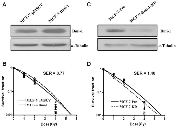
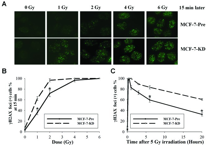
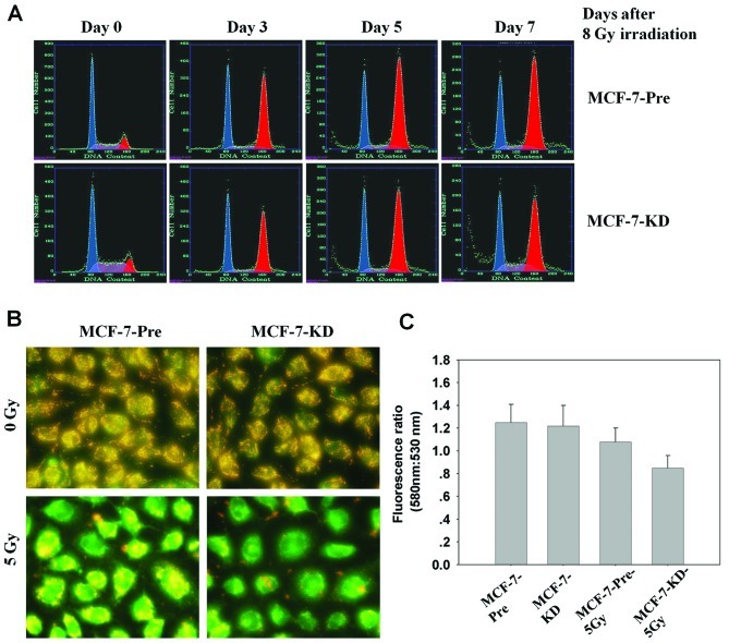
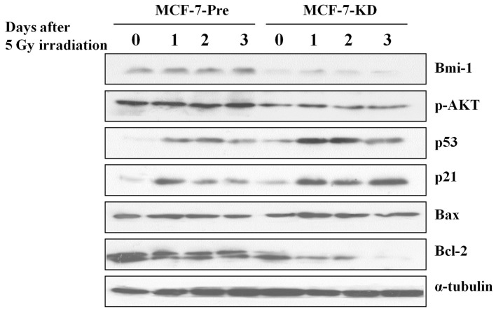
Verification
Claims the paper makes
The claims and statistics of this paper haven’t been checked yet.
Run the check — free, no account needed. It reads the open-access text and checks the sources it cites.
Claims other papers make about this one
Each is checked against this paper’s own text: does it say what the citing paper says it does?
The cited source says what the claim attributes to it.
In the citing paper
“Thus, the activation of PRCs, evident from increased expression of BMI1 and SUZ12,77 contributed to the radio-resistance of tumor cells,137,138 including glioblastoma.139,140”
From MiR-128-3p – a gray eminence of the human central nervous system (2024) · cites this paper as [138]
Self-check
Percentages, sums and reported differences re-computed from the paper’s own numbers.
The self-check needs the paper’s full text.
Citations
Cites
Jacobs, J. J. L., Kieboom, K., Marino, S., DePinho, R. A., & van Lohuizen, M. (1999) · Nature · doi:10.1038/16476 · Cited by 1,580
Glinsky, G. V., Berezovska, O., & Glinskii, A. B. (2005) · Journal of Clinical Investigation · doi:10.1172/jci23412 · Cited by 916
Research works printed but not linked: 41
- Jacobs, 1999, Cellular memory of transcriptional states by Polycomb-group proteins, Semin Cell Dev Biol, 10, 227, 10.1006/scdb.1999.0304 · DOI
- Schwartz, 2007, Polycomb silencing mechanisms and the management of genomic programmes, Nat Rev Genet, 8, 9, 10.1038/nrg1981 · DOI · Paper page
- Schuettengruber, 2007, Genome regulation by polycomb and trithorax proteins, Cell, 128, 735, 10.1016/j.cell.2007.02.009 · DOI
- Spivakov, 2007, Epigenetic signatures of stem-cell identity, Nat Rev Genet, 8, 263, 10.1038/nrg2046 · DOI
- Rajasekhar, 2007, Concise review: roles of polycomb group proteins in development and disease: a stem cell perspective, Stem Cells, 25, 2498, 10.1634/stemcells.2006-0608 · DOI
- Haupt, 1991, Novel zinc finger gene implicated as myc collaborator by retrovirally accelerated lymphomagenesis in E mu-myc transgenic mice, Cell, 65, 753, 10.1016/0092-8674(91)90383-A · DOI
- van Lohuizen, 1991, Identification of cooperating oncogenes in E mu-myc transgenic mice by provirus tagging, Cell, 65, 737, 10.1016/0092-8674(91)90382-9 · DOI
- Jacobs, 1999, Bmi-1 collaborates with c-Myc in tumorigenesis by inhibiting c-Myc-induced apoptosis via INK4a/ARF, Genes Dev, 13, 2678, 10.1101/gad.13.20.2678 · DOI
- Dimri, 2002, The Bmi-1 oncogene induces telomerase activity and immortalizes human mammary epithelial cells, Cancer Res, 62, 4736
- Guney, 2006, Cellular senescence, epigenetic switches and c-Myc, Cell Cycle, 5, 2319, 10.4161/cc.5.20.3348 · DOI
- Datta, 2007, Bmi-1 cooperates with H-Ras to transform human mammary epithelial cells via dysregulation of multiple growth-regulatory pathways, Cancer Res, 67, 10286, 10.1158/0008-5472.CAN-07-1636 · DOI
- Molofsky, 2003, Bmi-1 dependence distinguishes neural stem cell self-renewal from progenitor proliferation, Nature, 425, 962, 10.1038/nature02060 · DOI
- Raaphorst, 2003, Self-renewal of hematopoietic and leukemic stem cells: a central role for the Polycomb-group gene Bmi-1, Trends Immunol, 24, 522, 10.1016/S1471-4906(03)00241-2 · DOI
- Grinstein, 2007, Cellular signaling in normal and cancerous stem cells, Cell Signal, 19, 2428, 10.1016/j.cellsig.2007.06.021 · DOI
- Liu, 2006, Hedgehog signaling and Bmi-1 regulate self-renewal of normal and malignant human mammary stem cells, Cancer Res, 66, 6063, 10.1158/0008-5472.CAN-06-0054 · DOI
- Vrzalikova, 2008, Prognostic value of Bmi-1 oncoprotein expression in NSCLC patients: a tissue microarray study, J Cancer Res Clin Oncol, 134, 1037, 10.1007/s00432-008-0361-y · DOI
- Liu, 2008, Bmi-1 expression predicts prognosis for patients with gastric carcinoma, J Surg Oncol, 97, 267, 10.1002/jso.20934 · DOI
- Wang, 2008, Increased polycomb-group oncogene Bmi-1 expression correlates with poor prognosis in hepatocellular carcinoma, J Cancer Res Clin Oncol, 134, 535, 10.1007/s00432-007-0316-8 · DOI
- Chowdhury, 2007, Expression of Polycomb-group (PcG) protein BMI-1 predicts prognosis in patients with acute myeloid leukemia, Leukemia, 21, 1116, 10.1038/sj.leu.2404623 · DOI
- Arnes, 2008, Independent prognostic value of the basal-like phenotype of breast cancer and associations with EGFR and candidate stem cell marker BMI-1, Histopathology, 52, 370, 10.1111/j.1365-2559.2007.02957.x · DOI
- Song, 2006, Bmi-1 is a novel molecular marker of nasopharyngeal carcinoma progression and immortalizes primary human nasopharyngeal epithelial cells, Cancer Res, 66, 6225, 10.1158/0008-5472.CAN-06-0094 · DOI
- Qin, 2008, Expression and clinical significance of Bmi-1 protein in bladder cancer, Ai Zheng, 27, 1327
- Mani, 2008, The epithelial-mesenchymal transition generates cells with properties of stem cells, Cell, 133, 704, 10.1016/j.cell.2008.03.027 · DOI
- Yang, 2010, Bmi1 is essential in Twist1-induced epithelial-mesenchymal transition, Nat Cell Biol, 12, 982, 10.1038/ncb2099 · DOI
- Song, 2009, The polycomb group protein Bmi-1 represses the tumor suppressor PTEN and induces epithelial-mesenchymal transition in human nasopharyngeal epithelial cells, J Clin Invest, 119, 3626, 10.1172/JCI39374 · DOI
- Theys, 2011, E-Cadherin loss associated with EMT promotes radioresistance in human tumor cells, Radiother Oncol, 99, 392, 10.1016/j.radonc.2011.05.044 · DOI
- Kurrey, 2009, Snail and slug mediate radioresistance and chemoresistance by antagonizing p53-mediated apoptosis and acquiring a stem-like phenotype in ovarian cancer cells, Stem Cells, 27, 2059, 10.1002/stem.154 · DOI · Paper page
- Guo, 2011, Bmi-1 promotes invasion and metastasis, and its elevated expression is correlated with an advanced stage of breast cancer, Mol Cancer, 10, 10, 10.1186/1476-4598-10-10 · DOI
- Dimri, 2000, Regulation of a senescence checkpoint response by the E2F1 transcription factor and p14(ARF) tumor suppressor, Mol Cell Biol, 20, 273, 10.1128/MCB.20.1.273-285.2000 · DOI
- Liu, 2009, Relationship between methylation status of ERCC1 promoter and radiosensitivity in glioma cell lines, Cell Biol Int, 33, 1111, 10.1016/j.cellbi.2009.07.004 · DOI
- Reers, 1995, Mitochondrial membrane potential monitored by JC-1 dye, Methods Enzymol, 260, 406, 10.1016/0076-6879(95)60154-6 · DOI
- Lee, 2008, Expression of Bmi-1 in epidermis enhances cell survival by altering cell cycle regulatory protein expression and inhibiting apoptosis, J Invest Dermatol, 128, 9, 10.1038/sj.jid.5700949 · DOI
- Yan, 2008, Expression and clinical significance of DNA-PKcs in nasopharyngeal carcinoma, Ai Zheng, 27, 979
- Kraus, 2002, In vitro chemo- and radio-resistance in small cell lung cancer correlates with cell adhesion and constitutive activation of AKT and MAP kinase pathways, Oncogene, 21, 8683, 10.1038/sj.onc.1205939 · DOI
- Liang, 2003, Targeting the phosphatidylinositol 3-kinase/Akt pathway for enhancing breast cancer cells to radiotherapy, Mol Cancer Ther, 2, 353
- Soderlund, 2005, Activation of the phosphatidylinositol 3-kinase/Akt pathway prevents radiation-induced apoptosis in breast cancer cells, Int J Oncol, 26, 25
- LoPiccolo, 2007, Targeting Akt in cancer therapy, Anticancer Drugs, 18, 861, 10.1097/CAD.0b013e3280cc2c6f · DOI
- Lemasters, 1999, Confocal microscopy of the mitochondrial permeability transition in necrotic and apoptotic cell death, Biochem Soc Symp, 66, 205, 10.1042/bss0660205 · DOI
- Kang, 2004, Caspase-independent cell death by arsenic trioxide in human cervical cancer cells: reactive oxygen species-mediated poly(ADP-ribose) polymerase-1 activation signals apoptosis-inducing factor release from mitochondria, Cancer Res, 64, 8960, 10.1158/0008-5472.CAN-04-1830 · DOI
- Kim, 2004, Critical role of reactive oxygen species and mitochondrial membrane potential in Korean mistletoe lectin-induced apoptosis in human hepatocarcinoma cells, Mol Pharmacol, 66, 1383, 10.1124/mol.104.001347 · DOI
- Park, 2005, Phytosphingosine in combination with ionizing radiation enhances apoptotic cell death in radiation-resistant cancer cells through ROS-dependent and -independent AIF release, Blood, 105, 1724, 10.1182/blood-2004-07-2938 · DOI
Cited by
Sorted by citations
Nanoreporter PET predicts the efficacy of anti-cancer nanotherapy
Pérez-Medina, C., Abdel-Atti, D., Tang, J., Zhao, Y., Fayad, Z. A., Lewis, J. S., Mulder, W. J. M., & Reiner, T. (2016) · Nature Communications · doi:10.1038/ncomms11838 · Cited by 128
MiR-128-3p – a gray eminence of the human central nervous system
Kiel, K., Król, S. K., Bronisz, A., & Godlewski, J. (2024) · Molecular Therapy Nucleic Acids · doi:10.1016/j.omtn.2024.102141 · Cited by 15
Metadata
- Authors
- LIU, Z.-G., LIU, L., XU, L.-H., YI, W., TAO, Y.-L., TU, Z.-W., LI, M.-Z., ZENG, M.-S., & XIA, Y.-F.
- Venue
- Oncology Reports
- Publisher
- Spandidos Publications
- Published
- 2011-12-30
- Volume
- 27
- Issue
- 4
- Pages
- 1116-1122
- ISSN
- 1021-335X
- 1791-2431
- PMC
- PMC3583403
- Cited by
- 36
- References
- 2
- Language
- en
About these checks
Registry, retraction, Cites and statistics results are computed, not judged, with one exception: a printed reference that no index lists is read once by a model before it counts as a citation error. Claims are extracted by a model and kept only when their quote appears verbatim in the text; each cited source is then checked, with a model’s help, for whether it says what the claim relies on. Those verdicts can be wrong — read the claim’s quote and the cited source before relying on one.
Statistics and arithmetic are recomputed, not judged.
Confidence bands are shown only for fields calibrated on our annotated test set. None is calibrated yet, so no band is shown.
What counts as a claim ›How we measure confidence ›How we check this ›
Are you an author, or do you see a problem with this page? Request a review
