Cited by 16Mesenchymal stem cell researchLiver physiology and pathologyTissue Engineering and Regenerative Medicine

The DOI matches its Crossref record; 3 of the 3 references it cites were found in a registry or an index.

Effect of glycyrrhizic acid and 18β-glycyrrhetinic acid on the differentiation of human umbilical cord-mesenchymal stem cells into hepatocytes

Open access

Abstract

BACKGROUND: preconditioning strategies have been employed to strengthen the regenerative and differentiation potential of MSCs towards hepatic lineage. Chemical compounds of the triterpene class; glycyrrhizic acid (GA) and 18β-glycyrrhetinic acid (GT) possess diverse therapeutic properties including hepato-protection and anti-fibrosis characteristics. They are capable of modulating several signaling pathways that are crucial in hepatic regeneration. Preconditioning with hepato-protective triterpenes may stimulate MSC fate transition towards hepatocytes. AIM: To explore the effect of GA and GT on hepatic differentiation of human umbilical cord-MSCs (hUC-MSCs). METHODS: hUC-MSCs were isolated and characterized phenotypically by flow cytometry and immunocytochemistry for the expression of MSC-associated surface molecules. Isolated cells were treated with GA, GT, and their combination for 24 h and then analyzed at three time points; day 7, 14, and 21. qRT-PCR was performed for the expression of hepatic genes. Expression of hepatic proteins was analyzed by immunocytochemistry at day 21. Periodic acid Schiff staining was performed to determine the functional ability of treated cells. RESULTS: The fusiform-shaped morphology of MSCs in the treatment groups in comparison with the untreated control, eventually progressed towards the polygonal morphology of hepatocytes with the passage of time. The temporal transcriptional profile of preconditioned MSCs displayed significant expression of hepatic genes with increasing time of differentiation. Preconditioned cells showed positive expression of hepatocyte-specific proteins. The results were further corroborated by positive periodic acid Schiff staining, indicating the presence of glycogen in their cytoplasm. Moreover, bi-nucleated cells, which is the typical feature of hepatocytes, were also seen in the preconditioned cells. CONCLUSION: Preconditioning with glycyrrhizic acid, 18β-glycyrrhetinic acid and their combination, successfully differentiates hUC-MSCs into hepatic-like cells. These MSCs may serve as a better therapeutic option for degenerative liver diseases in future.

Overview

The claims and statistics of this paper haven’t been checked yet.

Run the check — free, no account needed. It reads the open-access text and checks the sources it cites.

A few free checks a day · papers already verified open instantly · results are public

Integrity signals

Registry

Crossref · 10.4252/wjsc.v13.i10.1580

Cites

3 of 3 references verified0 citation errors · 0 not checkable

Statistics

Figures

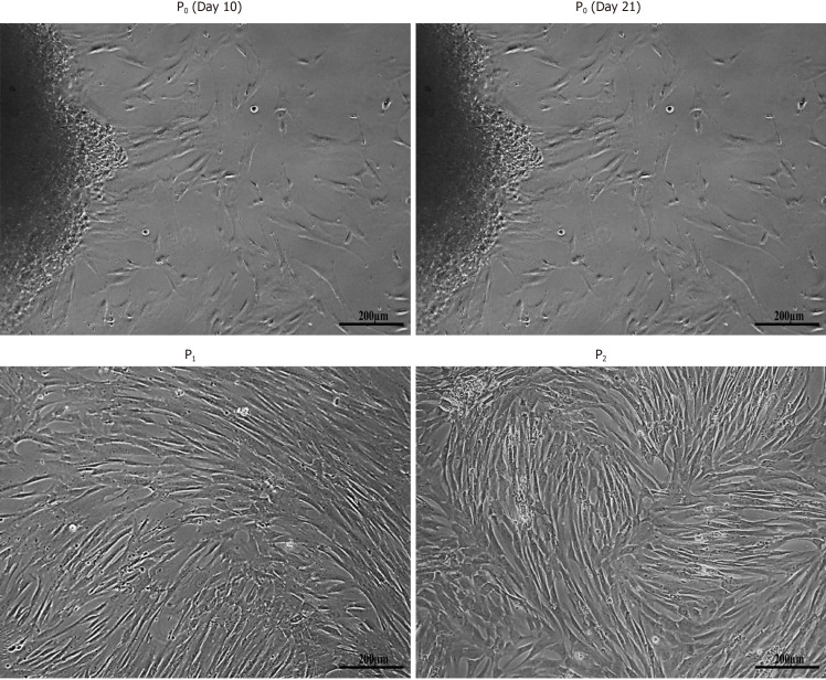
Figure 1 Isolation of human umbilical cord-mesenchymal stem cells. Morphological observations of human umbilical cord-mesenchymal stem cells showing passage 0 (P0) cells at day 10 and day 21 with cells migrating towards the periphery of the adherent tissue, and passage 1 (P1) and passage 2 (P2) cells with homogeneous population showing fusiform- and fibroblast-like appearance after sub-culturing. All images were taken under phase contrast at 10× magnification.
Figure 1 Isolation of human umbilical cord-mesenchymal stem cells. Morphological observations of human umbilical cord-mesenchymal stem cells showing passage 0 (P0) cells at day 10 and day 21 with cells migrating towards the periphery of the adherent tissue, and passage 1 (P1) and passage 2 (P2) cells with homogeneous population showing fusiform- and fibroblast-like appearance after sub-culturing. All images were taken under phase contrast at 10× magnification.CC BY-NC · PubMed Central
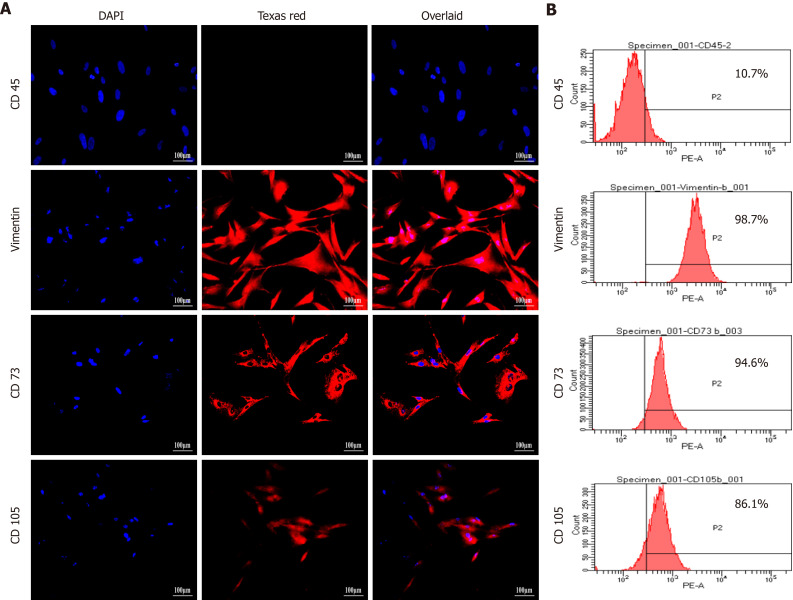
Figure 2 Characterization of human umbilical cord-mesenchymal stem cells by immunocytochemical analysis and flow cytometry. A: Immunocytochemical analysis showing positive expression of mesenchymal markers, CD73, CD105, and vimentin, and negative expression of hematopoietic marker CD45. Fluorescent micrographs were captured at 20 × magnification; B: Flow cytometric analysis showing mesenchymal markers CD73, CD105, and vimentin having more than 85% positive cells. Data were analyzed using Cell Quest Pro software.
Figure 2 Characterization of human umbilical cord-mesenchymal stem cells by immunocytochemical analysis and flow cytometry. A: Immunocytochemical analysis showing positive expression of mesenchymal markers, CD73, CD105, and vimentin, and negative expression of hematopoietic marker CD45. Fluorescent micrographs were captured at 20 × magnification; B: Flow cytometric analysis showing mesenchymal markers CD73, CD105, and vimentin having more than 85% positive cells. Data were analyzed using Cell Quest Pro software.CC BY-NC · PubMed Central
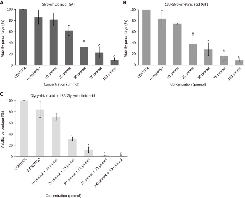
Figure 3 Cytotoxicity analysis of human umbilical cord-mesenchymal stem cells by MTT Assay. Bar graphs showing percent viability of human umbilical cord-mesenchymal stem cells treated with A: glycyrrhizic acid (GA), B: 18β-glycyrrhetinic acid (GT), and C: their combination (GA + GT) at different concentrations. A decrease in percent (%) cell viability is observed with increasing concentrations of GA, GT and their combination. Statistical analysis was conducted by One-way ANOVA and Post-Hoc Bonferroni tests. Results are expressed as mean ± SEM (n = 3); level of significance aP ≤ 0.05 (where aP ≤ 0.05, bP ≤ 0.01, and cP ≤ 0.001).
Figure 3 Cytotoxicity analysis of human umbilical cord-mesenchymal stem cells by MTT Assay. Bar graphs showing percent viability of human umbilical cord-mesenchymal stem cells treated with A: glycyrrhizic acid (GA), B: 18β-glycyrrhetinic acid (GT), and C: their combination (GA + GT) at different concentrations. A decrease in percent (%) cell viability is observed with increasing concentrations of GA, GT and their combination. Statistical analysis was conducted by One-way ANOVA and Post-Hoc Bonferroni tests. Results are expressed as mean ± SEM (n = 3); level of significance aP ≤ 0.05 (where aP ≤ 0.05, bP ≤ 0.01, and cP ≤ 0.001).CC BY-NC · PubMed Central
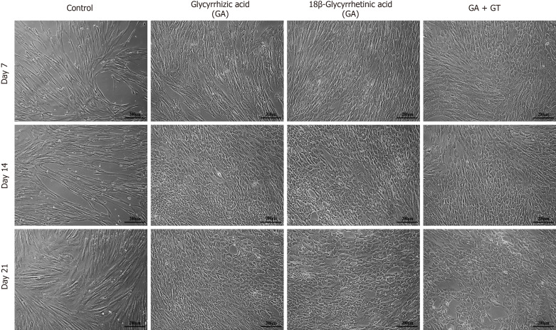
Figure 4 Morphological features of human umbilical cord-mesenchymal stem cells treated with glycyrrhizic acid (GA), 18β-glycyrrhetinic acid (GT), and their combination (GA + GT). At day 7, GA and GT treated MSCs became slightly shorter, flattened, and broader in appearance with retracted ends, while in the combination group, cells began to appear polygonal in shape. At day 14, changes were more apparent and cells progressively decreased in size and transformed into irregular or polygonal shape and by day 21, majority of the cells developed into more compact and polygonal morphology like that of hepatocytes exhibiting tight interactions between the cells in all treatment groups as compared to the untreated control. All images were taken under phase contrast at 10× magnification.
Figure 4 Morphological features of human umbilical cord-mesenchymal stem cells treated with glycyrrhizic acid (GA), 18β-glycyrrhetinic acid (GT), and their combination (GA + GT). At day 7, GA and GT treated MSCs became slightly shorter, flattened, and broader in appearance with retracted ends, while in the combination group, cells began to appear polygonal in shape. At day 14, changes were more apparent and cells progressively decreased in size and transformed into irregular or polygonal shape and by day 21, majority of the cells developed into more compact and polygonal morphology like that of hepatocytes exhibiting tight interactions between the cells in all treatment groups as compared to the untreated control. All images were taken under phase contrast at 10× magnification.CC BY-NC · PubMed Central
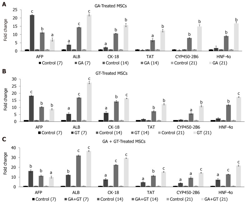
Figure 5 Expression of hepatic genes in human umbilical cord-mesenchymal stem cells treated with glycyrrhizic acid, 18β-glycyrrhetinic acid, and their combination. A and B: Glycyrrhizic acid (GA)- and 18β-glycyrrhetinic acid (GT)-treated mesenchymal stem cells showed significant expression of early hepatocyte marker AFP at all time points. Highest expression was observed at day 7 but it declined in a time-dependent manner. Significant expression levels of ALB and CK-18 were detected at all time points. Late hepatic markers, TAT, CYP450-2B6, and HNF-4α were not detectable at day 7 but were significantly up-regulated at day 14 and 21, as compared to the untreated control; C: GA + GT-treated MSCs revealed significant up-regulation of AFP at all time points. Highest expression was observed at day 7 but it declined with treatment time. Significant expression levels of ALB, CK-18, TAT, CYP450-2B6, and HNF-4α were observed at all time points and their expression increased with time of differentiation, as compared to the untreated control. Statistical analysis was conducted by independent sample t-test. Results are expressed as mean ± SEM (n = 3); level of significance aP ≤ 0.05 (where aP ≤ 0.05, bP ≤ 0.01, and cP ≤ 0.001).
Figure 5 Expression of hepatic genes in human umbilical cord-mesenchymal stem cells treated with glycyrrhizic acid, 18β-glycyrrhetinic acid, and their combination. A and B: Glycyrrhizic acid (GA)- and 18β-glycyrrhetinic acid (GT)-treated mesenchymal stem cells showed significant expression of early hepatocyte marker AFP at all time points. Highest expression was observed at day 7 but it declined in a time-dependent manner. Significant expression levels of ALB and CK-18 were detected at all time points. Late hepatic markers, TAT, CYP450-2B6, and HNF-4α were not detectable at day 7 but were significantly up-regulated at day 14 and 21, as compared to the untreated control; C: GA + GT-treated MSCs revealed significant up-regulation of AFP at all time points. Highest expression was observed at day 7 but it declined with treatment time. Significant expression levels of ALB, CK-18, TAT, CYP450-2B6, and HNF-4α were observed at all time points and their expression increased with time of differentiation, as compared to the untreated control. Statistical analysis was conducted by independent sample t-test. Results are expressed as mean ± SEM (n = 3); level of significance aP ≤ 0.05 (where aP ≤ 0.05, bP ≤ 0.01, and cP ≤ 0.001).CC BY-NC · PubMed Central
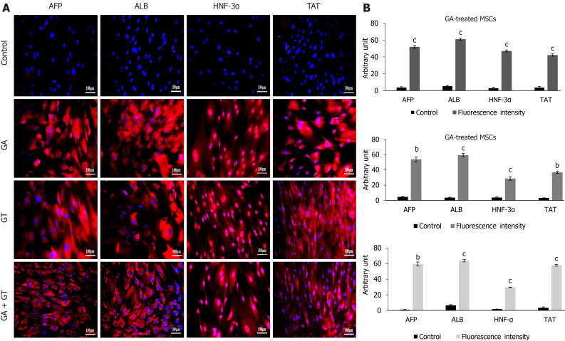
Figure 6 Immunocytochemical analysis and quantification of hepatic proteins in human umbilical cord-mesenchymal stem cells treated with glycyrrhizic acid, 18β-glycyrrhetinic acid and their combination. A: At day 21, treated mesenchymal stem cells exhibited positive expression for hepatocyte specific proteins including, alpha-fetoprotein, albumin, hepatocyte nuclear factor-3α, and tyrosine-aminotransferase in all treatment groups as compared to the untreated control; B: Fluorescence intensities of all treatment groups were measured using Image J software. Statistical analysis was conducted by independent sample t-test. Results are expressed as mean ± SEM (n = 3); level of significance aP ≤ 0.05 (where bP ≤ 0.01 and cP ≤ 0.001).
Figure 6 Immunocytochemical analysis and quantification of hepatic proteins in human umbilical cord-mesenchymal stem cells treated with glycyrrhizic acid, 18β-glycyrrhetinic acid and their combination. A: At day 21, treated mesenchymal stem cells exhibited positive expression for hepatocyte specific proteins including, alpha-fetoprotein, albumin, hepatocyte nuclear factor-3α, and tyrosine-aminotransferase in all treatment groups as compared to the untreated control; B: Fluorescence intensities of all treatment groups were measured using Image J software. Statistical analysis was conducted by independent sample t-test. Results are expressed as mean ± SEM (n = 3); level of significance aP ≤ 0.05 (where bP ≤ 0.01 and cP ≤ 0.001).CC BY-NC · PubMed Central
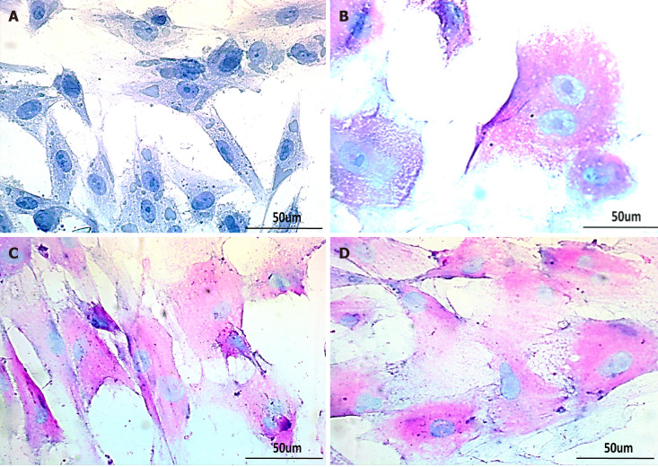
Figure 7 Glycogen detection in treated human umbilical cord-mesenchymal stem cells by periodic acid Schiff staining. Bright-field images of A: untreated control showing negative periodic acid Schiff staining (PAS); B: MSCs treated with glycyrrhizic acid (GA); C: 18β-glycyrrhetinic acid (GT); and D: GA+GT (combination group) at day 21 showing PAS positive cells stained as purple-pink, indicating the presence of glycogen in their cytoplasm. Bi-nucleated cells (a hepatocyte characteristic) were also observed. Images were taken at 40× magnification.
Figure 7 Glycogen detection in treated human umbilical cord-mesenchymal stem cells by periodic acid Schiff staining. Bright-field images of A: untreated control showing negative periodic acid Schiff staining (PAS); B: MSCs treated with glycyrrhizic acid (GA); C: 18β-glycyrrhetinic acid (GT); and D: GA+GT (combination group) at day 21 showing PAS positive cells stained as purple-pink, indicating the presence of glycogen in their cytoplasm. Bi-nucleated cells (a hepatocyte characteristic) were also observed. Images were taken at 40× magnification.CC BY-NC · PubMed Central

Verification

Claims the paper makes

The claims and statistics of this paper haven’t been checked yet.

Run the check — free, no account needed. It reads the open-access text and checks the sources it cites.

A few free checks a day · papers already verified open instantly · results are public

Claims other papers make about this one

Each is checked against this paper’s own text: does it say what the citing paper says it does?

Self-check

Percentages, sums and reported differences re-computed from the paper’s own numbers.

The self-check needs the paper’s full text.

Citations

Cites

Show all 3 references

Research works printed but not linked: 37

  1. 10.1016/j.stem.2014.04.010 · DOI · Paper page
  2. 10.1016/j.biocel.2003.11.001 · DOI
  3. 10.1155/2017/5259849 · DOI
  4. 10.1016/j.stem.2015.01.013 · DOI
  5. 10.1038/s41598-017-07633-7 · DOI
  6. 10.1007/s13273-013-0023-2 · DOI
  7. 10.1016/B978-0-12-805417-8.00028-7 · DOI
  8. 10.3390/molecules14062016 · DOI
  9. 10.3390/molecules22030400 · DOI
  10. 10.1016/j.intimp.2004.07.023 · DOI
  11. 10.1155/2014/872139 · DOI
  12. 10.1371/journal.pone.0068992 · DOI
  13. 10.4252/wjsc.v12.i12.1652 · DOI
  14. 10.2147/DDDT.S89658 · DOI
  15. 10.1101/pdb.prot095505 · DOI
  16. 10.1002/hep.20469 · DOI · Paper page
  17. 10.1111/j.1478-3231.2011.02469.x · DOI
  18. 10.1097/MAJ.0b013e31823e2867 · DOI
  19. 10.1016/j.jep.2016.06.011 · DOI
  20. 10.1590/s0100-879x2006005000173 · DOI
  21. 10.1016/s0304-3835(99)00225-6 · DOI
  22. 10.1007/BF02980047 · DOI
  23. 10.1016/j.intimp.2017.06.027 · DOI
  24. 10.1016/j.cbi.2017.04.009 · DOI
  25. 10.1007/978-1-60761-999-4_5 · DOI
  26. 10.1080/14653240600855905 · DOI · Paper page
  27. 10.4252/wjsc.v3.i12.113 · DOI
  28. 10.1155/2013/762196 · DOI
  29. 10.1371/journal.pone.0100417 · DOI
  30. 10.1016/s0014-5793(01)02423-1 · DOI
  31. 10.1155/2016/8648356 · DOI
  32. 10.3748/wjg.v16.i40.5092 · DOI
  33. Borhani-Haghighi M, Talaei-Khozani T, Ayatollahi M, Vojdani Z. Wharton's Jelly-derived Mesenchymal Stem Cells can Differentiate into Hepatocyte-like Cells by HepG2 Cell Line Extract. Iran J Med Sci 2015; 40: 143-151
  34. 10.1634/stemcells.2008-0963 · DOI
  35. 10.1016/s1043-2760(00)00271-x · DOI
  36. 10.1155/2019/5968236 · DOI
  37. 10.1016/j.tvjl.2012.05.013 · DOI

Metadata

Authors
Fatima, A., Malick, T. S., Khan, I., Ishaque, A., & Salim, A.
Affiliations
University of Karachi (PK)
Venue
World Journal of Stem Cells
Published
2021-10-26
Volume
13
Issue
10
Pages
1580-1594
ISSN
1948-0210 (ISSN-L)
OpenAlex
W3208339504
Cited by
16
References
3
Citations by year
2026: 1 · 2025: 2 · 2024: 6 · 2023: 4 · 2022: 3
FWCI
1.83
Type
journal article
Language
en
Open access
Licence
cc-by-nc
Locations
  • World Journal of Stem Cells (publishedVersion, cc-by-nc)
  • PubMed Central (publishedVersion, cc-by-nc)
  • PubMed (publishedVersion)
Topics
  • Life Sciences › Biochemistry, Genetics and Molecular Biology › Cell Biology › Mesenchymal stem cell research
  • Health Sciences › Medicine › Hepatology › Liver physiology and pathology
  • Health Sciences › Medicine › Surgery › Tissue Engineering and Regenerative Medicine
Keywords
glycyrrhizin, 18β-glycyrrhetinic acid, human umbilical cord mesenchymal stem cells, hepatocyte differentiation, triterpenoids, hepatic gene expression, hepatocyte-like cells, liver regeneration, preconditioning

About these checks

Registry, retraction, Cites and statistics results are computed, not judged, with one exception: a printed reference that no index lists is read once by a model before it counts as a citation error. Claims are extracted by a model and kept only when their quote appears verbatim in the text; each cited source is then checked, with a model’s help, for whether it says what the claim relies on. Those verdicts can be wrong — read the claim’s quote and the cited source before relying on one.

Statistics and arithmetic are recomputed, not judged.

Confidence bands are shown only for fields calibrated on our annotated test set. None is calibrated yet, so no band is shown.

What counts as a claim ›How we measure confidence ›How we check this ›

Are you an author, or do you see a problem with this page? Request a review

How to cite

Cite CoChat for the checks on this page, and the paper for its own findings.

This page
CoChat. (2026). Citation check: Effect of glycyrrhizic acid and 18β-glycyrrhetinic acid on the differentiation of human umbilical cord-mesenchymal stem cells into hepatocytes. https://app.cochat.ai/papers/10.4252/wjsc.v13.i10.1580
The paper
Fatima, A., Malick, T. S., Khan, I., Ishaque, A., & Salim, A. (2021). Effect of glycyrrhizic acid and 18β-glycyrrhetinic acid on the differentiation of human umbilical cord-mesenchymal stem cells into hepatocytes. World Journal of Stem Cells. https://doi.org/10.4252/wjsc.v13.i10.1580