

The DOI matches its Crossref record; 1 of the 1 references it cites were found in a registry or an index.
Ameliorative effect of transfersome gel of kepok banana peel extract (Musa balbisiana) against photoaging in Wistar rat skin
Open accessAbstract
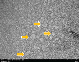
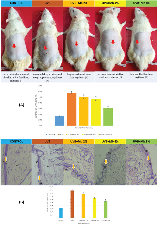
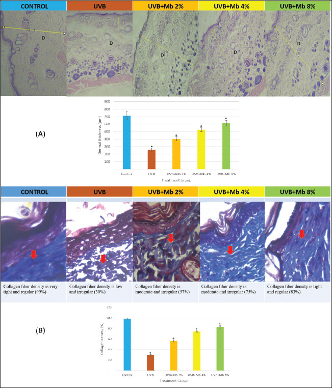
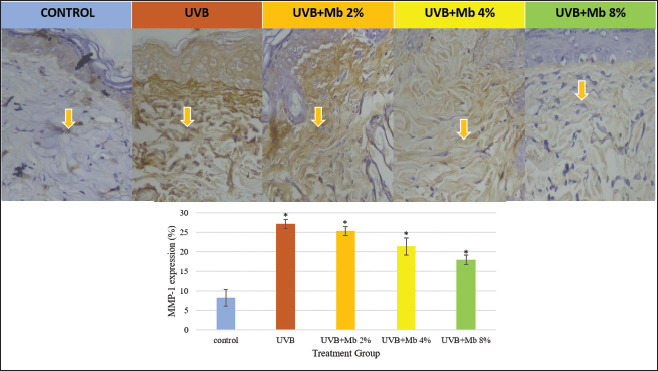
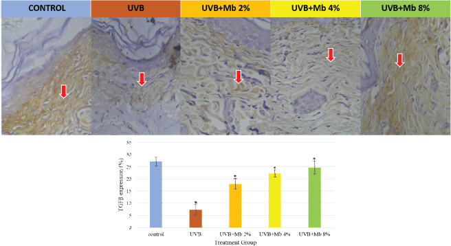
Background: Repeated acute exposure to UVB rays can cause photoaging. Musa balbisiana peel contains flavonoid compounds which act as antioxidants. But, the physicochemical of flavonoids are unstable, high molecular weight and easily oxidized, causing its use is still limited and transdermal delivery to be inefficient. Aim: To investigate the ameliorative effect of transfersome gel of Musa balbisiana peel against photoaging in Wistar rat skin. Methods: Transfersome gel was characterized by TEM. In vivo research was used to determine ameliorative effects of Musa balbisiana peel. The composition of transfersome consist of ethanol extracts of Musa balbisiana peel, SPC, and tween 80. The gel was applied three times a week for four weeks with a total UVB radiation dose of 840 mJ/cm2. To evaluate the repair mechanism by measuring the degree of wrinkles, epidermal thickening, dermal thinning, collagen fiber irregularity, matrix metalloproteinase 1 and TGF-β expression, MDA and TNFα levels. Results: TEM results show that gel transfersome Musa balbisiana peel has a round morphology with a diameter of ± 50 nm and no aggregation, which are defined as nanoparticles. Transfersome gel ameliorated degree of wrinkle, epidermal thickening, dermal thinning and irregularity of collagen fibers caused by UVB exposure, suppresses lipid peroxidation by decreasing MDA and TNFα level, also collagen imbalance by inhibiting MMP-1 expression and activating TGF-β expression, which were found statistically significantly different from non transfersome gel group. Conclusion: Transfersome gel of Musa balbisiana peel can act as an alternative medicine to ameliorate clinical photoaging due to exposure UVB.
Overview
The claims and statistics of this paper haven’t been checked yet.
Run the check — free, no account needed. It reads the open-access text and checks the sources it cites.
Integrity signals
Registry
Cites
Statistics
Figures
Verification
Claims the paper makes
The claims and statistics of this paper haven’t been checked yet.
Run the check — free, no account needed. It reads the open-access text and checks the sources it cites.
Claims other papers make about this one
Each is checked against this paper’s own text: does it say what the citing paper says it does?
The source backs part of the claim, not all of it — or backs it for a different population, condition or comparison.
In the citing paper
“For example, in 2024 a study reported the potent antioxidant activity and wound healing ability of banana peel transfer some gel and indicated that it plays an important role in skin regeneration and protect body against oxidative damage [9].”
From Formulation development, physical stability, and preliminary safety screening of cosmetic emulsions containing banana peel extract (2026) · cites this paper as [9]
Experimental — accuracy not yet measured
Self-check
Percentages, sums and reported differences re-computed from the paper’s own numbers.
The self-check needs the paper’s full text.
Citations
Cites
Potency antiinflammatory of ethanol extract gel of Kepok banana peel (Musa balbisiana).
Mayangsari, E., Mustika, A., Nurdiana, N., & Ardhayudicva, S. (2023) · PubMed · Cited by 2
Self-citation
Research works printed but not linked: 8
- Cheng Y.C Li T.S Su H.L Lee P.C Wang H.D Transdermal delivery systems of natural products applied to skin therapy and care Molecules (Basel, Switzerland) 2020 25 21 5051 33143260 PMC7662758
- Khan A Bai H Khan A Bai Z Neferine prevents ultraviolet radiationinduced skin photoaging Exp. Ther. Med 2020 31 89 96 PMC7132255 32266014
- Mayangsari E Mustika A Nurdiana N Samad N.A Comparison of UVA vs UVB photoaging rat models in short-term exposure Med. Arch 2024 78 2 88 91 38566862 PMC10983087
- Rahmi D Purnomo D Films B.P Chodijah S Husaini A Microemulgel formulation of Kepok banana peel extract (Musa paradisiaca L) as an antioxidant J. Phys. Conf. Ser 2019 1402 055090
- Rajabi M Mousa S.A Lipid nanoparticles and their application in nanomedicine Curr. Pharma. Biotechnol 2016 17 8 662 672 27087491
- Respati R.A Yusharyahya S.N Wibawa L.P Widaty S The dermoscopic features of photoaging and its association with Sun Index Score in the coastal population at Cilincing, Jakarta: a cross-sectional study Clin. Cosmet. Investing. Dermatol 2022 15 939 946 PMC9148576 35642176
- Surini S Leonyza A Suh C.W Formulation and in vitro penetration study of recombinant human epidermal growth factor-loaded transfersomal Emulgel Adv. Pharm. Bull 2020 10 4 586 594 33072536 PMC7539322
- Zhang Z Zhu H Zheng Y Zhang L Wang X Luo Z Tang J Lin L Du Z Dong C The effects and mechanism of collagen peptide and elastin peptide on skin aging induced by D-galactose combined with ultraviolet radiation J. Photochem. Photobiol. Biol 2020 210 111964 32717457
Cited by
Sorted by citations
Pribadi, A., Mustika, A., Qorib, M. F., Hamsidi, R., Puspita, D. I., & Priskila, O. (2024) · Research Journal of Pharmacy and Technology · doi:10.52711/0974-360x.2024.00850 · Cited by 3
Self-citationPimsee, P., Meenongwa, A., Rattanabun, W., Warinthip, N., Panomai, P., Keawbankrud, W., Sanseera, D., Khangkham, S., Kraipiyaset, P., Nakyai, W., et al. (2026) · Creative Science · doi:10.55674/cs.v18i3.265827
Adianingsih, O. R., Latifani, S., Fitriani, A. N., Wardani, A. D., Qurrotuain, I., Wahyudi, R. T., Dewi, A. M. P., Dhinata, J. A. W., Rumabutar, G. A., Puspita, O. E., et al. (2025) · Pharmacia · doi:10.3897/pharmacia.72.e174188
Metadata
- Authors
- Mayangsari, E., Mustika, A., Nurdiana, N., & Samad, N.
- Affiliations
- University of Brawijaya (ID)
- Universitas Airlangga (ID)
- Universiti Sains Malaysia (MY)
- Corresponding author
- Mustika, Arifa
- Venue
- Open Veterinary Journal
- Publisher
- ScopeMed
- Published
- 2024
- Volume
- 14
- Issue
- 8
- Pages
- 1990
- ISSN
- 2218-6050 (ISSN-L)
- 2226-4485
- PMC
- PMC11415925
- OpenAlex
- W4402333951
- Cited by
- 2
- References
- 1
- Citations by year
- 2025: 1 · 2024: 1
- FWCI
- 0.82
- Type
- journal article
- Language
- en
- Open access
- gold
- doi.org
- Licence
- Locations
- Open Veterinary Journal (publishedVersion, cc-by-nc)
- PubMed Central (publishedVersion, cc-by-nc)
- PubMed (publishedVersion)
- DOAJ (DOAJ: Directory of Open Access Journals) (submittedVersion, cc-by-sa)
- Funders
- Universiti Sains Malaysia
- Universitas Brawijaya
- Universitas Airlangga
- Lembaga Pengelola Dana Pendidikan
- Topics
- Health Sciences › Medicine › Dermatology › Skin Protection and Aging
- Life Sciences › Pharmacology, Toxicology and Pharmaceutics › Pharmaceutical Science › Advancements in Transdermal Drug Delivery
- Life Sciences › Biochemistry, Genetics and Molecular Biology › Cell Biology › melanin and skin pigmentation
- Keywords
- photoaging, kepok banana peel, Musa balbisiana, UV-B radiation, transdermal drug delivery, flavonoids, antioxidant activity, collagen degradation, MMP-1, TGF-beta, lipid peroxidation, wrinkles, epidermal thickening, dermal thinning, topical gel formulation
- MeSH terms
- Animals, Female, Gels, Plant Extracts, Skin, Ultraviolet Rays, Skin Aging, Rats, Wistar, Musa, Rats
About these checks
Registry, retraction, Cites and statistics results are computed, not judged, with one exception: a printed reference that no index lists is read once by a model before it counts as a citation error. Claims are extracted by a model and kept only when their quote appears verbatim in the text; each cited source is then checked, with a model’s help, for whether it says what the claim relies on. Those verdicts can be wrong — read the claim’s quote and the cited source before relying on one.
Statistics and arithmetic are recomputed, not judged.
Confidence bands are shown only for fields calibrated on our annotated test set. None is calibrated yet, so no band is shown.
What counts as a claim ›How we measure confidence ›How we check this ›
Are you an author, or do you see a problem with this page? Request a review
