

The DOI matches its Crossref record; 99 of the 100 references it cites were found in a registry or an index.
Disease model organism for Parkinson disease: Drosophila melanogaster
Open accessAbstract
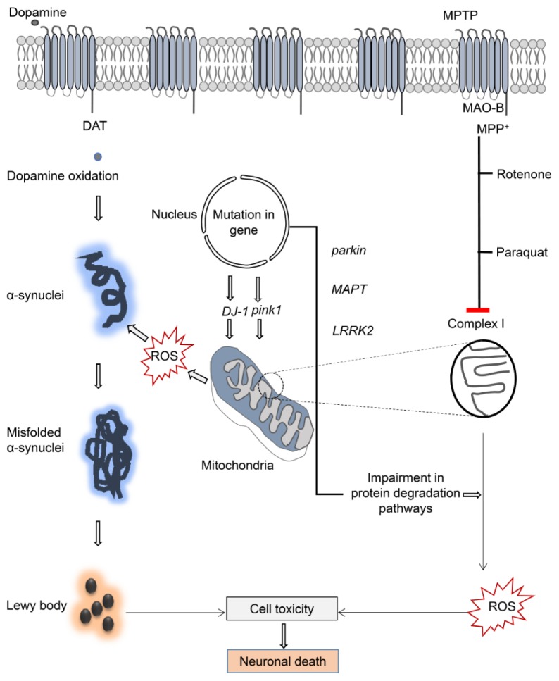
Parkinson's disease (PD) is a common neurodegenerative disorder characterized by selective and progressive loss of dopaminergic neurons. Genetic and environmental risk factors are associated with this disease. The genetic factors are composed of approximately 20 genes, such as SNCA, parkin, PTEN-induced kinase1 (pink1), leucine-rich repeat kinase 2 (LRRK2), ATP13A2, MAPT, VPS35, and DJ-1, whereas the environmental factors consist of oxidative stress-induced toxins such as 1-methyl-4-phenyl-1, 2, 3, 6-tetrahydropyridine (MPTP), rotenone, and paraquat. The analyses of their functions and mechanisms have provided important insights into the disease process, which has demonstrated that these factors cause oxidative damage and mitochondrial dysfunction. The most invaluable studies have been performed using disease model organisms, such as mice, fruit flies, and worms. Among them, Drosophila melanogaster has emerged as an excellent model organism to study both environmental and genetic factors and provide insights to the pathways relevant for PD pathogenesis, facilitating development of therapeutic strategies. In this review, we have focused on the fly model organism to summarize recent progress, including pathogenesis, neuroprotective compounds, and newer approaches. [BMB Reports 2019; 52(4): 250-258].
Overview
The claims and statistics of this paper haven’t been checked yet.
Run the check — free, no account needed. It reads the open-access text and checks the sources it cites.
Integrity signals
Registry
Cites
Statistics
Figures
Verification
Claims the paper makes
The claims and statistics of this paper haven’t been checked yet.
Run the check — free, no account needed. It reads the open-access text and checks the sources it cites.
Claims other papers make about this one
Each is checked against this paper’s own text: does it say what the citing paper says it does?
The source backs part of the claim, not all of it — or backs it for a different population, condition or comparison.
In the citing paper
“Another study that used Drosophila melanogaster showed a significant reduction of dopaminergic neurons, compared with the control group, under hypergravity conditions in a similar way to human’s Parkinson’s disease (PD)98.”
From Key points for the development of antioxidant cocktails to prevent cellular stress and damage caused by reactive oxygen species (ROS) during manned space missions (2021) · cites this paper as [98]
Experimental — accuracy not yet measured
Self-check
Percentages, sums and reported differences re-computed from the paper’s own numbers.
The self-check needs the paper’s full text.
Citations
Cites
Mutation in the α-Synuclein Gene Identified in Families with Parkinson's Disease
Polymeropoulos, M. H., Lavedan, C., Leroy, E., Ide, S. E., Dehejia, A., Dutra, A., Pike, B., Root, H., Rubenstein, J., Boyer, R., et al. (1997) · Science · doi:10.1126/science.276.5321.2045 · Cited by 8,240
Mutations in the parkin gene cause autosomal recessive juvenile parkinsonism
Kitada, T., Asakawa, S., Hattori, N., Matsumine, H., Yamamura, Y., Minoshima, S., Yokochi, M., Mizuno, Y., & Shimizu, N. (1998) · Nature · doi:10.1038/33416 · Cited by 5,227
α-Synuclein Locus Triplication Causes Parkinson's Disease
Singleton, A. B., Farrer, M., Johnson, J., Singleton, A., Hague, S., Kachergus, J., Hulihan, M., Peuralinna, T., Dutra, A., Nussbaum, R., et al. (2003) · Science · doi:10.1126/science.1090278 · Cited by 4,383
Parkin is recruited selectively to impaired mitochondria and promotes their autophagy
Narendra, D., Tanaka, A., Suen, D.-F., & Youle, R. J. (2008) · The Journal of Cell Biology · doi:10.1083/jcb.200809125 · Cited by 4,026
Chronic systemic pesticide exposure reproduces features of Parkinson's disease
Betarbet, R. S., Sherer, T., MacKenzie, G. M., Garcia‐Osuna, M., Panov, A. V., & Greenamyre, J. T. (2000) · Nature Neuroscience · doi:10.1038/81834 · Cited by 3,777
Mitochondrial membrane permeabilization in cell death
Kroemer, G., Galluzzi, L., & Brenner, C. (2007) · Physiological Reviews · doi:10.1152/physrev.00013.2006 · Cited by 3,598
Hereditary Early-Onset Parkinson's Disease Caused by Mutations in PINK1
Valente, E. M., Abou-Sleiman, P. M., Caputo, V., Muqit, M. M. K., Harvey, K., Gispert, S., Ali, Z., Del Turco, D., Bentivoglio, A. R., Healy, D. G., et al. (2004) · Science · doi:10.1126/science.1096284 · Cited by 3,481
Mutations in LRRK2 Cause Autosomal-Dominant Parkinsonism with Pleomorphic Pathology
Zimprich, A. A., Biskup, S., Leitner, P., Lichtner, P., Farrer, M. J., Lincoln, S., Kachergus, J. M., Hulihan, M., Uitti, R. J., Calne, D. B., et al. (2004) · Neuron · doi:10.1016/j.neuron.2004.11.005 · Cited by 3,087
A Drosophila model of Parkinson's disease
Feany, M. Β., & Bender, W. W. (2000) · Nature · doi:10.1038/35006074 · Cited by 2,156
Drosophila pink1 is required for mitochondrial function and interacts genetically with parkin
Clark, I. E., Dodson, M. W., Jiang, C., Cao, J. H., Huh, J. R., Seol, J. H., Yoo, S. J., Hay, B. A., & Guo, M. (2006) · Nature · doi:10.1038/nature04779 · Cited by 1,788
Research works printed but not linked: 1
- 10.1038/nature04779 · DOI · Paper page
Cited by
Sorted by citations
Teixeira, P. R., Dusi, R. G., Jimenez, P. C., Espíndola, L. S., & Costa-Lotufo, L. V. (2022) · Environmental Pollution · doi:10.1016/j.envpol.2022.118983 · Cited by 210
Disorders of the enteric nervous system — a holistic view
Niesler, B., Küerten, S., Demir, İ. E., & Schäfer, K. (2021) · Nature Reviews Gastroenterology & Hepatology · doi:10.1038/s41575-020-00385-2 · Cited by 209
Dovonou, A., Bolduc, C., Linan, V. S., Gora, C., Peralta, M. R., & Lévesque, M. (2023) · Translational Neurodegeneration · doi:10.1186/s40035-023-00368-8 · Cited by 218
Liu, W., Wei, S., Huang, G., Liu, L.-B., Gu, C., Shen, Y., Wang, X., Xia, S., Xie, A., Hu, L., et al. (2020) · The FASEB Journal · doi:10.1096/fj.201901565rr · Cited by 107
Gómez, X., Sanon, S., Zambrano, K., Asquel, S., Bassantes, M., Morales, J. E., Otáñez, G., Pomaquero, C., Villarroel, S., Zurita, A., et al. (2021) · npj Microgravity · doi:10.1038/s41526-021-00162-8 · Cited by 92
A Critical Review of Zebrafish Models of Parkinson’s Disease
Doyle, J. M., & Croll, R. P. (2022) · Frontiers in Pharmacology · doi:10.3389/fphar.2022.835827 · Cited by 94
Reversing insecticide resistance with allelic-drive in Drosophila melanogaster
Kaduskar, B., Kushwah, R. B. S., Auradkar, A., Guichard, A., Li, M., Bennett, J. B., Julio, A. H. F., Marshall, J. M., Montell, C., & Bier, E. (2022) · Nature Communications · doi:10.1038/s41467-021-27654-1 · Cited by 68
Ortega‐Arellano, H. F., Jiménez-Del-Río, M., & Velez‐Pardo, C. (2019) · Neurochemical Research · doi:10.1007/s11064-019-02835-z · Cited by 59
Musachio, E. A. S., Araújo, S. M., Bortolotto, V. C., Couto, S. D. F., Dahleh, M. M. M., Poetini, M. R., Fernandes, E. J., Meichtry, L. B., Ramborger, B. P., Roehrs, R., et al. (2020) · Food and Chemical Toxicology · doi:10.1016/j.fct.2020.111128 · Cited by 59
Neural shutdown under stress: an evolutionary perspective on spreading depolarization
Robertson, R. M., Dawson-Scully, K. D., & Andrew, R. D. (2020) · Journal of Neurophysiology · doi:10.1152/jn.00724.2019 · Cited by 51
Neuroinflammation in Parkinson’s Disease: From Gene to Clinic: A Systematic Review
Castillo-Rangel, C., Marín, G., Hernández-Contreras, K. A., Vichi-Ramírez, M. M., Zarate-Calderon, C., Torres-Pineda, O., Díaz-Chiguer, D. L., González, D. D. L. M., Apo, E. G., Teco-Cortes, J. A., et al. (2023) · International Journal of Molecular Sciences · doi:10.3390/ijms24065792 · Cited by 55
Sphingolipids in neurodegenerative diseases
Pan, X., Dutta, D., Lu, S., & Bellen, H. J. (2023) · Frontiers in Neuroscience · doi:10.3389/fnins.2023.1137893 · Cited by 50
Circadian clock disruption promotes the degeneration of dopaminergic neurons in male Drosophila
Dorcikova, M. M., Duret, L. C., Pottié, E., & Nagoshi, E. (2023) · Nature Communications · doi:10.1038/s41467-023-41540-y · Cited by 42
Sayyaed, A., Saraswat, N., Vyawahare, N. S., & Kulkarni, A. (2023) · Bulletin of the National Research Centre/Bulletin of the National Research Center · doi:10.1186/s42269-023-01047-4 · Cited by 41
Guo, Q., Wang, B., Wang, X., Smith, W. W., Zhu, Y., & Liu, Z. (2021) · Cells · doi:10.3390/cells10081850 · Cited by 34
Akinade, T. C., Babatunde, O. O., Adedara, A. O., Adeyemi, O., Otenaike, T. A., Ashaolu, O. P., Johnson, T. O., Terriente-Félix, A., Whitworth, A. J., & Abolaji, A. O. (2022) · Scientific Reports · doi:10.1038/s41598-022-08409-4 · Cited by 34
Drosophila as a Model for Human Disease: Insights into Rare and Ultra-Rare Diseases
Casas‐Tintó, S. (2024) · Insects · doi:10.3390/insects15110870 · Cited by 39
Sánchez-Giraldo, V., Monsalve, Y., Palacio, J., Mendivil‐Perez, M., Sierra, L., Velez‐Pardo, C., López, B. L., & Jiménez-Del-Río, M. (2019) · Journal of Drug Delivery Science and Technology · doi:10.1016/j.jddst.2019.101466 · Cited by 28
Metabolic Alterations in a Drosophila Model of Parkinson’s Disease Based on DJ-1 Deficiency
Solana‐Manrique, C., Sanz, F., Torregrosa, I., Palomino‐Schätzlein, M., Hernández-Oliver, C., Pineda‐Lucena, A., & Paricio, N. (2022) · Cells · doi:10.3390/cells11030331 · Cited by 24
Afsheen, S., Rehman, A. S., Jamal, A., Khan, N. H., & Parvez, S. (2024) · Ageing Research Reviews · doi:10.1016/j.arr.2024.102340 · Cited by 28
Animal Models of Human Disease
Lange, S., & Inal, J. M. (2023) · International Journal of Molecular Sciences · doi:10.3390/ijms242115821 · Cited by 25
Rasheed, M. Z., Khatoon, R., Talat, F., Alam, M. M., Tabassum, H., & Parvez, S. (2023) · ACS Omega · doi:10.1021/acsomega.2c03992 · Cited by 25
Idda, T., Bonas, C., Hoffmann, J., Bertram, J., Quinete, N., Schettgen, T., Fietkau, K., Esser, A., Stope, M. B., Leijs, M. M., et al. (2020) · Scientific Reports · doi:10.1038/s41598-020-78405-z · Cited by 23
Circadian rhythm disruption: a potential trigger in Parkinson’s disease pathogenesis
Xu, K., Zhang, Y., Shi, Y. X., Zhang, Y., Zhang, C., Wang, T., Lv, P., Bai, Y., & Wang, S. (2024) · Frontiers in Cellular Neuroscience · doi:10.3389/fncel.2024.1464595 · Cited by 24
Cell competition from development to neurodegeneration
RODRIGUES, M. C., Couceiro, J., & Moreno, E. (2021) · Disease Models & Mechanisms · doi:10.1242/dmm.048926 · Cited by 22
Thane, M., Paisios, E., Stöter, T., Krüger, A.-R., Gläß, S., Dahse, A.-K., Scholz, N., Gerber, B., Lehmann, D. J., & Schleyer, M. H. (2023) · Open Biology · doi:10.1098/rsob.220308 · Cited by 20
Chua, S. C. J. H., Tan, H. Q., Engelberg, D., & Lim, L. H. K. (2019) · Pharmaceuticals · doi:10.3390/ph12040147 · Cited by 19
A new metabolic model of Drosophila melanogaster and the integrative analysis of Parkinson’s disease
Cesur, M. F., Basile, A., Patil, K. R., & Çakır, T. (2023) · Life Science Alliance · doi:10.26508/lsa.202201695 · Cited by 18
Ahn, Y., Han, S., Kim, M. G., Hong, K., Kim, W. J., Suh, H. J., & Jo, K. (2021) · Pharmaceutical Biology · doi:10.1080/13880209.2021.1949356 · Cited by 18
Narwal, S., Singh, A., & Tare, M. (2024) · Frontiers in Cellular Neuroscience · doi:10.3389/fncel.2023.1295805 · Cited by 16
Poetini, M. R., Musachio, E. A. S., Araújo, S. M., Bortolotto, V. C., Meichtry, L. B., Silva, N. C., Janner, D. E., Novo, D. L. R., Mesko, M. F., Roehrs, R., et al. (2022) · NeuroToxicology · doi:10.1016/j.neuro.2022.02.004 · Cited by 16
Dithianon exposure induces dopaminergic neurotoxicity in Caenorhabditis elegans
Lee, Y., Choi, S., & Kim, K. W. (2023) · Ecotoxicology and Environmental Safety · doi:10.1016/j.ecoenv.2023.114752 · Cited by 15
Self-citationNunes, R. G. D. S., de Amorim, L. C., Bezerra, I. C., da Silva, A. J., dos Santos, C. A. L., Gubert, P., de Menezes, I. R. A., Duarte, A. E., Barros, L. M., Andrade‐da‐Costa, B. L. D. S., et al. (2024) · Journal of Toxicology and Environmental Health · doi:10.1080/15287394.2024.2338431 · Cited by 13
Drosophila Models Reveal Properties of Mutant Lamins That Give Rise to Distinct Diseases
Walker, S. G., Langland, C. J., Viles, J., Hecker, L. A., & Wallrath, L. L. (2023) · Cells · doi:10.3390/cells12081142 · Cited by 12
Ramos, V., Reis, M. A., Ferreira, L., Silva, A. M., Ferraz, R., Vieira, M., Vasconcelos, V. M., & Martins, R. F. (2023) · Biomolecules · doi:10.3390/biom13101444 · Cited by 13
Ganesan, S., & Parvathi, V. D. (2021) · Egyptian Journal of Medical Human Genetics · doi:10.1186/s43042-021-00208-2 · Cited by 12
Solana‐Manrique, C., Moltó, M. D., Calap-Quintana, P., Sanz, F., Llorens, J. V., & Paricio, N. (2019) · Springer eBooks · doi:10.1007/978-981-13-2218-1_15 · Cited by 12
Sankar, S., Chandrasekaran, N., Moovendhan, M., & Parvathi, V. D. (2024) · Environmental Quality Management · doi:10.1002/tqem.70021 · Cited by 11
How Rearing Systems for Various Species of Flies Benefit Humanity
Pascacio‐Villafán, C., & Cohen, A. C. (2023) · Insects · doi:10.3390/insects14060553 · Cited by 13
Plasticity of pheromone-mediated avoidance behavior in C. elegans
Cheon, Y., Hwang, H., & Kim, K. (2020) · Journal of neurogenetics · doi:10.1080/01677063.2020.1802723 · Cited by 10
Quintero-Espinosa, D. A., Jiménez-Del-Río, M., & Velez‐Pardo, C. (2024) · Neurochemical Research · doi:10.1007/s11064-024-04141-9 · Cited by 10
Erhardt, B., Marcora, M. S., Marcora, M. S., Frenkel, L., Bochicchio, P. A., Bodín, D. H., Silva, B. A., Dellepiane, M. R. B., Ferrari, C. C., Allo, M. Á., et al. (2021) · European Journal of Neuroscience · doi:10.1111/ejn.15401 · Cited by 9
Dumitrescu, E., Copeland, J. M., & Venton, B. J. (2022) · ACS Chemical Neuroscience · doi:10.1021/acschemneuro.2c00277 · Cited by 10
Progress in Parkinson's disease animal models of genetic defects: Characteristics and application
Zhang, C., Chen, S., Li, X., Xu, Q., Lin, Y., Lin, F., Yuan, M., Zi, Y., & Cai, J. (2022) · Biomedicine & Pharmacotherapy · doi:10.1016/j.biopha.2022.113768 · Cited by 9
Pedroso, A. L., Schonwald, M. K., Corte, C. L. D., Soares, F. A. A., Sperança, A., Godoi, B., & de Carvalho, N. R. (2023) · Toxicology Research · doi:10.1093/toxres/tfad081 · Cited by 9
Monoamine Oxidase Inhibitors in Toxic Models of Parkinsonism
Бунеева, О. А., & Medvedev, A. E. (2025) · International Journal of Molecular Sciences · doi:10.3390/ijms26031248 · Cited by 11
Unrevealing the molecular mechanisms of MPTP-induced Parkinson’s in experimental animals
Mondal, S., & Firdous, S. M. (2025) · Medicinal Chemistry Research · doi:10.1007/s00044-025-03409-1 · Cited by 9
The international exchange of Drosophila melanogaster strains
Cook, K. R., & Parks, A. L. (2022) · Scientific and Technical Review (World Organisation for Animal Health) · doi:10.20506/rst.41.1.3305 · Cited by 9
Asthana, J., & Shravage, B. V. (2022) · Frontiers in Aging Neuroscience · doi:10.3389/fnagi.2022.986849 · Cited by 10
Caffeine improves mitochondrial function in PINK1B9-null mutant Drosophila melanogaster
Gonçalves, D. F., Senger, L. R., Foletto, J. V. P., Michelotti, P., Soares, F. A. A., & Corte, C. L. D. (2022) · Journal of Bioenergetics and Biomembranes · doi:10.1007/s10863-022-09952-5 · Cited by 10
Bernardes, L. M. M., Malta, S. M., Rodrigues, T. S., Covizzi, L. F., Rosa, R. B., Justino, A. B., Teixeira, R. R., Espíndola, F. S., dos Santos, D. O., Ueira‐Vieira, C., et al. (2023) · PLoS ONE · doi:10.1371/journal.pone.0291242 · Cited by 9
Yang, P., Liu, Y., Tong, Z., Huang, Q., Xie, X., Mao, S.-Y., Ding, J., Lu, M., Tan, R. X., & Hu, G. (2024) · Acta Pharmacologica Sinica · doi:10.1038/s41401-024-01259-y · Cited by 9
Okonta, C., Ogunyemi, O. M., Olabuntu, B. A., & Abolaji, A. O. (2025) · Toxicology Reports · doi:10.1016/j.toxrep.2025.102004 · Cited by 10
Mo, L., Li, R., He, C., Chen, Q., Xu, C., Shen, L., Chen, K., & Wu, Y. (2022) · Human Cell · doi:10.1007/s13577-022-00796-0 · Cited by 8
Animal Models of Human Disease
(2011) · Progress in molecular biology and translational science · doi:10.1016/c2010-0-65612-4 · Cited by 8
Jung, B. C., Kim, S. H., Cho, Y., & Kim, Y. S. (2023) · BMB Reports · doi:10.5483/bmbrep.2023-0134 · Cited by 7
In vivo and in silico models of Drosophila for Parkinson's disease
Cesur, M. F., Strazdins, R., Sarkar, S., & Çakır, T. (2025) · FEBS Journal · doi:10.1111/febs.70140 · Cited by 7
Sanz, F., Solana‐Manrique, C., & Paricio, N. (2023) · ACS Chemical Neuroscience · doi:10.1021/acschemneuro.3c00026 · Cited by 7
The impact of Parkinson’s disease-associated gut microbiota on the transcriptome in Drosophila
Liu, X., Yang, M., Liu, R., Zhou, F., Zhu, H., & Wang, X. (2023) · Microbiology Spectrum · doi:10.1128/spectrum.00176-23 · Cited by 7
Use of invertebrates to model chemically induced parkinsonism-symptoms
Corrêa, C. L., Gomes, C. A. B. D. A., Mello, A. D. A., Nogueira, N. D. S., Medeiros, T. B., de Barros, C. M., & Allodi, S. (2023) · Biochemical Society Transactions · doi:10.1042/bst20221172 · Cited by 7
A Comprehensive Study of miRNAs in Parkinson’s Disease: Diagnostics and Therapeutic Approaches
Siddique, Y. H., & Owais, S. (2022) · CNS & Neurological Disorders - Drug Targets · doi:10.2174/1871527321666220111152756 · Cited by 6
Setzu, M. D., Mocci, I., Fabbri, D., Carta, P., Muroni, P., Diana, A., Dettori, M. A., & Casu, M. A. (2024) · Biomolecules · doi:10.3390/biom14030273 · Cited by 5
Experimental Models in Parkinson’s Disease: Advantages and Disadvantages
Altunlu, Ö., Topatan, E., Al-yaqoobi, Z., Burul, F., Bayram, C., Sezen, S., Okkay, I. F., Okkay, U., & Hacımüftüoğlu, A. (2024) · Ağrı Tıp Fakültesi Dergisi · doi:10.61845/agrimedical.1471854 · Cited by 6
Paraquat neurotoxicity: Oxidative stress and neuronal dysfunction in the ascidian brain
Silva, R. R. E., Portal, T. M., Nogueira, N. D. S., Nogueira, T. D. S., Mello, A. D. A., & de Barros, C. M. (2025) · Comparative Biochemistry and Physiology Part C Toxicology & Pharmacology · doi:10.1016/j.cbpc.2025.110128 · Cited by 6
Drug discovery and development for Parkinson’s disease: are preclinical models good enough?
Reinares-Sebastián, A., Esteban-García, N., Takada, M., & Trigo‐Damas, I. (2025) · Frontiers in Aging Neuroscience · doi:10.3389/fnagi.2025.1692592 · Cited by 5
Kasamo, Y., Kikuchi, K., Yamakuchi, M., Otsuka, S., Takada, S., Kambe, Y., Ito, T., Kawahara, K.-I., Arita, K., Yoshimoto, K., et al. (2021) · International Journal of Molecular Sciences · doi:10.3390/ijms22189941 · Cited by 5
Jellinger, K. A. (2019) · Expert Opinion on Drug Discovery · doi:10.1080/17460441.2019.1638908 · Cited by 6
Sexually dimorphic mechanisms of VGLUT-mediated protection from dopaminergic neurodegenerationPreprint
Buck, S. A., Rubin, S. A., Kunkhyen, T., Treiber, C. D., Xue, X., Fenno, L. E., Mabry, S. J., Sundar, V. R., Yang, Z., Shah, D., et al. (2023) · bioRxiv (Cold Spring Harbor Laboratory) · doi:10.1101/2023.10.02.560584 · Cited by 5
Buck, S. A., Mabry, S. J., Kunkhyen, T., Yang, Z., Rubin, S. A., Yang, J., Cheetham, C. E. J., & Freyberg, Z. (2025) · Aging Cell · doi:10.1111/acel.70096 · Cited by 6
Zhang, W., Ju, Y., Ren, Y., Miao, Y., & Wang, Y. (2024) · Current Drug Targets · doi:10.2174/0113894501281402231218071641 · Cited by 4
Diana, A., Collu, M., Casu, M. A., Mocci, I., Aguilar‐Santelises, M., & Setzu, M. D. (2020) · Brain Sciences · doi:10.3390/brainsci10010045 · Cited by 4
Modeling of Parkinson’s disease in Drosophila based on DJ-1 deficiency
Sanz, F., Solana‐Manrique, C., & Paricio, N. (2022) · Elsevier eBooks · doi:10.1016/b978-0-323-89833-1.00043-4 · Cited by 4
Ishola, I., Oloyo, A., Olubodun-Obadun, T., Godswill, O., Omilabu, S., & Adeyemi, O. (2022) · Metabolic brain disease · doi:10.1007/s11011-022-01120-3 · Cited by 3
Samson, J. S., Rajagopal, K., & Parvathi, V. D. (2025) · Brain Research · doi:10.1016/j.brainres.2025.149505 · Cited by 3
Giles, H. H., Petersen, S. C., Call, G. B., & Chaston, J. M. (2021) · Microbiology Resource Announcements · doi:10.1128/mra.01372-20 · Cited by 2
Boosting life sciences research in Brazil: building a case for a local Drosophila stock center
Oliveira, M. T., Anhezini, L., Araujo, H. M., de Oliveira, M. F., & Lima, C. A. C. (2024) · Genetics and Molecular Biology · doi:10.1590/1678-4685-gmb-2023-0202 · Cited by 2
In vivo Effects of Salicornia herbacea and Calystegia soldanella Extracts for Memory Improvement
Sang, J., Poudel, S., & Lee, Y. (2024) · Journal of Microbiology and Biotechnology · doi:10.4014/jmb.2312.12045 · Cited by 2
Self-citationLuan, H. (2025) · Immune discovery. · doi:10.70322/immune.2025.10001 · Cited by 2
The utility and caveat of split-GAL4s in the study of neurodegeneration
Stickley, L., Koch, R., & Nagoshi, E. (2023) · Fly · doi:10.1080/19336934.2023.2192847 · Cited by 2
Khvastunov, V. O., Goleva, T. N., Epremyan, K. K., & Rogov, A. G. (2024) · Nanobiotechnology Reports · doi:10.1134/s2635167624602377 · Cited by 1
Advancement in the animal model-based therapeutics in neurodegenerative diseases
Pal, D., Das, P., Ghosh, P., Gupta, S., Basak, P., & Nandi, S. K. (2025) · Elsevier eBooks · doi:10.1016/b978-0-443-33877-9.00007-0 · Cited by 1
Idowu, O. K., Oremosu, A. A., & Dosumu, O. O. (2026) · Metabolic Brain Disease · doi:10.1007/s11011-026-01795-y · Cited by 1
Baisgaard, A. E., Koldby, K. M., Kristensen, T., Nyegaard, M., & Rohde, P. D. (2023) · Insects · doi:10.3390/insects14020168 · Cited by 1
Absence of pretaporter restrains features of the parkin phenotype in Drosophila
Silvera, M. C., Cantera, R., & Ferreiro, M. J. (2024) · Experimental Neurology · doi:10.1016/j.expneurol.2024.114997 · Cited by 1
Thane, M., Paisios, E., Stöter, T., Krüger, A.-R., Gläß, S., Dahse, A.-K., Scholz, N., Gerber, B., Lehmann, D. J., & Schleyer, M. H. (2022) · bioRxiv (Cold Spring Harbor Laboratory) · doi:10.1101/2022.09.27.509663 · Cited by 1
Sanz, F., Solana‐Manrique, C., & Paricio, N. (2022) · bioRxiv (Cold Spring Harbor Laboratory) · doi:10.1101/2022.12.28.522104 · Cited by 1
Exhaustive benchmarking of de novo assembly methods for eukaryotic genomesPreprint
Southwood, D., Rane, R., Lee, S. F., Oakeshott, J. G., & Ranganathan, S. (2023) · bioRxiv (Cold Spring Harbor Laboratory) · doi:10.1101/2023.04.18.537422 · Cited by 1
Rai, J., Aswini, S., Paul, S., Shelke, T., & Gupta, I. (2025) · bioRxiv (Cold Spring Harbor Laboratory) · doi:10.1101/2025.04.04.647141 · Cited by 1
Modeling Mechanisms and Applications of Parkinson's Disease Animal ModelsPreprint
Cui, Y., LIANG, Y., Liu, Y., & Zhang, B. (2022) · SSRN Electronic Journal · doi:10.2139/ssrn.4175871
A genome-scale metabolic model of Drosophila melanogaster for integrative analysis of brain diseasesPreprint
Cesur, M. F., Patil, K. R., & Çakır, T. (2022) · bioRxiv (Cold Spring Harbor Laboratory) · doi:10.1101/2022.08.22.504778
Circadian clock disruption promotes the degeneration of dopaminergic neuronsPreprint
Dorcikova, M. M., Duret, L. C., Pottié, E., & Nagoshi, E. (2022) · bioRxiv (Cold Spring Harbor Laboratory) · doi:10.1101/2022.10.15.512376
Usefulness of Drosophila in studying Parkinson's disease and role of natural plant products
(2023) · Advances in neurotoxicology · doi:10.1016/bs.ant.2023.01.007
Dhande, S. R., & Pansare, N. (2024) · Research Journal of Pharmacy and Technology · doi:10.52711/0974-360x.2024.00491
Maintenance of Mitochondrial Dynamics for Healthy Brain Ageing
Mishra, E., & Thakur, M. K. (2024) · Healthy ageing and longevity · doi:10.1007/978-3-031-68513-2_10
Modeling of Parkinson's Disease in Different Models
Subhan, I., & Siddique, Y. H. (2024) · CNS & Neurological Disorders - Drug Targets · doi:10.2174/0118715273326866240922193029
Ethical regulations for induction and validation of PD models
Oraby, A. (2024) · Elsevier eBooks · doi:10.1016/b978-0-443-16128-5.00019-0
Sayed, F. E., Mahmoud, A. K., Mohammed, O. A. M., Soliman, R. I., & Abdur-Rahman, M. (2024) · Elsevier eBooks · doi:10.1016/b978-0-443-16128-5.00014-1
Neuroprotective role of ranolazine in Parkinson disease: Drosophila melanogaster model
Parvesh, P., Kumar, S., Singh, G., Khatri, R., Shkula, S., Kaushik, K., Lather, A., & Hooda, T. (2025) · Scripta Medica · doi:10.5937/scriptamed56-53259
AGAKWU, M. U., OGUNMEFUN, G. S., YUSUF, H. O., ONMONYA, Y. A., & ADUNBI, J. O. (2024) · Journal of Biochemistry and molecular biology · doi:10.36108/jbmb/4202.20.0140
Anadu, V. E., Jimoh-Abdulghaffaar, H. O., Gilbert, T. T., Aneke, O. V., Arietarhire, L. O., Ichipi-Ifukor, P. C., Abdullahi, A. M., Okah, L., Sabine, A. F. Y., Oritsetimeyin, B. U., et al. (2025) · Biology Open · doi:10.1242/bio.061998
Metadata
- Authors
- Aryal, B., & Lee, Y.
- Affiliations
- Kookmin University (KR)
- Venue
- BMB Reports
- Published
- 2019-04-30
- Volume
- 52
- Issue
- 4
- Pages
- 250-258
- ISSN
- 1976-6696 (ISSN-L)
- 1976-670X
- PMC
- PMC6507844
- OpenAlex
- W2942930317
- Cited by
- 111
- References
- 100
- Citations by year
- 2026: 7 · 2025: 16 · 2024: 21 · 2023: 25 · 2022: 20 · 2021: 9 · 2020: 7 · 2019: 5
- FWCI
- 6.94
- Type
- journal article
- Language
- en
- Open access
- gold
- doi.org
- Licence
- cc-by-nc
- Locations
- BMB Reports (publishedVersion, cc-by-nc)
- PubMed Central (publishedVersion, cc-by-nc)
- PubMed (publishedVersion)
- Funders
- National Research Foundation of Korea (NRF-2016R1D1A1B03931273, NRF-2018R1A2B6004202)
- Ministry of Education
- Kookmin University
- Topics
- Health Sciences › Medicine › Clinical Neurology › Parkinson's Disease Mechanisms and Treatments
- Life Sciences › Biochemistry, Genetics and Molecular Biology › Molecular Biology › Nuclear Receptors and Signaling
- Life Sciences › Neuroscience › Cellular and Molecular Neuroscience › Neurobiology and Insect Physiology Research
- Keywords
- Parkinson's disease, Drosophila melanogaster, dopaminergic neuron loss, neurodegeneration, genetic risk factors, environmental risk factors, oxidative stress, mitochondrial dysfunction, neuroprotective compounds, Parkin, LRRK2, MPTP, rotenone, paraquat, SNCA, PINK1, DJ-1, ATP13A2, MAPT, VPS35
- MeSH terms
- Leucine-Rich Repeat Serine-Threonine Protein Kinase-2, Protein Deglycase DJ-1, PTEN-Induced Putative Kinase, Adenosine Triphosphatases, Animals, Disease Models, Animal, Drosophila melanogaster, Humans, Membrane Proteins, Mitochondria, Parkinson Disease, Hazardous Substances, Protein Serine-Threonine Kinases, Oxidative Stress, Neurodegenerative Diseases, Drosophila Proteins, Vesicular Transport Proteins, Ubiquitin-Protein Ligases, alpha-Synuclein, Dopaminergic Neurons
About these checks
Registry, retraction, Cites and statistics results are computed, not judged, with one exception: a printed reference that no index lists is read once by a model before it counts as a citation error. Claims are extracted by a model and kept only when their quote appears verbatim in the text; each cited source is then checked, with a model’s help, for whether it says what the claim relies on. Those verdicts can be wrong — read the claim’s quote and the cited source before relying on one.
Statistics and arithmetic are recomputed, not judged.
Confidence bands are shown only for fields calibrated on our annotated test set. None is calibrated yet, so no band is shown.
What counts as a claim ›How we measure confidence ›How we check this ›
Are you an author, or do you see a problem with this page? Request a review
