

The DOI matches its Crossref record; 1 of the 1 references it cites were found in a registry or an index.
Propolis Inhibits UVA-Induced Apoptosis of Human Keratinocyte HaCaT Cells by Scavenging ROS
Open accessAbstract
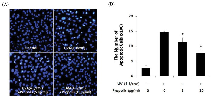
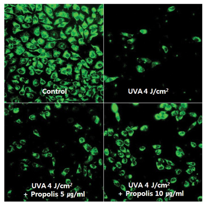
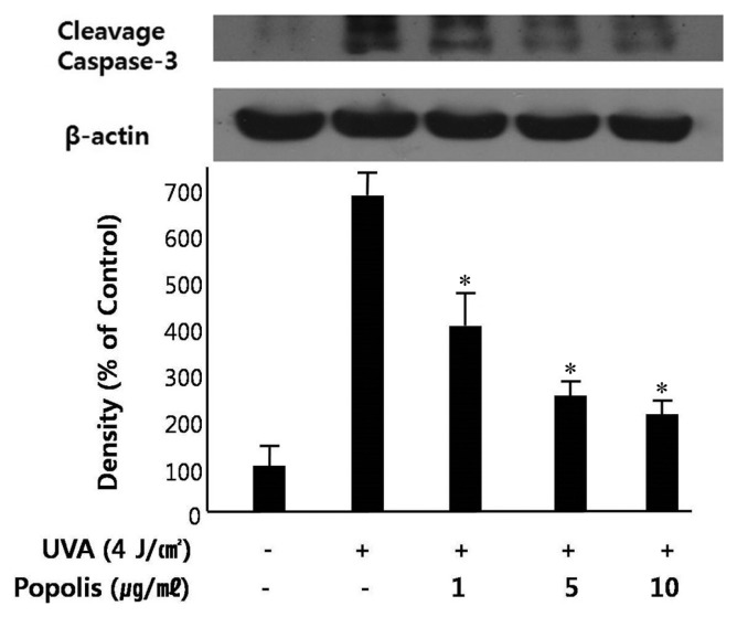
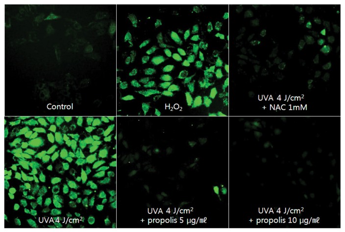
Propolis is a resinous material collected by honeybees from several plant sources. This research aimed at showing its protective effect against UVA-induced apoptosis of human keratinocyte HaCaT cells. Using Hoechst staining, it was demonstrated that propolis (5 and 10 μg/mL) significantly inhibited the apoptosis of HaCaT cells induced by UVA-irradiation. Propolis also showed the protective effect against loss of mitochondrial membrane potential induced by UVA-irradiaiton in HaCaT cells. Propolis also inhibited the expression of activated caspase-3 induced by UVA-irradiation. To investigate the role of ROS in UVA-induced apoptosis and protection by propolis, the generation of ROS was determined in cells. The results showed that the generation of ROS was markedly reduced in cells pretreated with propolis. Consequently, propolis protected human keratinocyte HaCaT cells against UVA-induced apoptosis, which might be related to the reduction of ROS generation by UVA-irradiation.
Overview
The claims and statistics of this paper haven’t been checked yet.
Run the check — free, no account needed. It reads the open-access text and checks the sources it cites.
Integrity signals
Registry
Cites
Statistics
Figures
Verification
Claims the paper makes
The claims and statistics of this paper haven’t been checked yet.
Run the check — free, no account needed. It reads the open-access text and checks the sources it cites.
Claims other papers make about this one
Each is checked against this paper’s own text: does it say what the citing paper says it does?
The check wasn’t confident either way, so it gives no verdict — never disputed.
In the citing paper
“and to promote inflammatory disorders, aging, androgenetic alopecia, and cancers of the skin (Kim et al. 2016).”
From Particulate matter 2.5 damages skin cells by inducing oxidative stress, subcellular organelle dysfunction, and apoptosis (2018) · cites this paper as Kim et al. 2016
Experimental — accuracy not yet measured
Self-check
Percentages, sums and reported differences re-computed from the paper’s own numbers.
The self-check needs the paper’s full text.
Citations
Cites
Sunburn and p53 in the onset of skin cancer
Ziegler, A., Jonason, A. S., Leffellt, D. J., Simon, J. A., Sharma, H. W., Kimmelman, J., Remington, L., Jacks, T., & Brash, D. E. (1994) · Nature · doi:10.1038/372773a0 · Cited by 1,497
Research works printed but not linked: 48
- 1998, J. Cell Biol., 140, 171, 10.1083/jcb.140.1.171 · DOI · Paper page
- 1987, Photochem. Photobiol., 45, 683, 10.1111/j.1751-1097.1987.tb07401.x · DOI
- 2004, Toxicol. Appl. Pharmacol., 195, 288, 10.1016/j.taap.2003.09.028 · DOI
- 2001, Phytother. Res., 15, 561, 10.1002/ptr.1029 · DOI
- 1998, Food Chem. Toxicol., 36, 347, 10.1016/S0278-6915(97)00145-2 · DOI
- 2005, J. Ethnopharmacol., 100, 114, 10.1016/j.jep.2005.05.004 · DOI
- 2000, J. Ethnopharmacol., 73, 243, 10.1016/S0378-8741(00)00320-2 · DOI
- 1999, J. Ethnopharmacol., 64, 235, 10.1016/S0378-8741(98)00131-7 · DOI
- 1997, Zhongguo Yao Li Xue Bao, 18, 274
- 2003, J. Nat. Prod., 66, 503, 10.1021/np0203180 · DOI
- 2002, J. Nutr. Biochem., 13, 572, 10.1016/S0955-2863(02)00208-5 · DOI
- 2003, J. Ethnopharmacol., 84, 265, 10.1016/S0378-8741(02)00329-X · DOI
- 2004, Biol. Pharm. Bull., 27, 727, 10.1248/bpb.27.727 · DOI
- 2002, Pharmazie, 57, 852
- 2007, Biol. Pharm. Bull., 30, 946, 10.1248/bpb.30.946 · DOI
- 2002, Fitoterapia, 73, S1, 10.1016/S0367-326X(02)00185-5 · DOI
- 2004, J. Ethnopharmacol., 94, 59, 10.1016/j.jep.2004.04.026 · DOI
- 1999, Radiat. Res., 151, 74, 10.2307/3579750 · DOI
- 2005, J. Environ. Pathol. Toxicol. Oncol., 24, 299, 10.1615/JEnvironPatholToxicolOncol.v24.i4.70 · DOI
- 2007, Biomed. Pharmacother., 61, 292, 10.1016/j.biopha.2007.02.012 · DOI
- 2008, Phytomedicine, 15, 851, 10.1016/j.phymed.2008.02.010 · DOI
- 2010, FEBS Lett., 584, 4019, 10.1016/j.febslet.2010.08.014 · DOI
- 2009, Biomed. Pharmacother., 63, 86, 10.1016/j.biopha.2008.08.001 · DOI
- 1995, Biochim. Biophys. Acta, 1241, 139, 10.1016/0304-4157(95)00003-A · DOI
- 1997, Cell, 91, 627, 10.1016/S0092-8674(00)80450-X · DOI
- 1999, Oncogene, 18, 7719, 10.1038/sj.onc.1203249 · DOI
- 2002, Biochimie, 84, 131, 10.1016/S0300-9084(02)01369-X · DOI
- 1999, Cell. Signal., 1, 77
- 2000, J. Immunol., 164, 6199, 10.4049/jimmunol.164.12.6199 · DOI
- 2001, Curr. Probl. Dermatol., 29, 83
- 2002, Oncogene, 21, 5844, 10.1038/sj.onc.1205743 · DOI
- 1998, J. Cell. Physiol., 177, 324, 10.1002/(SICI)1097-4652(199811)177:2<324::AID-JCP14>3.0.CO;2-9 · DOI
- 2000, Circ. Res., 87, 179, 10.1161/01.RES.87.3.179 · DOI
- 2000, J. Ethnopharmacol., 71, 109, 10.1016/S0378-8741(99)00189-0 · DOI
- 2004, Food Chem., 84, 329, 10.1016/S0308-8146(03)00216-4 · DOI
- 2001, Phytomedicine, 8, 16, 10.1078/0944-7113-00004 · DOI
- 2007, Food Chem., 103, 729, 10.1016/j.foodchem.2006.09.014 · DOI
- 1995, Cancer Res., 55, 3576
- 1995, Cancer Res., 55, 2310
- 2011, Food Chem. Toxicol., 49, 78, 10.1016/j.fct.2010.09.034 · DOI
- 2010, Nat. Prod. Res., 24, 375, 10.1080/14786410903370908 · DOI
- 2007, Cancer Lett., 245, 218, 10.1016/j.canlet.2006.01.016 · DOI
- 2009, Oncol. Rep., 22, 349
- 2007, J. Agric. Food Chem., 55, 7366, 10.1021/jf0710579 · DOI
- 2007, J. Agric. Food Chem., 55, 5289, 10.1021/jf070201n · DOI
- 2007, J. Ethnopharmacol., 113, 1, 10.1016/j.jep.2007.05.012 · DOI
- 2012, Journal of Apiculture, 27, 243
- 2006, Toxicol. In Vitro, 20, 1154, 10.1016/j.tiv.2006.02.009 · DOI
Cited by
Sorted by citations
Piao, M. J., Ahn, M. J., Kang, K. A., Ryu, Y. S., Hyun, Y. J., Shilnikova, K., Zhen, A. X., Jeong, J. W., Choi, Y. H., Kang, H. K., et al. (2018) · Archives of Toxicology · doi:10.1007/s00204-018-2197-9 · Cited by 339
“Images of stained cells were generated by confocal microscopy as previously described (Kim and Yoo 2016; Soeur et al. 2017).”
Natural Antioxidants: Multiple Mechanisms to Protect Skin From Solar Radiation
Dunaway, S., Odin, R., Zhou, L., Ji, L., Zhang, Y., & Kadekaro, A. L. (2018) · Frontiers in Pharmacology · doi:10.3389/fphar.2018.00392 · Cited by 280
Metadata
- Authors
- Kim, H. B., & Yoo, B. S.
- Affiliations
- Kyonggi University (KR)
- Corresponding author
- Yoo, Byung Sun
- Venue
- Toxicological Research
- Published
- 2016-10-30
- Volume
- 32
- Issue
- 4
- Pages
- 345-351
- ISSN
- 1976-8257 (ISSN-L)
- 2234-2753
- PMC
- PMC5080852
- OpenAlex
- W2531157978
- Cited by
- 42
- References
- 1
- Citations by year
- 2026: 1 · 2025: 1 · 2024: 5 · 2023: 1 · 2022: 7 · 2021: 7 · 2020: 1 · 2019: 4 · 2018: 9 · 2017: 5
- FWCI
- 6.51
- Type
- journal article
- Language
- en
- Open access
- hybrid
- doi.org
- Licence
- Locations
- Toxicological Research (publishedVersion, cc-by-nc)
- PubMed Central (publishedVersion, cc-by-nc)
- PubMed (publishedVersion)
- Funders
- Kyonggi University(KGU)
- Topics
- Life Sciences › Agricultural and Biological Sciences › Insect Science › Bee Products Chemical Analysis
- Health Sciences › Medicine › Dermatology › Skin Protection and Aging
- Health Sciences › Medicine › Pharmacology (medical) › Healthcare and Venom Research
- Keywords
- propolis, HaCaT keratinocytes, reactive oxygen species, mitochondrial membrane potential, caspase-3, photoprotection, photodamaged skin
About these checks
Registry, retraction, Cites and statistics results are computed, not judged, with one exception: a printed reference that no index lists is read once by a model before it counts as a citation error. Claims are extracted by a model and kept only when their quote appears verbatim in the text; each cited source is then checked, with a model’s help, for whether it says what the claim relies on. Those verdicts can be wrong — read the claim’s quote and the cited source before relying on one.
Statistics and arithmetic are recomputed, not judged.
Confidence bands are shown only for fields calibrated on our annotated test set. None is calibrated yet, so no band is shown.
What counts as a claim ›How we measure confidence ›How we check this ›
Are you an author, or do you see a problem with this page? Request a review
How to cite
Cite CoChat for the checks on this page, and the paper for its own findings.
- This page
- CoChat. (2026). Citation check: Propolis Inhibits UVA-Induced Apoptosis of Human Keratinocyte HaCaT Cells by Scavenging ROS. https://app.cochat.ai/papers/10.5487/tr.2016.32.4.345
- The paper
- Kim, H. B., & Yoo, B. S. (2016). Propolis Inhibits UVA-Induced Apoptosis of Human Keratinocyte HaCaT Cells by Scavenging ROS. Toxicological Research. https://doi.org/10.5487/tr.2016.32.4.345
