

Deep learning enhanced magnetocardiography enables multi-task detection of coronary, ventricular, and rhythm disorders
Open accessThe DOI matches its Crossref record; 33 of the 33 references it cites were found in a registry or an index.
Abstract
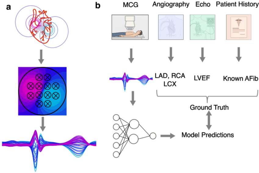
Background: Magnetocardiography (MCG) captures the magnetic fields generated by myocardial currents, theoretically preserving electrophysiological details lost in surface potentials. However, its clinical application has been hindered by small datasets and the complexity of analysing high-dimensional magnetic field data. We sought to develop a self-supervised deep learning framework to detect high burden coronary artery disease (CAD), left ventricular dysfunction, and arrhythmia risk from resting MCG recordings. Methods: We developed MCG2Vec, a contrastive deep learning encoder trained on raw 64-channel MCG signals. The model was pre-trained on unlabeled 10-second segments to learn generalizable signal morphology, then fine-tuned for clinical tasks in a retrospective cohort of 1,732 consecutive patients. The primary endpoints were the detection of significant CAD ( ≥ 70% stenosis), reduced left ventricular ejection fraction (LVEF <55%), and atrial fibrillation (AF) risk, all derived from sinus-rhythm recordings. Model performance was evaluated using patient-stratified five-fold cross-validation and interpreted using Grad-CAM activation mapping. Results: In the validation analysis, the model detected significant CAD with an area under the curve (AUC) of 0.89 (95% CI 0.82 - 0.93) and successfully localized ischemia to the left anterior descending (AUC 0.88), right coronary (0.82), and left circumflex arteries (0.82). Reduced LVEF was identified with an AUC of 0.81 (95% CI 0.71 - 0.91). Furthermore, the model predicted a history of paroxysmal AF from sinus-rhythm recordings with an AUC of 0.77 (95% CI 0.69 - 0.85). Explainability analysis confirmed that the model relied on physiologically distinct phases of the cardiac cycle (repolarization heterogeneity for ischemia and P-wave/S-wave morphology for arrhythmia risk) rather than non-specific noise. Conclusion: Deep learning-enhanced magnetocardiography enables the accurate, non-invasive detection of ischemia, ventricular dysfunction, and arrhythmia risk from a single resting scan. By unlocking the latent diagnostic information within the cardiac magnetic field, this approach offers a scalable, radiation-free adjunct to standard electrocardiography for precision cardiac diagnostics.
Overview
The claims and statistics of this paper haven’t been checked yet.
Free: it reads the open-access text and checks the first 5 claims that cite a source we can check. Create a free account to check every claim.
Integrity signals
Registry
Cites
Statistics
Figures


Verification
Claims the paper makes
The claims and statistics of this paper haven’t been checked yet.
Free: it reads the open-access text and checks the first 5 claims that cite a source we can check. Create a free account to check every claim.
Claims other papers make about this one
Each is checked against this paper’s own text: does it say what the citing paper says it does?
The source backs part of the claim, not all of it — or backs it for a different population, condition or comparison.
In the citing paper
“Despite these advances [75,76,77], common structural constraints persist, including limited external validation, modest dataset scale, absence of calibration analyses, and restricted prospective outcome-based evaluation.”
From The Heart’s Electromagnetic Field in Emotions, Empathy and Human Connection: Biosensor-Derived Insights into Heart–Brain Axis Mechanisms and a Basis for Novel BioMagnetoTherapies (2026) · cites this paper as [75]
Experimental — accuracy not yet measuredThe cited source says what the claim attributes to it.
In the citing paper
“MCG2Vec advances the field through self-supervised pretraining on raw multi-channel MCG signals, eliminating reliance on engineered features and enabling scalable, data-driven representation learning.”
Self-check
Percentages, sums and reported differences re-computed from the paper’s own numbers.
The self-check needs the paper’s full text.
Citations
Cites
2021 ESC guidelines for the diagnosis and treatment of acute and chronic heart failure
McDonagh, T. A., Metra, M., Adamo, M., Gardner, R. S., Baumbach, A., Böhm, M., Burri, H., Butler, J., Čelutkienė, J., Chioncel, O., et al. (2021) · European Heart Journal · doi:10.1093/eurheartj/ehab368 · Cited by 13,344
Hindricks, G., Potpara, T., Dagres, N., Arbelo, E., Bax, J. J., Blomström-Lundqvist, C., Boriani, G., Castella, M., Dan, G.-A., Dilaveris, P. E., et al. (2021) · European Heart Journal · doi:10.1093/eurheartj/ehaa612 · Cited by 9,971
2019 ESC Guidelines for the diagnosis and management of chronic coronary syndromes
Knuuti, J., Wijns, W., Saraste, A., Capodanno, D., Barbato, E., Funck-Brentano, C., Prescott, E., Storey, R. F., Deaton, C., Cuisset, T., et al. (2020) · European Heart Journal · doi:10.1093/eurheartj/ehz425 · Cited by 7,166
Hämäläinen, M., Hari, R., Ilmoniemi, R. J., Knuutila, J., & Lounasmaa, O. V. (1993) · Reviews of Modern Physics · doi:10.1103/revmodphys.65.413 · Cited by 4,684
Global burden of heart failure: a comprehensive and updated review of epidemiology
Savarese, G., Becher, P. M., Lund, L. H., Seferovic, P., Rosano, G. M. C., & Coats, A. J. S. (2023) · Cardiovascular Research · doi:10.1093/cvr/cvac013 · Cited by 3,315
Hannun, A. Y., Rajpurkar, P., Haghpanahi, M., Tison, G. H., Bourn, C., Turakhia, M. P., & Ng, A. Y. (2019) · Nature Medicine · doi:10.1038/s41591-018-0268-3 · Cited by 3,148
wav2vec 2.0: A Framework for Self-Supervised Learning of Speech Representations
Baevski, A., Zhou, H., Mohamed, A., & Auli, M. (2020) · arXiv · doi:10.48550/arxiv.2006.11477 · Cited by 2,369
Attia, Z. I., Noseworthy, P. A., Lopez-Jimenez, F., Asirvatham, S. J., Deshmukh, A. J., Gersh, B. J., Carter, R. E., Yao, X., Rabinstein, A. A., Erickson, B. J., et al. (2019) · The Lancet · doi:10.1016/s0140-6736(19)31721-0 · Cited by 1,613
Standardized cardiovascular magnetic resonance imaging (CMR) protocols: 2020 update
Kramer, C. M., Barkhausen, J., Bucciarelli-Ducci, C., Flamm, S. D., Kim, R. J., & Nagel, E. (2020) · Journal of Cardiovascular Magnetic Resonance · doi:10.1186/s12968-020-00607-1 · Cited by 1,245
Roberts, M., Driggs, D., Thorpe, M., Gilbey, J., Yeung, M., Ursprung, S., Aviles-Rivero, A. I., Etmann, C., McCague, C., Beer, L., et al. (2021) · Nature Machine Intelligence · doi:10.1038/s42256-021-00307-0 · Cited by 1,044
Research works printed but not linked: 2
- 10.48550/arXiv.2505.18185 · DOI
- C. Yang , M. Westover , and J. Sun , “BIOT: Biosignal Transformer for Cross-data Learning in the Wild,” in Advances in Neural Information Processing Systems, A. Oh , T. Naumann , A. Globerson , K. Saenko , M. Hardt , and S. Levine , Eds., Curran Associates, Inc., 2023, pp. 78240–78260. [Online]. Available: https://proceedings.neurips.cc/paper_files/paper/2023/file/f6b30f3e2dd9cb53bbf2024402d02295-Paper-Conference.pdf
Cited by
Palantzas, A., & Anagnostouli, M. (2026) · Sensors · doi:10.3390/s26051738 · Cited by 1
“Despite these advances [75–77], common structural constraints persist, including limited external validation, modest dataset scale, absence of calibration analyses, and restricted prospective outcome-based evaluation.”
Metadata
- Authors
- Kranz, D. D., Kahriman, O., Dischl, D., Treskatsch, S., Sander, A., Brachmann, J., Park, J.-W., & Wessel, N.
- Affiliations
- Humboldt-Universität zu Berlin (DE)
- Deutsches Herzzentrum München (DE)
- Charité – Universitätsmedizin Berlin (DE)
- University of Split (HR)
- Deutsches Herzzentrum der Charité (DE)
- Corresponding author
- Wessel, Niels
- Venue
- medRxiv
- Publisher
- openRxiv
- Published
- 2025-12-02
- OpenAlex
- W4417151796
- Cited by
- 1
- References
- 33
- Citations by year
- 2026: 1
- Type
- posted content
- Language
- en
- Open access
- green
- doi.org
- Licence
- Locations
- medRxiv (acceptedVersion, cc-by-nc-nd)
- Topics
- Health Sciences › Medicine › Cardiology and Cardiovascular Medicine › ECG Monitoring and Analysis
- Health Sciences › Health Professions › Health Information Management › Artificial Intelligence in Healthcare
- Health Sciences › Medicine › Radiology, Nuclear Medicine and Imaging › Cardiac Imaging and Diagnostics
- Keywords
- magnetocardiography, coronary artery disease detection, left ventricular dysfunction, atrial fibrillation risk, ischemia localization, self-supervised contrastive learning, cardiac magnetic field, noninvasive cardiac assessment
About these checks
Registry, retraction, Cites and statistics results are computed, not judged, with one exception: a printed reference that no index lists is read once by a model before it counts as a citation error. Claims are extracted by a model and kept only when their quote appears verbatim in the text; each cited source is then checked, with a model’s help, for whether it says what the claim relies on. Those verdicts can be wrong — read the claim’s quote and the cited source before relying on one.
Statistics and arithmetic are recomputed, not judged.
Confidence bands are shown only for fields calibrated on our annotated test set. None is calibrated yet, so no band is shown.
What counts as a claim ›How we measure confidence ›How we check this ›
Are you an author, or do you see a problem with this page? Request a review
How to cite
Cite CoChat for the checks on this page, and the paper for its own findings.
- This page
- CoChat. (2026). Citation check: Deep learning enhanced magnetocardiography enables multi-task detection of coronary, ventricular, and rhythm disorders. https://app.cochat.ai/papers/10.64898/2025.11.30.25341301
- The paper
- Kranz, D. D., Kahriman, O., Dischl, D., Treskatsch, S., Sander, A., Brachmann, J., Park, J.-W., & Wessel, N. (2025). Deep learning enhanced magnetocardiography enables multi-task detection of coronary, ventricular, and rhythm disorders. medRxiv. https://doi.org/10.64898/2025.11.30.25341301
Data sources
Bibliographic data: Semantic Scholar, OpenAlex, Crossref.
Contains information from the Semantic Scholar Academic Graph Datasets, made available under the ODC Attribution License (ODC-BY 1.0).
Semantic Scholar’s data is described in: Kinney, R., et al. (2023). The Semantic Scholar Open Data Platform. arXiv:2301.10140.
