Cited by 97

The DOI matches its Crossref record.

Radiation-induced small extracellular vesicles as “carriages” promote tumor antigen release and trigger antitumor immunity

Lin, Wanzun, Xu, Yanyan, Chen, Xiaochuan, Liu, Jun, Weng, Youliang, Zhuang, Qingyang, Lin, Feifei, Huang, Zongwei, Wu, Shihong, Ding, Jianming, Chen, Long, Qiu, Xianxin, Zhang, Lurong, Wu, Junxin, Lin, Duo, Qiu, Sufang·2020·Theranostics

Overview

The claims and statistics of this paper haven’t been checked yet.

Run the check — free, no account needed. It reads the open-access text and checks the sources it cites.

A few free checks a day · papers already verified open instantly · results are public

Integrity signals

Registry

Crossref · 10.7150/thno.43539

Cites

Statistics

Figures

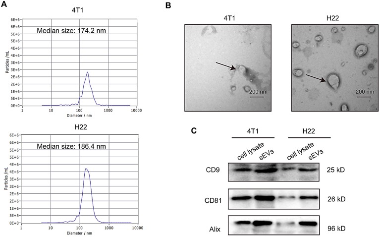
Figure 1 Characterization of small extracellular vesicles (sEVs). (A) Particle size distribution measured via nanoparticle tracking analysis (NTA). (B) sEV morphology revealed via transmission electron microscopy (TEM). (C) Western blot analysis of specific sEV-specific surface markers.
Figure 1 Characterization of small extracellular vesicles (sEVs). (A) Particle size distribution measured via nanoparticle tracking analysis (NTA). (B) sEV morphology revealed via transmission electron microscopy (TEM). (C) Western blot analysis of specific sEV-specific surface markers.CC BY · PubMed Central
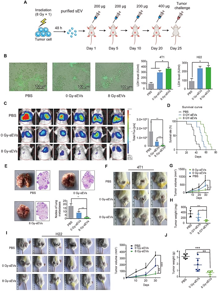
Figure 2 Antitumor immunity triggered by radiation-induced sEVs against primary tumor and experimental lung metastasis. (A) Experimental schema depicting sEV purification and immunization. H22 cells and 4T1 cells were subjected to 8 Gy irradiation, and sEVs derived from 8 Gy-irradiated cells (8 Gy-sEVs) or non-irradiated cells (0 Gy-sEVs) were purified via ultracentrifugation. ICR and BALB/c mice were immunized with 200 μg of each type of sEV four times at 5-day intervals and challenged by injection of tumor cells. (B) Cell-cell contact between green fluorescent protein (GFP)-positive 4T1 cells and lymphocytes was imaged under fluorescent reverse-microscopy. In a co-culture of 4T1 tumor cells and lymphocytes, 4T1 tumor cells were closely surrounded by lymphocytes from mice immunized with 8 Gy-sEVs, although this was not observed in lymphocytes from mice immunized with 0 Gy-sEVs or the control group. The killing effect was measured as the amount of lactate dehydrogenase (LDH) released (n = 4/group). (C) In vivo bioluminescence imaging. GFP-luciferase-4T1 cells were intravenously injected into the tail vein of BALB/c mice after immunization. Two weeks later, the growth of 4T1 experimental lung metastases was measured using an IVIS Lumina III in vivo imaging system. Luciferase signals from mice immunized with 8 Gy-sEVs were lower than those obtained from the control or 0 Gy group (n = 6/group). (D) BALB/c mice were intravenously injected with 4T1 cells via the tail vein. Differences in survival were observed between BALB/c mice immunized with 0 Gy-sEVs, 8 Gy-sEVs, and the control group (n = 10/group). (E) Images and hematoxylin & eosin staining of lung metastases and statistical analysis of lung metastatic nodules (n = 6/group). (F-G) Primary tumor growth and growth curve of 4T1 breast cancer cells (n = 6/group). (H) Tumor weights of 4T1 breast cancer cells at the end of the experiment (n = 6/group). (I) Primary tumor growth and growth curve of H22 hepatoma cells (n = 6/group). (J) Tumor weights of H22 hepatoma cells at the end of the experiment (n = 6/group). P-values were determined using a two-tailed Student's t-test (*P < 0.05, **P < 0.01, ***P < 0.001).
Figure 2 Antitumor immunity triggered by radiation-induced sEVs against primary tumor and experimental lung metastasis. (A) Experimental schema depicting sEV purification and immunization. H22 cells and 4T1 cells were subjected to 8 Gy irradiation, and sEVs derived from 8 Gy-irradiated cells (8 Gy-sEVs) or non-irradiated cells (0 Gy-sEVs) were purified via ultracentrifugation. ICR and BALB/c mice were immunized with 200 μg of each type of sEV four times at 5-day intervals and challenged by injection of tumor cells. (B) Cell-cell contact between green fluorescent protein (GFP)-positive 4T1 cells and lymphocytes was imaged under fluorescent reverse-microscopy. In a co-culture of 4T1 tumor cells and lymphocytes, 4T1 tumor cells were closely surrounded by lymphocytes from mice immunized with 8 Gy-sEVs, although this was not observed in lymphocytes from mice immunized with 0 Gy-sEVs or the control group. The killing effect was measured as the amount of lactate dehydrogenase (LDH) released (n = 4/group). (C) In vivo bioluminescence imaging. GFP-luciferase-4T1 cells were intravenously injected into the tail vein of BALB/c mice after immunization. Two weeks later, the growth of 4T1 experimental lung metastases was measured using an IVIS Lumina III in vivo imaging system. Luciferase signals from mice immunized with 8 Gy-sEVs were lower than those obtained from the control or 0 Gy group (n = 6/group). (D) BALB/c mice were intravenously injected with 4T1 cells via the tail vein. Differences in survival were observed between BALB/c mice immunized with 0 Gy-sEVs, 8 Gy-sEVs, and the control group (n = 10/group). (E) Images and hematoxylin & eosin staining of lung metastases and statistical analysis of lung metastatic nodules (n = 6/group). (F-G) Primary tumor growth and growth curve of 4T1 breast cancer cells (n = 6/group). (H) Tumor weights of 4T1 breast cancer cells at the end of the experiment (n = 6/group). (I) Primary tumor growth and growth curve of H22 hepatoma cells (n = 6/group). (J) Tumor weights of H22 hepatoma cells at the end of the experiment (n = 6/group). P-values were determined using a two-tailed Student's t-test (*P < 0.05, **P < 0.01, ***P < 0.001).CC BY · PubMed Central
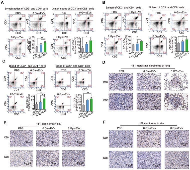
Figure 3 CD8 and CD4 cell infiltration in 8 Gy-sEV-immunized mice. (A-C) The lymph nodes, spleen, and blood of BALB/c mice were double-stained using anti-mouse fluorescein isothiocyanate (FITC)-anti-mouse CD3, peridinin chlorophyll protein complex (PerCP)-anti-mouse CD8, or allophycocyanin (APC)-anti-mouse CD4, and analyzed using flow cytometry to detect CD4 and CD8. (D-F) 4T1 lung metastases, 4T1 primary tumors, and H22 primary tumors were harvested at the end of the experiment and stained with anti-mouse CD8 (imaged at 200× magnification). P-values were determined using a two-tailed Student's t-test (*P < 0.05, **P < 0.01, ***P < 0.001).
Figure 3 CD8 and CD4 cell infiltration in 8 Gy-sEV-immunized mice. (A-C) The lymph nodes, spleen, and blood of BALB/c mice were double-stained using anti-mouse fluorescein isothiocyanate (FITC)-anti-mouse CD3, peridinin chlorophyll protein complex (PerCP)-anti-mouse CD8, or allophycocyanin (APC)-anti-mouse CD4, and analyzed using flow cytometry to detect CD4 and CD8. (D-F) 4T1 lung metastases, 4T1 primary tumors, and H22 primary tumors were harvested at the end of the experiment and stained with anti-mouse CD8 (imaged at 200× magnification). P-values were determined using a two-tailed Student's t-test (*P < 0.05, **P < 0.01, ***P < 0.001).CC BY · PubMed Central
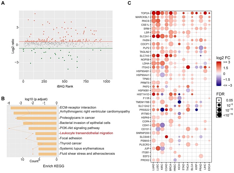
Figure 4 Proteomic and bioinformatic analysis of altered cargos in radiation-induced sEVs. (A) Scatter plot showing the distribution of 146 differentially expressed proteins quantified in 8 Gy-sEVs and 0 Gy-sEVs (fold-change > 1.5 and P < 0.05). Red dots indicate up-regulated proteins and green dots indicate down-regulated proteins. (B) Kyoto Encyclopedia of Genes and Genomes (KEGG) pathway analysis of differentially expressed proteins. (C) Expression of up-regulated proteins in various cancers in the Cancer Genome Atlas (TCGA). The intensity of the color represents log2 fold-change (tumor vs normal). Y-axis shows protein name and X-axis shows tumor type, including lung adenocarcinoma (LUAD), lung squamous cell carcinoma (LUSC), kidney renal clear cell carcinoma (KIRC), colon adenocarcinoma (COAD), breast invasive carcinoma (BRCA), head and neck squamous cell carcinoma (HNSC), kidney chromophobe (KICH), stomach adenocarcinoma (STAD), thyroid carcinoma (THCA), bladder urothelial carcinoma (BLCA), kidney renal papillary cell carcinoma (KIRP), prostate adenocarcinoma (PRAD), liver hepatocellular carcinoma (LIHC), and esophageal carcinoma (ESCA).
Figure 4 Proteomic and bioinformatic analysis of altered cargos in radiation-induced sEVs. (A) Scatter plot showing the distribution of 146 differentially expressed proteins quantified in 8 Gy-sEVs and 0 Gy-sEVs (fold-change > 1.5 and P < 0.05). Red dots indicate up-regulated proteins and green dots indicate down-regulated proteins. (B) Kyoto Encyclopedia of Genes and Genomes (KEGG) pathway analysis of differentially expressed proteins. (C) Expression of up-regulated proteins in various cancers in the Cancer Genome Atlas (TCGA). The intensity of the color represents log2 fold-change (tumor vs normal). Y-axis shows protein name and X-axis shows tumor type, including lung adenocarcinoma (LUAD), lung squamous cell carcinoma (LUSC), kidney renal clear cell carcinoma (KIRC), colon adenocarcinoma (COAD), breast invasive carcinoma (BRCA), head and neck squamous cell carcinoma (HNSC), kidney chromophobe (KICH), stomach adenocarcinoma (STAD), thyroid carcinoma (THCA), bladder urothelial carcinoma (BLCA), kidney renal papillary cell carcinoma (KIRP), prostate adenocarcinoma (PRAD), liver hepatocellular carcinoma (LIHC), and esophageal carcinoma (ESCA).CC BY · PubMed Central
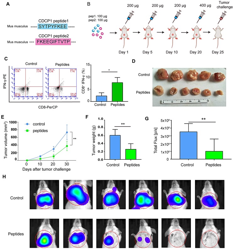
Figure 5 Peptides derived from up-regulated proteins in sEVs triggered antitumor immunity. (A) Two peptides derived from CUB domain-containing protein 1 (CDCP1) were synthesized. (B) Experiment design. (C) Flow cytometry (FCM) analysis of differences of CD8+-interferon (IFN)γ+ T lymphocytes in the control group (phosphate-buffered saline (PBS) with Freund's adjuvant) and peptide group (CDCP1 peptides with Freund's adjuvant; n = 3/group). (D) Primary 4T1 tumor sizes in the control group (immunization with PBS four times, at 5-day intervals) and peptide group (immunization with 200 µg CDCP1 peptides four times, at 5-day intervals; n = 6/group). (E) Primary 4T1 tumor growth curve in the control (immunization with PBS four times, at 5-day intervals) and peptide-treated group (immunization with 200 µg CDCP1 peptides four times, at 5-day intervals; n = 6/group). (F) Primary 4T1 tumor weights (n = 6/group). (G-H) Experimental metastasis model and in vivo imaging of the lungs in the control group (immunization with PBS four times, at 5-day intervals) and peptide group (immunization with 200 µg CDCP1 peptides four times, at 5-day intervals; n = 6/group). P-values were determined using a two-tailed Student's t-test (*P < 0.05, **P < 0.01, ***P < 0.001).
Figure 5 Peptides derived from up-regulated proteins in sEVs triggered antitumor immunity. (A) Two peptides derived from CUB domain-containing protein 1 (CDCP1) were synthesized. (B) Experiment design. (C) Flow cytometry (FCM) analysis of differences of CD8+-interferon (IFN)γ+ T lymphocytes in the control group (phosphate-buffered saline (PBS) with Freund's adjuvant) and peptide group (CDCP1 peptides with Freund's adjuvant; n = 3/group). (D) Primary 4T1 tumor sizes in the control group (immunization with PBS four times, at 5-day intervals) and peptide group (immunization with 200 µg CDCP1 peptides four times, at 5-day intervals; n = 6/group). (E) Primary 4T1 tumor growth curve in the control (immunization with PBS four times, at 5-day intervals) and peptide-treated group (immunization with 200 µg CDCP1 peptides four times, at 5-day intervals; n = 6/group). (F) Primary 4T1 tumor weights (n = 6/group). (G-H) Experimental metastasis model and in vivo imaging of the lungs in the control group (immunization with PBS four times, at 5-day intervals) and peptide group (immunization with 200 µg CDCP1 peptides four times, at 5-day intervals; n = 6/group). P-values were determined using a two-tailed Student's t-test (*P < 0.05, **P < 0.01, ***P < 0.001).CC BY · PubMed Central
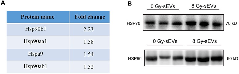
Figure 6 Heat shock protein (HSP)70 and HSP90 expression in radiation-enriched sEVs. (A) Proteomic analysis showing up-regulation of the HSP family in sEVs from irradiated 4T1 cells. (B) Western blot confirming that Hsp70 and Hsp90 were up-regulated in sEVs from irradiated 4T1 cells.
Figure 6 Heat shock protein (HSP)70 and HSP90 expression in radiation-enriched sEVs. (A) Proteomic analysis showing up-regulation of the HSP family in sEVs from irradiated 4T1 cells. (B) Western blot confirming that Hsp70 and Hsp90 were up-regulated in sEVs from irradiated 4T1 cells.CC BY · PubMed Central
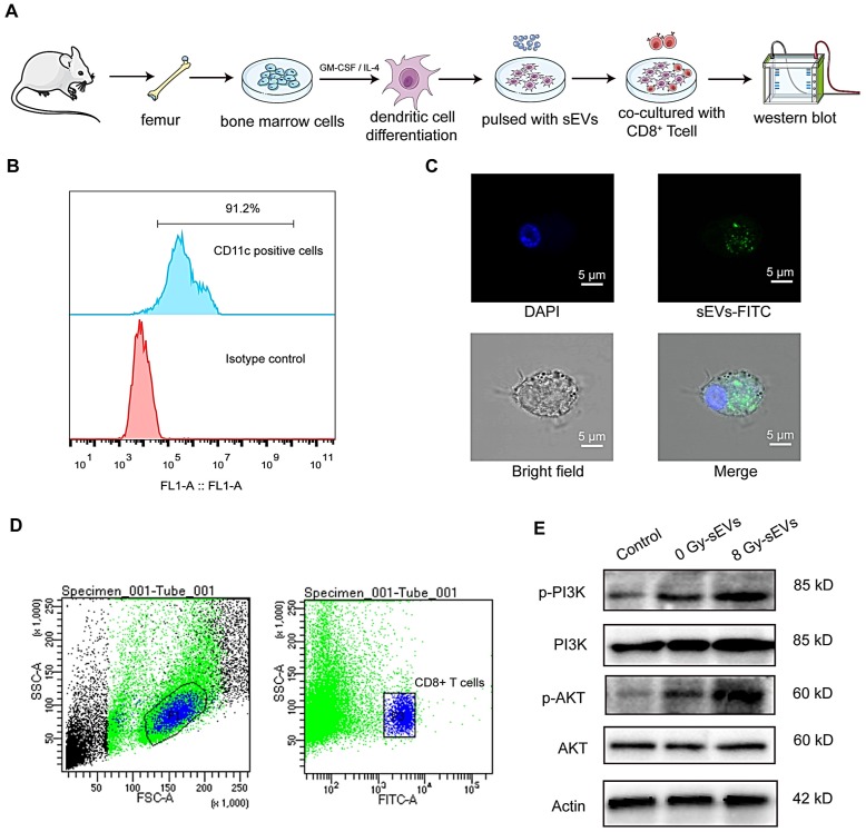
Figure 7 the underlying mechanism on how sEVs achieve antitumor immunity after radiation. (A) Experimental design. Bone marrow cells were isolated from the femurs of mice and stimulated with GM-CSF and IL-4 for DCs differentiation. Then, DCs were pulsed with sEVs and co-culture with CD8+ T cell. Western bolt was used to analyze activated signaling in CD8+ T cell. (B) Detection of CD11c+ dendritic cells by FCM after stimulation with granulocyte-macrophage colony-stimulating factor (GM-CSF) and interleukin (IL)-4. (C) Uptake of sEVs by DCs. sEVs were first stained with PKH67 dye before 24-h culture with DCs and analysis via confocal microscopy. (D) Panels illustrate the gating strategy used for isolation of CD8+ T cell subsets via fluorescence-activated cell sorting. (E) Western blot revealed the activated signaling in CD8+ T cell.
Figure 7 the underlying mechanism on how sEVs achieve antitumor immunity after radiation. (A) Experimental design. Bone marrow cells were isolated from the femurs of mice and stimulated with GM-CSF and IL-4 for DCs differentiation. Then, DCs were pulsed with sEVs and co-culture with CD8+ T cell. Western bolt was used to analyze activated signaling in CD8+ T cell. (B) Detection of CD11c+ dendritic cells by FCM after stimulation with granulocyte-macrophage colony-stimulating factor (GM-CSF) and interleukin (IL)-4. (C) Uptake of sEVs by DCs. sEVs were first stained with PKH67 dye before 24-h culture with DCs and analysis via confocal microscopy. (D) Panels illustrate the gating strategy used for isolation of CD8+ T cell subsets via fluorescence-activated cell sorting. (E) Western blot revealed the activated signaling in CD8+ T cell.CC BY · PubMed Central
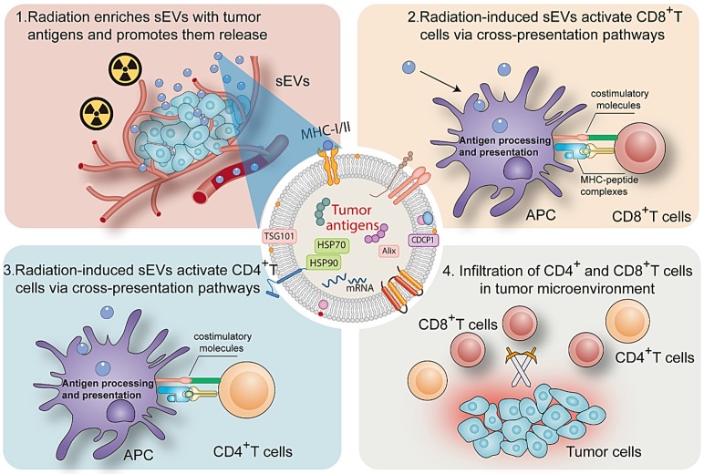
Figure 8 Mechanism by which radiation-induced sEVs trigger antitumor immunity.
Figure 8 Mechanism by which radiation-induced sEVs trigger antitumor immunity.CC BY · PubMed Central

Verification

Claims the paper makes

The claims and statistics of this paper haven’t been checked yet.

Run the check — free, no account needed. It reads the open-access text and checks the sources it cites.

A few free checks a day · papers already verified open instantly · results are public

Claims other papers make about this one

Each is checked against this paper’s own text: does it say what the citing paper says it does?

Self-check

Percentages, sums and reported differences re-computed from the paper’s own numbers.

The self-check needs the paper’s full text.

Citations

Cites

No references could be resolved yet.

Research works printed but not linked: 31

  1. [1]Ko EC Benjamin KT Formenti SC Generating antitumor immunity by targeted radiation therapy: Role of dose and fractionation Adv Radiat Oncol 2018 3 486 93 30370347 PMC6200901
  2. [2]Liu Y Dong Y Kong L Shi F Zhu H Yu J Abscopal effect of radiotherapy combined with immune checkpoint inhibitors J Hematol Oncol 2018 11 104 30115069 PMC6097415
  3. [3]Formenti SC Demaria S Radiation therapy to convert the tumor into an in situ vaccine Int J Radiat Oncol Biol Phys 2012 84 879 80 23078897 PMC3811126
  4. [4]Hessvik NP Llorente A Current knowledge on exosome biogenesis and release Cell Mol Life Sci 2018 75 193 208 28733901 PMC5756260
  5. [5]Becker A Thakur BK Weiss JM Kim HS Peinado H Lyden D Extracellular Vesicles in Cancer: Cell-to-Cell Mediators of Metastasis Cancer cell 2016 30 836 48 27960084 PMC5157696
  6. [6]He C Zheng S Luo Y Wang B Exosome Theranostics: Biology and Translational Medicine Theranostics 2018 8 237 55 29290805 PMC5743472
  7. [7]André F Schartz NEC Chaput N Flament C Raposo G Amigorena S Tumor-derived exosomes: a new source of tumor rejection antigens Vaccine 2002 20 Suppl 4 A28 A31 12477425
  8. [8]Rao Q Zuo B Lu Z Gao X You A Wu C Tumor-derived exosomes elicit tumor suppression in murine hepatocellular carcinoma models and humans in vitro Hepatology 2016 64 456 72 26990897
  9. [9]Billiau A Matthys P Modes of action of Freund's adjuvants in experimental models of autoimmune diseases J Leukoc Biol 2001 70 849 60 11739546
  10. [10]El-Andaloussi S Lee Y Lakhal-Littleton S Li J Seow Y Gardiner C Exosome-mediated delivery of siRNA in vitro and in vivo Nat Protoc 2012 7 2112 26 23154783
  11. [11]Alvarez-Erviti L Seow Y Yin H Betts C Lakhal S Wood MJA Delivery of siRNA to the mouse brain by systemic injection of targeted exosomes Nat Biotechnol 2011 29 341 5 21423189
  12. [12]Rammensee H Bachmann J Emmerich NP Bachor OA Stevanović S SYFPEITHI: database for MHC ligands and peptide motifs Immunogenetics 1999 50 213 9 10602881
  13. [13]Vita R Overton JA Greenbaum JA Ponomarenko J Clark JD Cantrell JR The immune epitope database (IEDB) 3.0 Nucleic Acids Res 2015 43 D405 D12 25300482 PMC4384014
  14. [14]Hirayama M Nishimura Y The present status and future prospects of peptide-based cancer vaccines Int Immunol 2016 28 319 28 27235694
  15. [15]Wortmann A He Y Deryugina EI Quigley JP Hooper JD The cell surface glycoprotein CDCP1 in cancer-insights, opportunities, and challenges IUBMB Life 2009 61 723 30 19514048
  16. [16]Figueiredo C Wittmann M Wang D Dressel R Seltsam A Blasczyk R Heat shock protein 70 (HSP70) induces cytotoxicity of T-helper cells Blood 2009 113 3008 16 19018093
  17. [17]Lv L-H Wan Y-L Lin Y Zhang W Yang M Li G-L Anticancer drugs cause release of exosomes with heat shock proteins from human hepatocellular carcinoma cells that elicit effective natural killer cell antitumor responses in vitro The Journal of biological chemistry 2012 287 15874 85 22396543 PMC3346092
  18. [18]Mellman I Steinman RM Dendritic cells: specialized and regulated antigen processing machines Cell 2001 106 255 8 11509172
  19. [19]van der Pol E Böing AN Harrison P Sturk A Nieuwland R Classification, functions, and clinical relevance of extracellular vesicles Pharmacol Rev 2012 64 676 705 22722893
  20. [20]Liu C Su C Design strategies and application progress of therapeutic exosomes Theranostics 2019 9 1015 28 30867813 PMC6401399
  21. [21]Lumniczky K Candéias SM Gaipl US Frey B Editorial: Radiation and the Immune System: Current Knowledge and Future Perspectives Front Immunol 2017 8 1933 29410662 PMC5787080
  22. [22]Siva S MacManus MP Martin RF Martin OA Abscopal effects of radiation therapy: a clinical review for the radiobiologist Cancer letters 2015 356 82 90 24125863
  23. [23]Ozpiskin OM Zhang L Li JJ Immune targets in the tumor microenvironment treated by radiotherapy Theranostics 2019 9 1215 31 30867826 PMC6401500
  24. [24]Buchwald ZS Wynne J Nasti TH Zhu S Mourad WF Yan W Radiation, Immune Checkpoint Blockade and the Abscopal Effect: A Critical Review on Timing, Dose and Fractionation Front Oncol 2018 8 612 30619752 PMC6306034
  25. [25]Popp I Grosu AL Niedermann G Duda DG Immune modulation by hypofractionated stereotactic radiation therapy: Therapeutic implications Radiother Oncol 2016 120 185 94 27495145
  26. [26]Zhang T Yu H Ni C Zhang T Liu L Lv Q Hypofractionated stereotactic radiation therapy activates the peripheral immune response in operable stage I non-small-cell lung cancer Scientific reports 2017 7 4866 28687760 PMC5501824
  27. [27]Frey B Rubner Y Wunderlich R Weiss EM Pockley AG Fietkau R Induction of abscopal anti-tumor immunity and immunogenic tumor cell death by ionizing irradiation - implications for cancer therapies Curr Med Chem 2012 19 1751 64 22414083
  28. [28]Zhou J Wang G Chen Y Wang H Hua Y Cai Z Immunogenic cell death in cancer therapy: Present and emerging inducers J Cell Mol Med 2019 23 4854 65 31210425 PMC6653385
  29. [29]de Charette M Marabelle A Houot R Turning tumour cells into antigen presenting cells: The next step to improve cancer immunotherapy? Eur J Cancer 2016 68 134 47 27755997
  30. [30]Pistoia V Morandi F Pezzolo A Raffaghello L Prigione I MYCN: from oncoprotein to tumor-associated antigen Front Oncol 2012 2 174 23162796 PMC3499703
  31. [31]Lin W Liu J Chen J Li J Qiu S Ma J Peptides of tetraspanin oncoprotein CD151 trigger active immunity against primary tumour and experimental lung metastasis EBioMedicine 2019 49 133 44 31668880 PMC6945203

Cited by

Sorted by citations

Metadata

Authors
Lin, W., Xu, Y., Chen, X., Liu, J., Weng, Y., Zhuang, Q., Lin, F., Huang, Z., Wu, S., Ding, J., Chen, L., Qiu, X., Zhang, L., Wu, J., Lin, D., & Qiu, S.
Venue
Theranostics
Published
2020
Volume
10
Issue
11
Pages
4871-4884
ISSN
1838-7640
Cited by
97
References
0
Language
en

About these checks

Registry, retraction, Cites and statistics results are computed, not judged, with one exception: a printed reference that no index lists is read once by a model before it counts as a citation error. Claims are extracted by a model and kept only when their quote appears verbatim in the text; each cited source is then checked, with a model’s help, for whether it says what the claim relies on. Those verdicts can be wrong — read the claim’s quote and the cited source before relying on one.

Statistics and arithmetic are recomputed, not judged.

Confidence bands are shown only for fields calibrated on our annotated test set. None is calibrated yet, so no band is shown.

What counts as a claim ›How we measure confidence ›How we check this ›

Are you an author, or do you see a problem with this page? Request a review