Student or educator? Sign up with your school email and get 6 months of Pro free.
Cited by 3Cellular transport and secretionMicrotubule and mitosis dynamicsFungal and yeast genetics research

The DOI matches its Crossref record; 156 of the 156 references it cites were found in a registry or an index.

Septins function in exocytosis via physical interactions with the exocyst complex in fission yeast cytokinesis

Open access
Singh, Davinder, Liu, Yajun, Zhu, Yi-Hua, Zhang, Sha, Naegele, Shelby M, Wu, Jian-Qiu·2025·eLife

Published version of a preprint

Abstract

Septins can function as scaffolds for protein recruitment, membrane-bound diffusion barriers, or membrane curvature sensors. Septins are important for cytokinesis, but their exact roles are still obscure. In fission yeast, four septins (Spn1–Spn4) accumulate at the rim of the division plane as rings. The octameric exocyst complex, which tethers exocytic vesicles to the plasma membrane, exhibits a similar localization and is essential for plasma membrane deposition during cytokinesis. Without septins, the exocyst spreads across the division plane but is absent from the rim during septum formation. These results suggest that septins and the exocyst physically interact for proper localization and function. Indeed, we predicted six pairs of interactions between septin and exocyst subunits by AlphaFold, most of them are confirmed by co-immunoprecipitation and yeast two-hybrid assays. Exocyst mislocalization results in mistargeting of secretory vesicles and their cargos, which leads to cell-separation delay in septin mutants. Our results indicate that septins guide the targeting of the exocyst complex on the plasma membrane for vesicle tethering during cytokinesis through physical interactions.

Overview

The claims and statistics of this paper haven’t been checked yet.

Run the check — free, no account needed. It reads the open-access text and checks the sources it cites.

A few free checks a day · papers already verified open instantly · results are public

Integrity signals

Registry

Crossref · 10.7554/elife.101113.3

Cites

156 of 156 references verified0 citation errors · 0 not checkable

Statistics

Figures

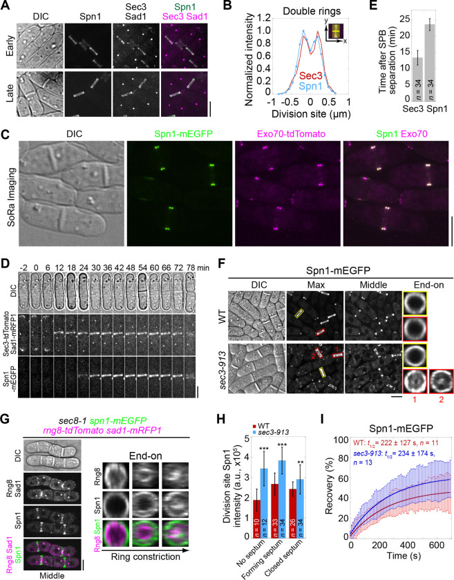
Figure 1. Septins and the exocyst colocalize at the division site and septins partially depend on the exocyst for localization. (A) Co-localization of Spn1-mEGFP and Sec3-tdTomato at the division site in cells without (early) and with (late) septa. Sad1-mRFP1 marks the spindle pole body (SPB). (B) Line scans showing Spn1 and Sec3 intensities at the division site along the cell long axis in septated cells as in (A). (C) SoRa (Super resolution by Optical Re-Assignment) confocal microscopy of cells expressing both Spn1-mEGFP and Exo70-tdTomato showing their perfect colocalization in the middle focal plane. (D) Time course and (E) quantification (in minutes) of Sec3 and Spn1 localizations and appearance timing at the division site. SPB separation is defined as time 0. (F) Localization of Spn1 (Max intensity projection, Middle focal plane, and End-on view of the division site) in WT and sec3–913 cells grown at 36°C for 4 h. Yellow boxes, cells without septa; Red boxes, cells with septa. (G) Localization of Spn1 and the contractile-ring marker Rng8 in sec8–1 cells grown at 36°C for 4 h. (H) Spn1 intensities at the division site in WT and sec3–913 cells grown at 36°C for 4 h. Cells were grouped into no septum, forming septum, and closed septum stages. **, P < 0.01; ***, P < 0.001. (I) FRAP analyses (photobleached at time 0) of Spn1 at the division site in WT and sec3–913 cells grown at 36°C for 4 h. Mean ± SD. Bars, 5 μm.
Figure 1. Septins and the exocyst colocalize at the division site and septins partially depend on the exocyst for localization. (A) Co-localization of Spn1-mEGFP and Sec3-tdTomato at the division site in cells without (early) and with (late) septa. Sad1-mRFP1 marks the spindle pole body (SPB). (B) Line scans showing Spn1 and Sec3 intensities at the division site along the cell long axis in septated cells as in (A). (C) SoRa (Super resolution by Optical Re-Assignment) confocal microscopy of cells expressing both Spn1-mEGFP and Exo70-tdTomato showing their perfect colocalization in the middle focal plane. (D) Time course and (E) quantification (in minutes) of Sec3 and Spn1 localizations and appearance timing at the division site. SPB separation is defined as time 0. (F) Localization of Spn1 (Max intensity projection, Middle focal plane, and End-on view of the division site) in WT and sec3–913 cells grown at 36°C for 4 h. Yellow boxes, cells without septa; Red boxes, cells with septa. (G) Localization of Spn1 and the contractile-ring marker Rng8 in sec8–1 cells grown at 36°C for 4 h. (H) Spn1 intensities at the division site in WT and sec3–913 cells grown at 36°C for 4 h. Cells were grouped into no septum, forming septum, and closed septum stages. **, P < 0.01; ***, P < 0.001. (I) FRAP analyses (photobleached at time 0) of Spn1 at the division site in WT and sec3–913 cells grown at 36°C for 4 h. Mean ± SD. Bars, 5 μm.CC BY · PubMed Central
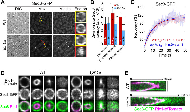
Figure 2. Septin rings recruit or anchor the exocyst complex to the rim of the division plane during late stage of cytokinesis. (A) Localization of Sec3 at the division site in WT and spn1Δ cells. Yellow boxes, cells without a septum; Red boxes, cells with a closed septum. (B) Sec3 intensity at the division site in WT and spn1Δ cells. ***, P < 0.001. (C) FRAP analyses of Sec3 at the division site in WT and spn1Δ cells. Mean ± SEM. (D and E) End-on views (D) and kymographs (E) of Sec8 and the contractile ring marker Rlc1 at the division site in WT and spn1Δ cells. Bars, 5 μm.
Figure 2. Septin rings recruit or anchor the exocyst complex to the rim of the division plane during late stage of cytokinesis. (A) Localization of Sec3 at the division site in WT and spn1Δ cells. Yellow boxes, cells without a septum; Red boxes, cells with a closed septum. (B) Sec3 intensity at the division site in WT and spn1Δ cells. ***, P < 0.001. (C) FRAP analyses of Sec3 at the division site in WT and spn1Δ cells. Mean ± SEM. (D and E) End-on views (D) and kymographs (E) of Sec8 and the contractile ring marker Rlc1 at the division site in WT and spn1Δ cells. Bars, 5 μm.CC BY · PubMed Central
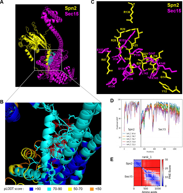
Figure 3. The 3D structural model of predicted interactions between Spn2 and Sec15 generated by AlphaFold. (A-C) AlphaFold2_advanced predicted interaction between Spn2 and Sec15 in rank 1 model with pLDDT score of 81.6. The pTM value = 0.51. Spn2 is colored in yellow and Sec15 in magenta in (A) and (C). (B) Inset of enlarged view of the predicted interactions, contacts between interface residues with distance <4 Å are colored in red (those in cyan in A). Residues are colored corresponding to their pLDDT scores as indicated in the legends below. (C) The key interacting residues and their side chains are shown. (D) Residue position scores of five predicted models for Spn2 and Sec15 interactions ranked according to pLDDT scores. (E) PAE (Predicted Alignment Error) plot for the top ranked model shown in (A-D), where colors represent confidence in the relative positioning of residues across the two proteins. Lower values (blue) represent high confidence while higher values (red) show low confidence in domain-domain interactions.
Figure 3. The 3D structural model of predicted interactions between Spn2 and Sec15 generated by AlphaFold. (A-C) AlphaFold2_advanced predicted interaction between Spn2 and Sec15 in rank 1 model with pLDDT score of 81.6. The pTM value = 0.51. Spn2 is colored in yellow and Sec15 in magenta in (A) and (C). (B) Inset of enlarged view of the predicted interactions, contacts between interface residues with distance <4 Å are colored in red (those in cyan in A). Residues are colored corresponding to their pLDDT scores as indicated in the legends below. (C) The key interacting residues and their side chains are shown. (D) Residue position scores of five predicted models for Spn2 and Sec15 interactions ranked according to pLDDT scores. (E) PAE (Predicted Alignment Error) plot for the top ranked model shown in (A-D), where colors represent confidence in the relative positioning of residues across the two proteins. Lower values (blue) represent high confidence while higher values (red) show low confidence in domain-domain interactions.CC BY · PubMed Central
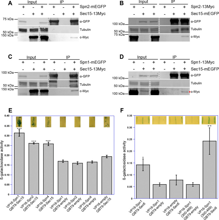
Figure 4. Septins and the exocyst interact physically. Reciprocal co-immunoprecipitation of Sec15 with Spn2 (A and B) and Spn1 (C and D). Septin or exocyst subunits tagged with mEGFP or 13Myc were immunoprecipitated using antibodies against GFP from cell lysates, separated on SDS-PAGE, and incubated with appropriate antibodies. Tubulin was used as a loading control. Asterisk (*) in (D) marks Spn1–13Myc. The vertical dashed lines mark the positions of protein ladders that were excised out. (E and F) Septins and the exocyst subunits may interact directly revealed by the yeast two-hybrid assays. X-gal overlay results (insets on the top of the columns) and quantification of β-galactosidase activities using ONPG showing interactions between (E) Sec15 with Spn1, Spn2, and Spn4; and (F) Sec6 with Spn1 and its coil-coil motif Spn1(300–469). Data is shown in Mean ± SD, n = 3 (in E) or 4 (in F). ***p ≤ 0.0001, **p ≤ 0.001, *p ≤ 0.01 compared with their respective controls in one-way ANOVA with Tukey’s post hoc test.
Figure 4. Septins and the exocyst interact physically. Reciprocal co-immunoprecipitation of Sec15 with Spn2 (A and B) and Spn1 (C and D). Septin or exocyst subunits tagged with mEGFP or 13Myc were immunoprecipitated using antibodies against GFP from cell lysates, separated on SDS-PAGE, and incubated with appropriate antibodies. Tubulin was used as a loading control. Asterisk (*) in (D) marks Spn1–13Myc. The vertical dashed lines mark the positions of protein ladders that were excised out. (E and F) Septins and the exocyst subunits may interact directly revealed by the yeast two-hybrid assays. X-gal overlay results (insets on the top of the columns) and quantification of β-galactosidase activities using ONPG showing interactions between (E) Sec15 with Spn1, Spn2, and Spn4; and (F) Sec6 with Spn1 and its coil-coil motif Spn1(300–469). Data is shown in Mean ± SD, n = 3 (in E) or 4 (in F). ***p ≤ 0.0001, **p ≤ 0.001, *p ≤ 0.01 compared with their respective controls in one-way ANOVA with Tukey’s post hoc test.CC BY · PubMed Central
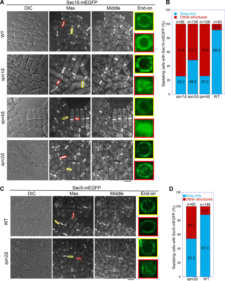
Figure 5. Localization patterns of both Sec15 and Sec5 at the division site depend on septins. Localization of (A, B) Sec15 and (C, D) Sec5 at the division site in WT and septin mutant cells. Yellow boxes, cells without a septum; Red boxes, cells with a closed septum in (A, C). (B, D) Quantification of cells with intact and mislocalized Sec15 (B) and Sec5 (D) signals in WT and septin mutant cells with obvious septa. Scale bars, 5 μm.
Figure 5. Localization patterns of both Sec15 and Sec5 at the division site depend on septins. Localization of (A, B) Sec15 and (C, D) Sec5 at the division site in WT and septin mutant cells. Yellow boxes, cells without a septum; Red boxes, cells with a closed septum in (A, C). (B, D) Quantification of cells with intact and mislocalized Sec15 (B) and Sec5 (D) signals in WT and septin mutant cells with obvious septa. Scale bars, 5 μm.CC BY · PubMed Central
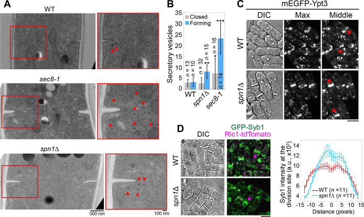
Figure 6. Septins are important for proper localization and distribution of secretory vesicles. (A and B) EM thin-section images (A) and quantifications of secretory vesicles (B) in WT, sec8–1, and spn1Δ cells with forming or closed septa. Cells were grown at 36°C for 4 h. Red boxes indicate the enlarged regions on the right. Arrowheads mark secretory vesicles. *, P < 0.05; **, P < 0.001; ***, P < 0.0001 compared to WT. n = numbers of thin sections. (C and D) Localizations of the Rab11 GTPase Ypt3 (C) and the v-SNARE Syb1 and Rlc1 (D) in WT and spn1Δ cells. Arrows mark examples of cells with closed septa. Syb1 intensities at the division site (D, right) from line scans at the middle focal plane of cells with closed septa (at the end of ring constriction indicated by an Rlc1 dot at the center of the division plane). Bars, 500 nm (A, left), 100 nm (A, right), and 5 μm (C and D).
Figure 6. Septins are important for proper localization and distribution of secretory vesicles. (A and B) EM thin-section images (A) and quantifications of secretory vesicles (B) in WT, sec8–1, and spn1Δ cells with forming or closed septa. Cells were grown at 36°C for 4 h. Red boxes indicate the enlarged regions on the right. Arrowheads mark secretory vesicles. *, P < 0.05; **, P < 0.001; ***, P < 0.0001 compared to WT. n = numbers of thin sections. (C and D) Localizations of the Rab11 GTPase Ypt3 (C) and the v-SNARE Syb1 and Rlc1 (D) in WT and spn1Δ cells. Arrows mark examples of cells with closed septa. Syb1 intensities at the division site (D, right) from line scans at the middle focal plane of cells with closed septa (at the end of ring constriction indicated by an Rlc1 dot at the center of the division plane). Bars, 500 nm (A, left), 100 nm (A, right), and 5 μm (C and D).CC BY · PubMed Central
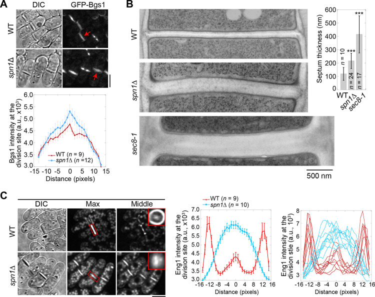
Figure 7. Septins are important for localization and distribution of secretory cargos Bgs1 and Eng1. (A) Localization (top) and intensity (bottom) of the glucan synthase Bgs1 in WT and spn1Δ cells. Arrows mark examples of cells with a closed septum. Bgs1 intensities from line scans across the division site at the middle focal plane were compared in cells with closed septa. (B) EM thin-section images (left) and septum thickness (right) of WT, spn1Δ, and sec8–1 cells with closed septa. Cells were grown at 36°C for 4 h. ***, P < 0.0001 compared to WT. (C) Localization (left) and intensity (middle and right) of Eng1-GFP in WT and spn1Δ cells. The end-on views of Eng1 at the division site in cells with closed septa are shown as insets. Eng1 intensities (Middle, mean intensities; and Right, individual cells) are from line scans at the middle focal plane. Bars, 5 μm (A and C) and 500 nm (B).
Figure 7. Septins are important for localization and distribution of secretory cargos Bgs1 and Eng1. (A) Localization (top) and intensity (bottom) of the glucan synthase Bgs1 in WT and spn1Δ cells. Arrows mark examples of cells with a closed septum. Bgs1 intensities from line scans across the division site at the middle focal plane were compared in cells with closed septa. (B) EM thin-section images (left) and septum thickness (right) of WT, spn1Δ, and sec8–1 cells with closed septa. Cells were grown at 36°C for 4 h. ***, P < 0.0001 compared to WT. (C) Localization (left) and intensity (middle and right) of Eng1-GFP in WT and spn1Δ cells. The end-on views of Eng1 at the division site in cells with closed septa are shown as insets. Eng1 intensities (Middle, mean intensities; and Right, individual cells) are from line scans at the middle focal plane. Bars, 5 μm (A and C) and 500 nm (B).CC BY · PubMed Central

Verification

Claims the paper makes

The claims and statistics of this paper haven’t been checked yet.

Run the check — free, no account needed. It reads the open-access text and checks the sources it cites.

A few free checks a day · papers already verified open instantly · results are public

Self-check

Percentages, sums and reported differences re-computed from the paper’s own numbers.

The self-check needs the paper’s full text.

Citations

Cites

Show all 156 references

Metadata

Authors
Singh, D., Liu, Y., Zhu, Y.-H., Zhang, S., Naegele, S. M., & Wu, J.-Q.
Affiliations
  • The Ohio State University (US)
  • Department of Molecular Genetics, The Ohio State University
Corresponding author
Wu, Jian-Qiu
Venue
eLife
Published
2025-10-31
Volume
13
ISSN
2050-084X (ISSN-L)
OpenAlex
W4400590789
Cited by
3
References
156
Citations by year
2026: 1 · 2025: 2
FWCI
1.50
Type
journal article
Language
en
Open access
Locations
  • eLife (publishedVersion, cc-by)
  • bioRxiv (Cold Spring Harbor Laboratory) (acceptedVersion, cc-by)
  • PubMed (publishedVersion)
  • PubMed Central (submittedVersion, cc-by)
Funders
  • National Institutes of Health (GM118746)
  • The Ohio State University (Pelotonia Graduate Fellowship, Pelotonia Undergraduate Fellowship)
Topics
  • Life Sciences › Biochemistry, Genetics and Molecular Biology › Cell Biology › Cellular transport and secretion
  • Life Sciences › Biochemistry, Genetics and Molecular Biology › Cell Biology › Microtubule and mitosis dynamics
  • Life Sciences › Biochemistry, Genetics and Molecular Biology › Molecular Biology › Fungal and yeast genetics research
Keywords
septins, exocyst complex, exocytosis, cytokinesis, Schizosaccharomyces pombe, protein-protein interaction, vesicle tethering, division plane, septum formation, cell isolation

About these checks

Registry, retraction, Cites and statistics results are computed, not judged, with one exception: a printed reference that no index lists is read once by a model before it counts as a citation error. Claims are extracted by a model and kept only when their quote appears verbatim in the text; each cited source is then checked, with a model’s help, for whether it says what the claim relies on. Those verdicts can be wrong — read the claim’s quote and the cited source before relying on one.

Statistics and arithmetic are recomputed, not judged.

Confidence bands are shown only for fields calibrated on our annotated test set. None is calibrated yet, so no band is shown.

What counts as a claim ›How we measure confidence ›How we check this ›

Are you an author, or do you see a problem with this page? Request a review

How to cite

Cite CoChat for the checks on this page, and the paper for its own findings.

This page
CoChat. (2026). Citation check: Septins function in exocytosis via physical interactions with the exocyst complex in fission yeast cytokinesis. https://app.cochat.ai/papers/10.7554/elife.101113.3
The paper
Singh, D., Liu, Y., Zhu, Y.-H., Zhang, S., Naegele, S. M., & Wu, J.-Q. (2025). Septins function in exocytosis via physical interactions with the exocyst complex in fission yeast cytokinesis. eLife. https://doi.org/10.7554/elife.101113.3

Data sources

Bibliographic data: Semantic Scholar, OpenAlex, Crossref.

Contains information from the Semantic Scholar Academic Graph Datasets, made available under the ODC Attribution License (ODC-BY 1.0).

Semantic Scholar’s data is described in: Kinney, R., et al. (2023). The Semantic Scholar Open Data Platform. arXiv:2301.10140.