

The DOI matches its Crossref record; 4 of the 4 references it cites were found in a registry or an index.
Structure-guided secretome analysis of gall-forming microbes offers insights into effector diversity and evolution
Open accessPublished version of a preprint
Abstract
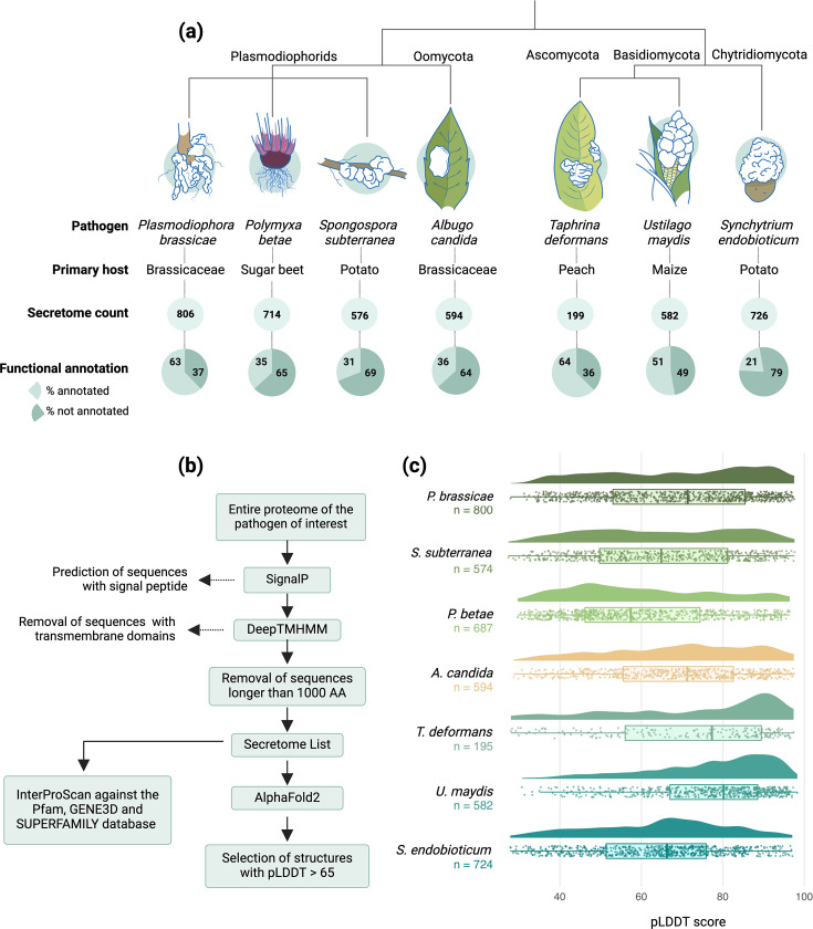
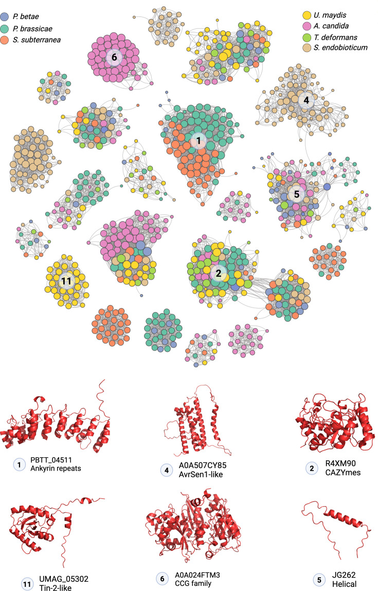
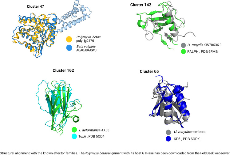
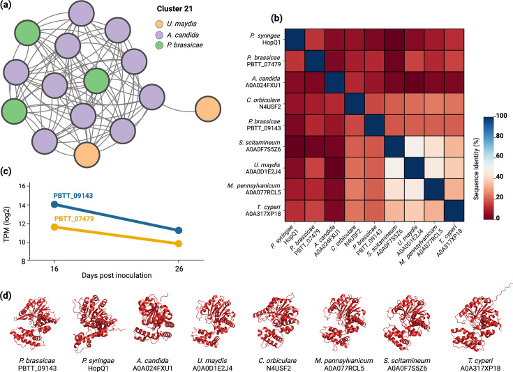
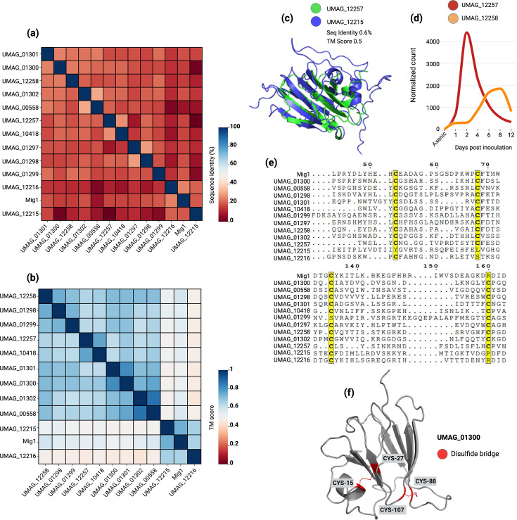
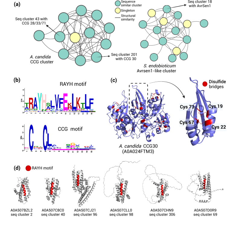
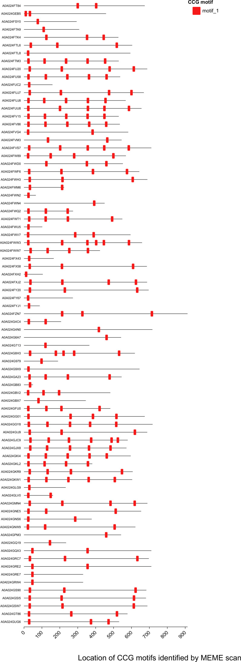
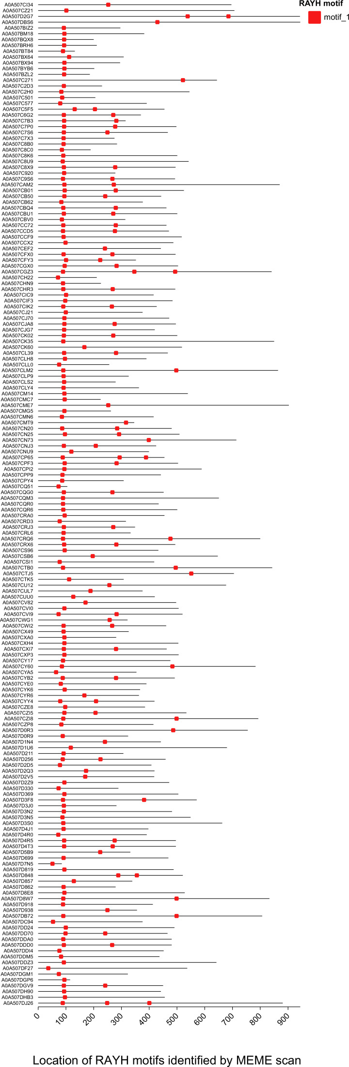
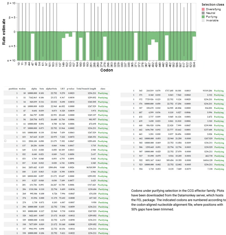
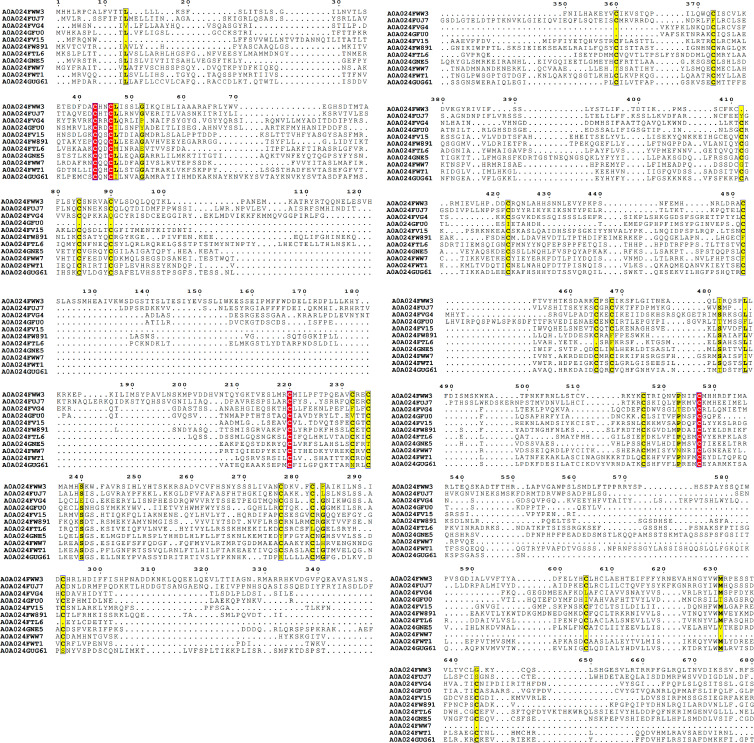
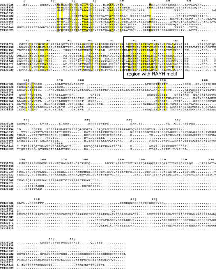
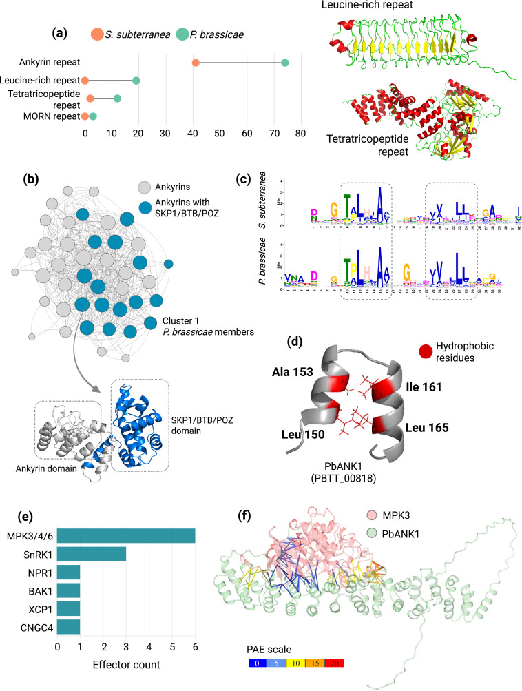
Phytopathogens secrete effector molecules to manipulate host immunity and metabolism. Recent advances in structural genomics have identified fungal effector families whose members adopt similar folds despite sequence divergence, highlighting their importance in virulence and immune evasion. To extend the scope of comparative structure-guided analysis to more evolutionarily distant phytopathogens with similar lifestyles, we used AlphaFold2 to predict the 3D structures of the secretome from selected plasmodiophorid, oomycete, and fungal gall-forming pathogens. Clustering protein folds based on structural homology revealed species-specific expansions and a low abundance of known orphan effector families. We identified novel sequence-unrelated but structurally similar (SUSS) effector clusters, rich in conserved motifs such as 'CCG' and 'RAYH'. We demonstrate that these motifs likely play a central role in maintaining the overall fold. We also identified a SUSS cluster adopting a nucleoside hydrolase-like fold conserved among various gall-forming microbes. Notably, ankyrin proteins (ANK) were significantly expanded in gall-forming plasmodiophorids, with most being highly expressed during clubroot disease, suggesting a role in pathogenicity. Subsequently, we screened ANK proteins against Arabidopsis immunity hubs using AlphaFold-Multimer and verified one of the positive results by Y2H and BiFC assays to show that the ankyrin effector PbANK1 targets host MPK3 and a zinc-binding dehydrogenase. These findings suggest a potential new mechanism in which ANK effectors target multiple host proteins involved in stress sensing, opening a novel avenue to study the role of ANK in host–pathogen interactions. Altogether, this study advances our understanding of secretome landscapes in gall-forming microbes and provides a valuable resource for broadening structural phylogenomic studies across diverse phytopathogens.
Overview
The claims and statistics of this paper haven’t been checked yet.
Run the check — free, no account needed. It reads the open-access text and checks the sources it cites.
Integrity signals
Registry
Cites
Statistics
Figures
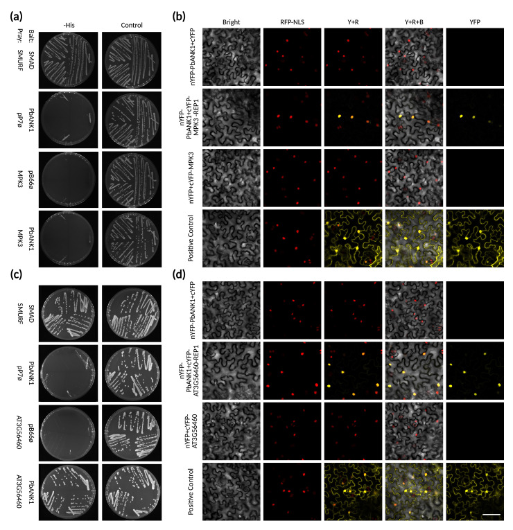
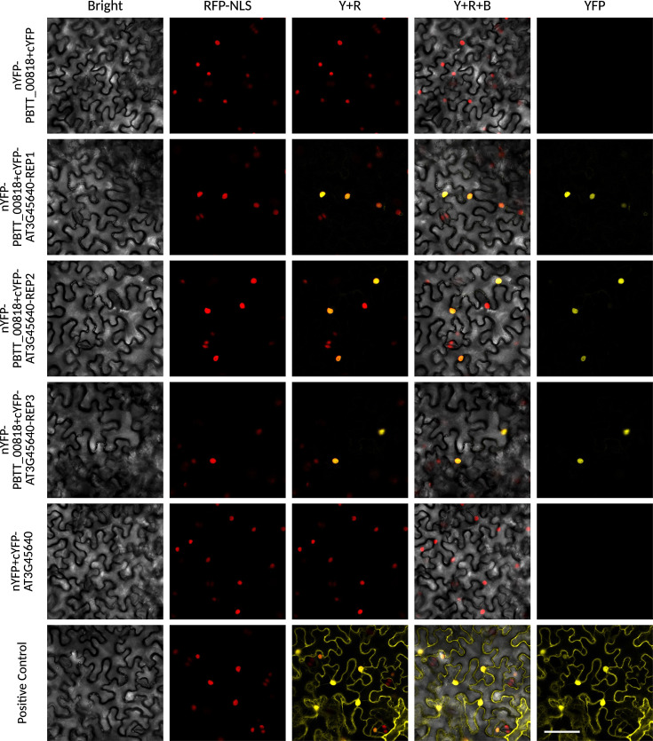
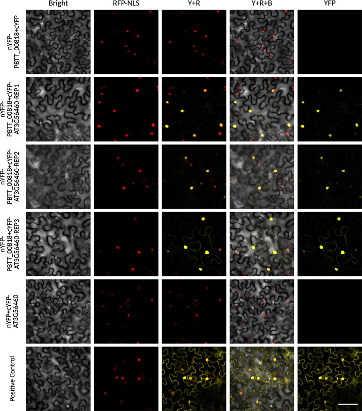
Verification
Claims the paper makes
The claims and statistics of this paper haven’t been checked yet.
Run the check — free, no account needed. It reads the open-access text and checks the sources it cites.
Self-check
Percentages, sums and reported differences re-computed from the paper’s own numbers.
The self-check needs the paper’s full text.
Citations
Cites
Highly accurate protein structure prediction with AlphaFold
Jumper, J., Evans, R., Pritzel, A., Green, T., Figurnov, M., Ronneberger, O., Tunyasuvunakool, K., Bates, R., Žídek, A., Potapenko, A., et al. (2021) · Nature · doi:10.1038/s41586-021-03819-2 · Cited by 43,764
Fast and accurate protein structure search with Foldseek
van Kempen, M., Kim, S. S., Tumescheit, C., Mirdita, M., Lee, J., Gilchrist, C. L. M., Söding, J., & Steinegger, M. (2022) · doi:10.1101/2022.02.07.479398 · Cited by 2,598
Fast and accurate protein structure search with Foldseek
van Kempen, M., Kim, S. S., Tumescheit, C., Mirdita, M., Lee, J., Gilchrist, C. L. M., Söding, J., & Steinegger, M. (2024) · Nature Biotechnology · doi:10.1038/s41587-023-01773-0 · Cited by 2,596
Clustering predicted structures at the scale of the known protein universe
Barrio-Hernandez, I., Yeo, J., Jänes, J., Mirdita, M., Gilchrist, C. L. M., Wein, T., Varadi, M., Velankar, S., Beltrao, P., & Steinegger, M. (2023) · Nature · doi:10.1038/s41586-023-06510-w · Cited by 362
Research works printed but not linked: 87
- Bailey, 1998, Combining evidence using p-values: application to sequence homology searches, Bioinformatics, 14, 48, 10.1093/bioinformatics/14.1.48 · DOI
- Basse, 2000, Characterization of a Ustilago maydis gene specifically induced during the biotrophic phase: evidence for negative as well as positive regulation, Molecular and Cellular Biology, 20, 329, 10.1128/MCB.20.1.329-339.2000 · DOI
- Bradley, 2022, Secreted glycoside hydrolase proteins as effectors and invasion patterns of plant-associated fungi and oomycetes, Frontiers in Plant Science, 13, 10.3389/fpls.2022.853106 · DOI
- Camacho, 2009, BLAST+: architecture and applications, BMC Bioinformatics, 10, 10.1186/1471-2105-10-421 · DOI · Paper page
- Camoni, 2018, 14-3-3 proteins in plant hormone signaling: doing several things at once, Frontiers in Plant Science, 9, 10.3389/fpls.2018.00297 · DOI
- Cao, 2023, Structural polymorphisms within a common powdery mildew effector scaffold as a driver of coevolution with cereal immune receptors, PNAS, 120, 10.1073/pnas.2307604120 · DOI
- Chen, 2022, Direct recognition of pathogen effectors by plant NLR immune receptors and downstream signalling, Essays in Biochemistry, 66, 471, 10.1042/EBC20210072 · DOI
- Ciaghi, 2018, Draft genome resource for the potato powdery scab pathogen Spongospora subterranea, Molecular Plant-Microbe Interactions, 31, 1227, 10.1094/MPMI-06-18-0163-A · DOI
- Cissé, 2013, Genome sequencing of the plant pathogen Taphrina deformans, the causal agent of peach leaf curl, mBio, 4, 10.1128/mBio.00055-13 · DOI
- Colland, 2004, Functional proteomics mapping of a human signaling pathway, Genome Research, 14, 1324, 10.1101/gr.2334104 · DOI
- Combier, 2022, Candidate effector proteins from the oomycetes Plasmopara viticola and Phytophthora parasitica share similar predicted structures and induce cell death in Nicotiana species, PLOS ONE, 17, 10.1371/journal.pone.0278778 · DOI
- Decroës, 2019, First Draft genome sequence of a polymyxa genus member, polymyxa betae, the protist vector of rhizomania, Microbiol Resour Announc, 8, 10.1128/MRA.01509-18 · DOI
- Decroës, 2022, Rhizomania: Hide and seek of polymyxa betae and the beet necrotic yellow vein virus with Beta vulgaris, Molecular Plant-Microbe Interactions, 35, 989, 10.1094/MPMI-03-22-0063-R · DOI
- Decroës, 2022, Metagenomics approach for Polymyxa betae genome assembly enables comparative analysis towards deciphering the intracellular parasitic lifestyle of the plasmodiophorids, Genomics, 114, 9, 10.1016/j.ygeno.2021.11.018 · DOI
- de Guillen, 2015, Structure analysis uncovers a highly diverse but structurally conserved effector family in phytopathogenic fungi, PLOS Pathogens, 11, 10.1371/journal.ppat.1005228 · DOI
- Derbyshire, 2023, Surface frustration re-patterning underlies the structural landscape and evolvability of fungal orphan candidate effectors, Nature Communications, 14, 10.1038/s41467-023-40949-9 · DOI
- Dong, 2018, mTM-align: an algorithm for fast and accurate multiple protein structure alignment, Bioinformatics, 34, 1719, 10.1093/bioinformatics/btx828 · DOI
- Eichhorn, 1997, Characterization of alpha-ketoglutarate-dependent taurine dioxygenase from Escherichia coli, The Journal of Biological Chemistry, 272, 23031, 10.1074/jbc.272.37.23031 · DOI · Paper page
- Enright, 2002, An efficient algorithm for large-scale detection of protein families, Nucleic Acids Research, 30, 1575, 10.1093/nar/30.7.1575 · DOI
- Erdős, 2021, IUPred3: prediction of protein disorder enhanced with unambiguous experimental annotation and visualization of evolutionary conservation, Nucleic Acids Research, 49, W297, 10.1093/nar/gkab408 · DOI
- Evans, 2022, Protein Complex Prediction with AlphaFold-Multimer, 10.1101/2021.10.04.463034 · DOI · Paper page
- Fromont-Racine, 1997, Toward a functional analysis of the yeast genome through exhaustive two-hybrid screens, Nature Genetics, 16, 277, 10.1038/ng0797-277 · DOI
- Furzer, 2022, An improved assembly of the Albugo candida Ac2V genome reveals the expansion of the “CCG” class of effectors, Molecular Plant-Microbe Interactions, 35, 39, 10.1094/MPMI-04-21-0075-R · DOI
- Gao, 2019, Deacetylation of chitin oligomers increases virulence in soil-borne fungal pathogens, Nature Plants, 5, 1167, 10.1038/s41477-019-0527-4 · DOI
- Geyer, 2003, BTB/POZ domain proteins are putative substrate adaptors for cullin 3 ubiquitin ligases, Molecular Cell, 12, 783, 10.1016/s1097-2765(03)00341-1 · DOI
- González-García, 2021, Looking for a cultured surrogate for effectome studies of the clubroot pathogen, Frontiers in Microbiology, 12, 10.3389/fmicb.2021.650307 · DOI
- Gupta, 2007, Quantifying similarity between motifs, Genome Biology, 8, 10.1186/gb-2007-8-2-r24 · DOI · Paper page
- Hallgren, 2022, DeepTMHMM predicts alpha and beta transmembrane proteins using deep neural networks, 10.1101/2022.04.08.487609 · DOI
- Harmon, 2018, Bacterial Gall of Loropetalum chinense caused by Pseudomonas amygdali pv. loropetali pv. nov, Plant Disease, 102, 799, 10.1094/PDIS-04-17-0505-RE · DOI
- Hofer, 2023, Targeting the nucleotide metabolism of Trypanosoma brucei and other trypanosomatids, FEMS Microbiology Reviews, 47, 1, 10.1093/femsre/fuad020 · DOI
- Homma, 2023, AlphaFold-Multimer predicts cross-kingdom interactions at the plant-pathogen interface, Nature Communications, 14, 10.1038/s41467-023-41721-9 · DOI · Paper page
- Javed, 2023, The clubroot pathogen Plasmodiophora brassicae: A profile update, Molecular Plant Pathology, 24, 89, 10.1111/mpp.13283 · DOI
- Javed, 2024, Telomere-to-telomere genome assembly of the clubroot pathogen plasmodiophora brassicae, Genome Biology and Evolution, 16, 10.1093/gbe/evae122 · DOI
- Jones, 2014, InterProScan 5: genome-scale protein function classification, Bioinformatics, 30, 1236, 10.1093/bioinformatics/btu031 · DOI
- Kämper, 2006, Insights from the genome of the biotrophic fungal plant pathogen Ustilago maydis, Nature, 444, 97, 10.1038/nature05248 · DOI
- Kim, 2019, Graph-based genome alignment and genotyping with HISAT2 and HISAT-genotype, Nature Biotechnology, 37, 907, 10.1038/s41587-019-0201-4 · DOI · Paper page
- Kosakovsky Pond, 2005, Not so different after all: a comparison of methods for detecting amino acid sites under selection, Molecular Biology and Evolution, 22, 1208, 10.1093/molbev/msi105 · DOI
- Lanver, 2018, The biotrophic development of Ustilago maydis Studied by RNA-Seq Analysis, The Plant Cell, 30, 300, 10.1105/tpc.17.00764 · DOI
- Lassmann, 2020, Kalign 3: multiple sequence alignment of large datasets, Bioinformatics, 36, 1928, 10.1093/bioinformatics/btz795 · DOI
- Lazar, 2022, A new family of structurally conserved fungal effectors displays epistatic interactions with plant resistance proteins, PLOS Pathogens, 18, 10.1371/journal.ppat.1010664 · DOI
- Li, 2006, Ankyrin repeat: a unique motif mediating protein-protein interactions, Biochemistry, 45, 15168, 10.1021/bi062188q · DOI
- Li, 2013, The Pseudomonas syringae effector HopQ1 promotes bacterial virulence and interacts with tomato 14-3-3 proteins in a phosphorylation-dependent manner, Plant Physiology, 161, 2062, 10.1104/pp.112.211748 · DOI
- Li, 2023, Pathogen protein modularity enables elaborate mimicry of a host phosphatase, Cell, 186, 3196, 10.1016/j.cell.2023.05.049 · DOI · Paper page
- Liu, 2019, Arms race: diverse effector proteins with conserved motifs, Plant Signaling & Behavior, 14, 10.1080/15592324.2018.1557008 · DOI
- Lovelace, 2023, Effector identification in plant pathogens, Phytopathology, 113, 637, 10.1094/PHYTO-09-22-0337-KD · DOI
- Martel, 2021, The ETS-ETI cycle: evolutionary processes and metapopulation dynamics driving the diversification of pathogen effectors and host immune factors, Current Opinion in Plant Biology, 62, 10.1016/j.pbi.2021.102011 · DOI
- McMullan, 2015, Evidence for suppression of immunity as a driver for genomic introgressions and host range expansion in races of Albugo candida, a generalist parasite, eLife, 4, 10.7554/eLife.04550 · DOI
- Meng, 2023, UCSF ChimeraX: Tools for structure building and analysis, Protein Science, 32, 10.1002/pro.4792 · DOI
- Mesarich, 2015, Repeat-containing protein effectors of plant-associated organisms, Frontiers in Plant Science, 6, 10.3389/fpls.2015.00872 · DOI
- Mine, 2017, Pathogen exploitation of an abscisic acid- and jasmonate-inducible MAPK phosphatase and its interception by Arabidopsis immunity, PNAS, 114, 7456, 10.1073/pnas.1702613114 · DOI
- Mukhopadhyay, 2024, Decoding the arsenal: protist effectors and their impact on photosynthetic hosts, Molecular Plant-Microbe Interactions, 37, 498, 10.1094/MPMI-11-23-0196-CR · DOI
- Nomura, 2023, Bacterial pathogens deliver water- and solute-permeable channels to plant cells, Nature, 621, 586, 10.1038/s41586-023-06531-5 · DOI
- Ochoa, 2023, Natural variation in Arabidopsis responses to Plasmodiophora brassicae reveals an essential role for Resistance to Plasmodiophora brasssicae 1 (RPB1), The Plant Journal, 116, 1421, 10.1111/tpj.16438 · DOI
- Pan, 2008, Ankyrin repeat proteins comprise a diverse family of bacterial type IV effectors, Science, 320, 1651, 10.1126/science.1158160 · DOI
- Patro, 2017, Salmon provides fast and bias-aware quantification of transcript expression, Nature Methods, 14, 417, 10.1038/nmeth.4197 · DOI · Paper page
- Pérez-López, 2018, Identification of Plasmodiophora brassicae effectors - A challenging goal, Virulence, 9, 1344, 10.1080/21505594.2018.1504560 · DOI
- Perez-Lopez E. 2025. AlphaFold_effector_paper. Software Heritage. swh:1:rev:ab6a6377b9a29ac3d6a31516e9e55e3d3ecf2d7e. https://archive.softwareheritage.org/swh:1:dir:4518d9f1e26cf8523701c5b130bdfffbf5556ab1;origin=https://github.com/Edelab/AlphaFold_effector_paper;visit=swh:1:snp:2d0370d8fd67b81c29c193bd63e1211bfa044aa8;anchor=swh:1:rev:ab6a6377b9a29ac3d6a31516e9e55e3d3ecf2d7e.
- Petit, 1983, Chemical analysis of the wall of the yeast form of Taphrina deformans, Archives of Microbiology, 135, 141, 10.1007/BF00408024 · DOI
- Pond, 2005, HyPhy: hypothesis testing using phylogenies, Bioinformatics, 21, 676, 10.1093/bioinformatics/bti079 · DOI · Paper page
- Ramos, 2012, Pseudomonas savastanoi pv. savastanoi: some like it knot, Molecular Plant Pathology, 13, 998, 10.1111/j.1364-3703.2012.00816.x · DOI
- Redkar, 2023, The Arabidopsis WRR4A and WRR4B paralogous NLR proteins both confer recognition of multiple Albugo candida effectors, The New Phytologist, 237, 532, 10.1111/nph.18378 · DOI
- Reumann, 2007, Proteome analysis of Arabidopsis leaf peroxisomes reveals novel targeting peptides, metabolic pathways, and defense mechanisms, The Plant Cell, 19, 3170, 10.1105/tpc.107.050989 · DOI
- Rice, 2000, EMBOSS: the European molecular biology open software suite, Trends in Genetics, 16, 276, 10.1016/s0168-9525(00)02024-2 · DOI · Paper page
- Robert, 2014, Deciphering key features in protein structures with the new ENDscript server, Nucleic Acids Research, 42, W320, 10.1093/nar/gku316 · DOI · Paper page
- Schrödinger LLC. 2015. The pymol molecular graphics system. Pymol. 1.8. https://www.pymol.org/.
- Seong, 2021, Computational structural genomics unravels common folds and novel families in the secretome of fungal phytopathogen Magnaporthe oryzae, Molecular Plant-Microbe Interactions, 34, 1267, 10.1094/MPMI-03-21-0071-R · DOI
- Seong, 2023, Prediction of effector protein structures from fungal phytopathogens enables evolutionary analyses, Nature Microbiology, 8, 174, 10.1038/s41564-022-01287-6 · DOI
- Sievers, 2018, Clustal Omega for making accurate alignments of many protein sequences, Protein Science, 27, 135, 10.1002/pro.3290 · DOI · Paper page
- Steenwyk, 2020, ClipKIT: A multiple sequence alignment trimming software for accurate phylogenomic inference, PLOS Biology, 18, 10.1371/journal.pbio.3001007 · DOI
- Tanaka, 2019, Neofunctionalization of the secreted Tin2 effector in the fungal pathogen Ustilago maydis, Nature Microbiology, 4, 251, 10.1038/s41564-018-0304-6 · DOI
- Teufel, 2022, SignalP 6.0 predicts all five types of signal peptides using protein language models, Nature Biotechnology, 40, 1023, 10.1038/s41587-021-01156-3 · DOI
- Teulet, 2023, A pathogen effector FOLD diversified in symbiotic fungi, The New Phytologist, 239, 1127, 10.1111/nph.18996 · DOI · Paper page
- Thynne, 2024, An array of Zymoseptoria tritici effectors suppress plant immune responses, Molecular Plant Pathology, 25, 10.1111/mpp.13500 · DOI
- Toruño, 2016, Plant-pathogen effectors: cellular probes interfering with plant defenses in spatial and temporal manners, Annual Review of Phytopathology, 54, 419, 10.1146/annurev-phyto-080615-100204 · DOI
- van de Vossenberg, 2019, The Synchytrium endobioticum AvrSen1 triggers a hypersensitive response in Sen1 potatoes while natural variants evade detection, Molecular Plant-Microbe Interactions, 32, 1536, 10.1094/MPMI-05-19-0138-R · DOI
- van de Vossenberg, 2019, Comparative genomics of chytrid fungi reveal insights into the obligate biotrophic and pathogenic lifestyle of Synchytrium endobioticum, Scientific Reports, 9, 10.1038/s41598-019-45128-9 · DOI
- Van Dongen, 2008, Graph clustering via a discrete uncoupling process, SIAM Journal on Matrix Analysis and Applications, 30, 121, 10.1137/040608635 · DOI
- Wickham, 2016, Ggplot2, 10.1007/978-3-319-24277-4 · DOI
- Win, 2012, Sequence divergent RXLR effectors share a structural fold conserved across plant pathogenic oomycete species, PLOS Pathogens, 8, 10.1371/journal.ppat.1002400 · DOI
- Xin, 2015, Pseudomonas syringae effector avirulence protein E localizes to the host plasma membrane and down-regulates the expression of the nonrace-specific disease resistance1/harpin-induced1-like13 gene required for antibacterial immunity in arabidopsis, Plant Physiology, 169, 793, 10.1104/pp.15.00547 · DOI
- Xu, 2016, Pathogen-responsive MPK3 and MPK6 reprogram the biosynthesis of indole glucosinolates and their derivatives in arabidopsis immunity, The Plant Cell, 28, 1144, 10.1105/tpc.15.00871 · DOI
- Yu, 2023, AlphaPulldown-a python package for protein-protein interaction screens using AlphaFold-Multimer, Bioinformatics, 39, 10.1093/bioinformatics/btac749 · DOI · Paper page
- Yu, 2024, The structural repertoire of Fusarium oxysporum f. sp. lycopersici effectors revealed by experimental and computational studies, eLife, 12, 10.7554/eLife.89280 · DOI
- Zhang, 2005, TM-align: a protein structure alignment algorithm based on the TM-score, Nucleic Acids Research, 33, 2302, 10.1093/nar/gki524 · DOI · Paper page
- Zhong, 2022, ParaFold: paralleling AlphaFold for large-scale predictions, 10.48550/arXiv.2111.06340 · DOI
- Schwelm A. 2018. Spongospora subterranea, whole genome shotgun sequencing project. NCBI Nucleotide. https://www.ncbi.nlm.nih.gov/nuccore/OUQQ00000000. OUQQ00000000.
- Kamper J, Kahmann R, Bolker M, Brefort T, Saville BJ, Banuett F, Kronstad JW, Gold SE, Muller O, Perlin MH, Wosten HA, de Vries R, Ruiz-Herrera J, Reynaga-Pena CG, Snetselaar K, McCann M, Perez-Martin J, Feldbrugge M, Basse CW, Steinberg G, Ibeas JI, Holloman W, Guzman P, Farman M, Stajich JE, Sentandreu R, Gonzalez-Prieto JM, Kennell JC, Molina L, Schirawski J, Mendoza-Mendoza A, Greilinger D, Munch K, Rossel N, Scherer M, Vranes M, Ladendorf O, Vincon V, Fuchs U, Sandrock B, Meng S, Cahill MJ, Boyce KJ, Klose J, Klosterman SJ, Deelstra HJ, Ortiz-Castellanos L, Li W, Sanchez-Alonso P, Schreier PH, Hauser-Hahn I, Vaupel M, Koopmann E, Friedrich G, Voss H, Schluter T, Margolis J, Platt D, Swimmer C, Gnirke A, Chen F, Vysotskaia V, Mannhaupt G, Guldener U, Munsterkotter M, Haase D, Oesterheld M, Mewes HW, Mauceli EW, DeCaprio D, Wade CM, Butler J, Young S, Jaffe DB, Calvo S, Nusbaum C, Galagan J, Birren BW, Ma LJ, Ho EC. 2006. Mycosarcoma maydis strain 521, whole genome shotgun sequencing project. NCBI Nucleotide. https://www.ncbi.nlm.nih.gov/nuccore/AACP00000000. AACP00000000.
Metadata
- Authors
- Mukhopadhyay, S., Javed, M. A., Wu, J., & Perez-Lopez, E.
- Affiliations
- Université Laval (CA)
- Départment de Phytologie, Faculté des sciences de l’agriculture et de l’alimentation, Université Laval
- Corresponding author
- Mukhopadhyay, Soham
- Venue
- eLife
- Publisher
- eLife Sciences Publications, Ltd
- Published
- 2025-10-07
- Volume
- 14
- ISSN
- 2050-084X (ISSN-L)
- PMC
- PMC12503489
- OpenAlex
- W4407840168
- Cited by
- 5
- References
- 4
- Citations by year
- 2026: 4 · 2025: 1
- Type
- posted content
- Language
- en
- Open access
- gold
- doi.org
- Licence
- Locations
- eLife (publishedVersion, cc-by)
- Europe PMC (PubMed Central) (publishedVersion, cc-by)
- PubMed (publishedVersion)
- PubMed Central (publishedVersion, cc-by)
- DOAJ (DOAJ: Directory of Open Access Journals) (submittedVersion, cc-by-sa)
- Funders
- Canola Council of Canada (2021.4)
- Natural Sciences and Engineering Research Council of Canada (RGPIN-2021-02518)
- Western Grain Research Foundation
- Manitoba Canola Growers Association
- Alberta Canola Producers Commission
- Fonds de recherche du Québec (Doctoral scholarships)
- Topics
- Life Sciences › Agricultural and Biological Sciences › Plant Science › Plant Disease Resistance and Genetics
- Life Sciences › Agricultural and Biological Sciences › Plant Science › Plant-Microbe Interactions and Immunity
- Life Sciences › Agricultural and Biological Sciences › Plant Science › Plant Pathogens and Resistance
- Keywords
- secretome, effector proteins, oomycete, plant pathogenic fungi, structural genomics, clubroot disease, MPK3, host-pathogen interaction, effector evolution
- MeSH terms
- Secretome, Fungal Proteins, Fungi, Oomycetes, Plant Diseases, Plant Tumors, Arabidopsis, Evolution, Molecular
About these checks
Registry, retraction, Cites and statistics results are computed, not judged, with one exception: a printed reference that no index lists is read once by a model before it counts as a citation error. Claims are extracted by a model and kept only when their quote appears verbatim in the text; each cited source is then checked, with a model’s help, for whether it says what the claim relies on. Those verdicts can be wrong — read the claim’s quote and the cited source before relying on one.
Statistics and arithmetic are recomputed, not judged.
Confidence bands are shown only for fields calibrated on our annotated test set. None is calibrated yet, so no band is shown.
What counts as a claim ›How we measure confidence ›How we check this ›
Are you an author, or do you see a problem with this page? Request a review
