

The DOI matches its Crossref record; 64 of the 64 references it cites were found in a registry or an index.
Biochemical adaptations of the retina and retinal pigment epithelium support a metabolic ecosystem in the vertebrate eye
Open accessPublished version of a preprint
Abstract
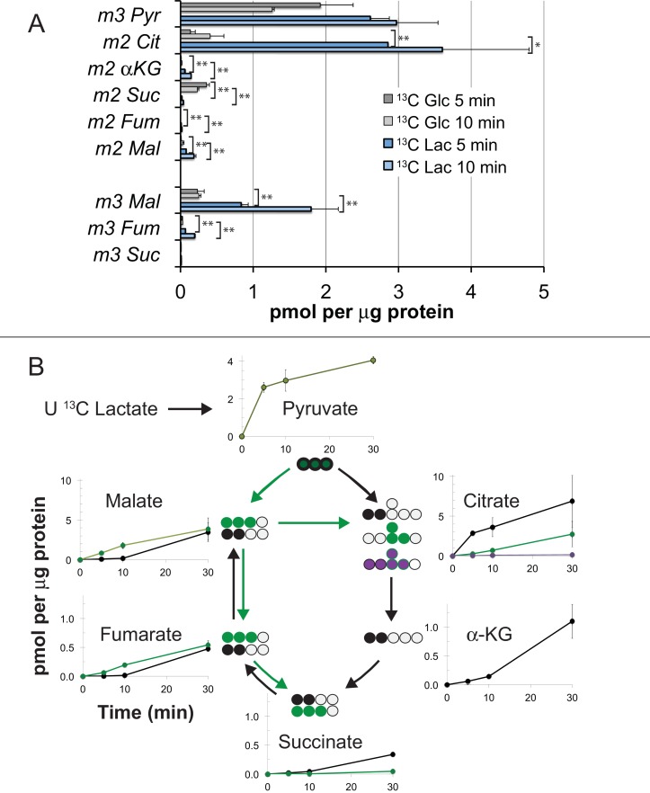
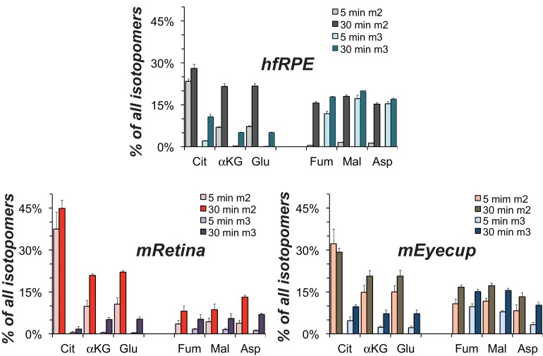
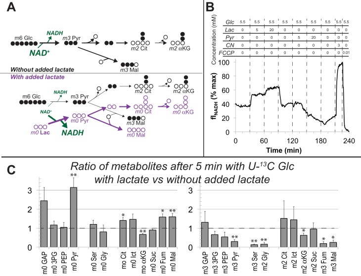
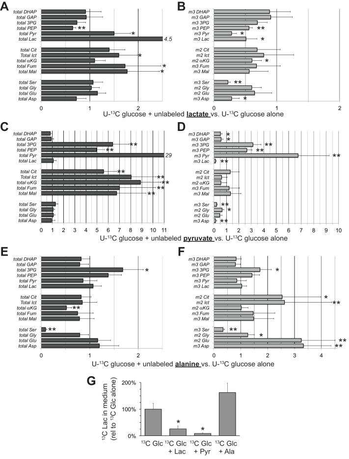
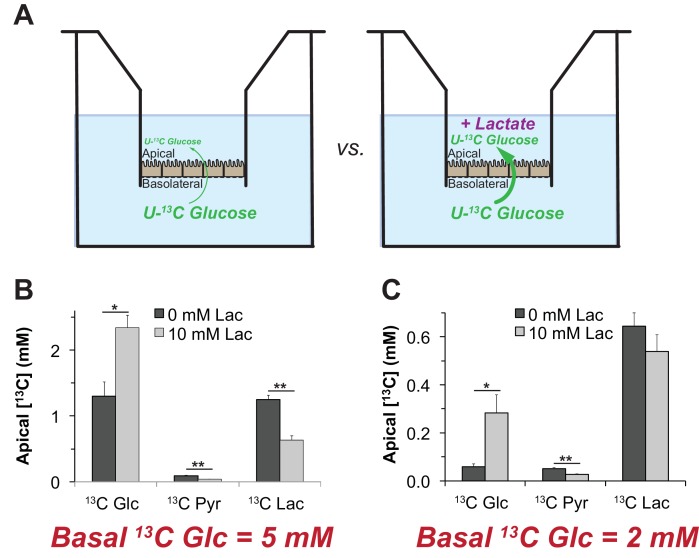
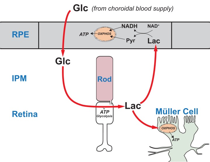
Here we report multiple lines of evidence for a comprehensive model of energy metabolism in the vertebrate eye. Metabolic flux, locations of key enzymes, and our finding that glucose enters mouse and zebrafish retinas mostly through photoreceptors support a conceptually new model for retinal metabolism. In this model, glucose from the choroidal blood passes through the retinal pigment epithelium to the retina where photoreceptors convert it to lactate. Photoreceptors then export the lactate as fuel for the retinal pigment epithelium and for neighboring Müller glial cells. We used human retinal epithelial cells to show that lactate can suppress consumption of glucose by the retinal pigment epithelium. Suppression of glucose consumption in the retinal pigment epithelium can increase the amount of glucose that reaches the retina. This framework for understanding metabolic relationships in the vertebrate retina provides new insights into the underlying causes of retinal disease and age-related vision loss.
Overview
The claims and statistics of this paper haven’t been checked yet.
Run the check — free, no account needed. It reads the open-access text and checks the sources it cites.
Integrity signals
Registry
Cites
Statistics
Figures
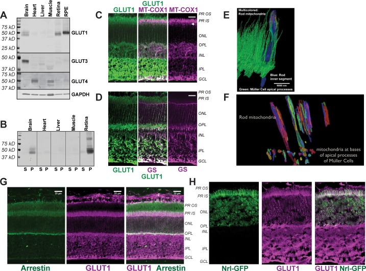
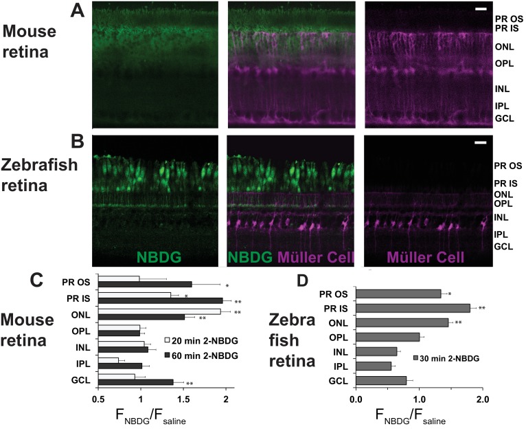
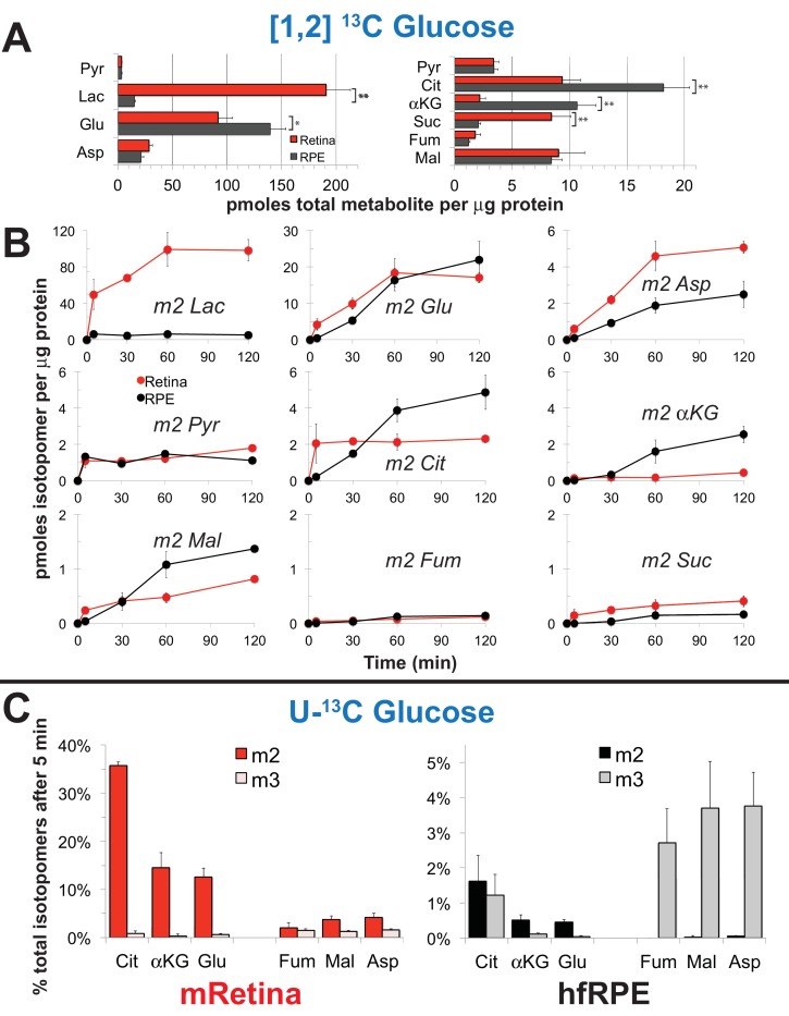
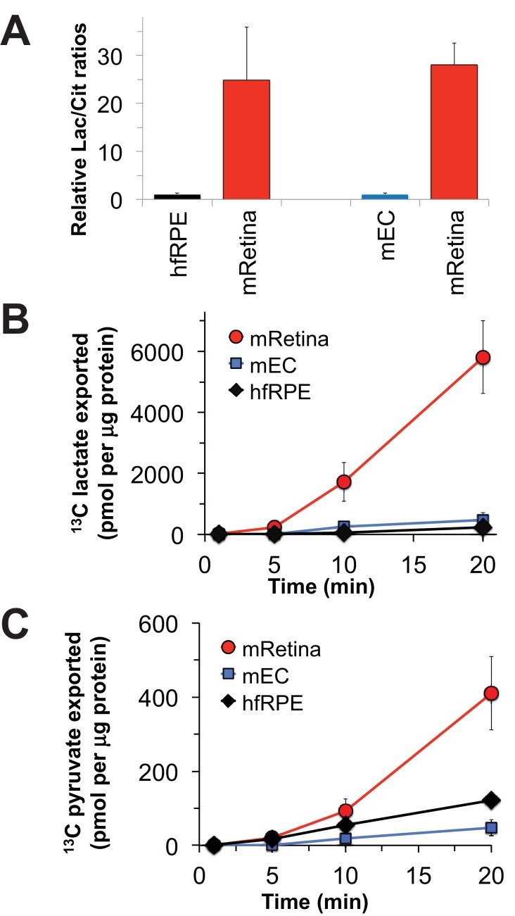
Verification
Claims the paper makes
The claims and statistics of this paper haven’t been checked yet.
Run the check — free, no account needed. It reads the open-access text and checks the sources it cites.
Claims other papers make about this one
Each is checked against this paper’s own text: does it say what the citing paper says it does?
In the citing paper
“In contrast, the RPE is oxidative and uses lactate produced by the photoreceptors and fatty acids from ingested OSs to fuel mitochondrial metabolism (Adijanto et al., 2014; Adijanto and Philp, 2014; Kanow et al., 2017; Reyes-Reveles et al., 2017).”
From The cell biology of the retinal pigment epithelium (2020) · cites this paper as Kanow et al., 2017
In the citing paper
“the opposite appears to be true: photoreceptors perform aerobic glycolysis and export lactate, which is then oxidized by Müller cells (Hurley et al., 2015; Kanow et al., 2017).”
From Fueling thought: Management of glycolysis and oxidative phosphorylation in neuronal metabolism (2018) · cites this paper as Kanow et al., 2017
Self-check
Percentages, sums and reported differences re-computed from the paper’s own numbers.
The self-check needs the paper’s full text.
Citations
Cites
Tissue-based map of the human proteome
Uhlén, M., Fagerberg, L., Hallström, B. M., Lindskog, C., Oksvold, P., Mardinoglu, A., Sivertsson, Å., Kampf, C., Sjöstedt, E., Asplund, A., et al. (2015) · Science · doi:10.1126/science.1260419 · Cited by 15,286
Prevalence of Age-Related Macular Degeneration in the United States
The Eye Diseases Prevalence Research Group* (2004) · Archives of Ophthalmology · doi:10.1001/archopht.122.4.564 · Cited by 2,783
The Metabolism of Carcinoma Cells
Warburg, O. H. (1925) · The Journal of Cancer Research · doi:10.1158/jcr.1925.148 · Cited by 1,005
Curcio, C. A., Millican, C. L., Allen, K. A., & Kalina, R. E. (1993) · PubMed · Cited by 678
Mattapallil, M. J., Wawrousek, E. F., Chan, C.-C., Zhao, H., Roychoudhury, J., Ferguson, T. A., & Caspi, R. R. (2012) · Investigative Opthalmology & Visual Science · doi:10.1167/iovs.12-9662 · Cited by 678
Stimulation of the insulin/mTOR pathway delays cone death in a mouse model of retinitis pigmentosa
Punzo, C., Kornacker, K., & Cepko, C. L. (2008) · Nature Neuroscience · doi:10.1038/nn.2234 · Cited by 515
Imaging cytosolic NADH-NAD(+) redox state with a genetically encoded fluorescent biosensor
Hung, Y. P., Albeck, J. G., Tantama, M., & Yellen, G. (2011) · Cell Metabolism · doi:10.1016/j.cmet.2011.08.012 · Cited by 509
Rod-Derived Cone Viability Factor Promotes Cone Survival by Stimulating Aerobic Glycolysis
Aït-Ali, N., Fridlich, R., Millet-Puel, G., Clérin, E., Delalande, F., Jaillard, C., Blond, F., Perrocheau, L., Reichman, S., Byrne, L. C., et al. (2015) · Cell · doi:10.1016/j.cell.2015.03.023 · Cited by 423
Glycolytic and oxidative metabolism in relation to retinal function.
Winkler, B. S. (1981) · The Journal of General Physiology · doi:10.1085/jgp.77.6.667 · Cited by 376
Akimoto, M., Cheng, H., Zhu, D., Brzezinski, J. A., Khanna, R., Filippova, E. V., Oh, E. C. T., Jing, Y., Linares, J., Brooks, M., et al. (2006) · Proceedings of the National Academy of Sciences · doi:10.1073/pnas.0508214103 · Cited by 321
Research works printed but not linked: 1
- Krebs, 1927, On the metabolism of the retina, Biochemische Zeitschrift, 189, 57
Cited by
Sorted by citations
The cell biology of the retinal pigment epithelium
Lakkaraju, A., Umapathy, A., Tan, L. X., Daniele, L., Philp, N. J., Boesze-Battaglia, K., & Williams, D. S. (2020) · Progress in Retinal and Eye Research · doi:10.1016/j.preteyeres.2020.100846 · Cited by 422
Fueling thought: Management of glycolysis and oxidative phosphorylation in neuronal metabolism
Yellen, G. (2018) · Journal of Cell Biology · doi:10.1083/jcb.201803152 · Cited by 371
Brown, E. E., DeWeerd, A. J., Ildefonso, C. J., Lewin, A. S., & Ash, J. D. (2019) · Redox Biology · doi:10.1016/j.redox.2019.101201 · Cited by 259
Lactate activates the mitochondrial electron transport chain independently of its metabolism
Cai, X., Ng, C., Jones, O., Fung, T. S., Ryu, K. W., Li, D., & Thompson, C. B. (2023) · Molecular Cell · doi:10.1016/j.molcel.2023.09.034 · Cited by 263
Photoreceptor cells and RPE contribute to the development of diabetic retinopathy
Tonade, D., & Kern, T. S. (2020) · Progress in Retinal and Eye Research · doi:10.1016/j.preteyeres.2020.100919 · Cited by 217
Metadata
- Authors
- Kanow, M. A., Giarmarco, M. M., Jankowski, C. S., Tsantilas, K., Engel, A. L., Du, J., Linton, J. D., Farnsworth, C. C., Sloat, S. R., Rountree, A., Sweet, I. R., Lindsay, K. J., Parker, E. D., Brockerhoff, S. E., Sadilek, M., Chao, J. R., & Hurley, J. B.
- Affiliations
- University of Washington (US)
- West Virginia University (US)
- Department of Chemistry, University of Washington, Seattle, United States
- Corresponding author
- Hurley, James B
- ORCID
- Venue
- eLife
- Publisher
- eLife Sciences Publications, Ltd
- Published
- 2017-09-13
- Volume
- 6
- ISSN
- 2050-084X (ISSN-L)
- PMC
- PMC5617631
- OpenAlex
- W2617421506
- Cited by
- 377
- References
- 64
- Citations by year
- 2026: 44 · 2025: 36 · 2024: 59 · 2023: 48 · 2022: 48 · 2021: 53 · 2020: 48 · 2019: 23 · 2018: 15 · 2017: 2
- FWCI
- 9.83
- Type
- journal article
- Language
- en
- Open access
- gold
- doi.org
- Licence
- Locations
- eLife (publishedVersion, cc-by)
- bioRxiv (Cold Spring Harbor Laboratory) (acceptedVersion, cc-by)
- Europe PMC (PubMed Central) (publishedVersion, cc-by)
- PubMed Central (publishedVersion, cc-by)
- PubMed (publishedVersion)
- The Research Repository @ WVU (West Virginia University) (submittedVersion, cc-by)
- DOAJ (DOAJ: Directory of Open Access Journals) (submittedVersion, cc-by-sa)
- Funders
- National Science Foundation (GRFP 2013158531)
- National Eye Institute (5T32EY007031, EY026030, EY026020, EY06641, EY017863, EY001730)
- National Institute of Diabetes and Digestive and Kidney Diseases (DK 17047)
- Topics
- Health Sciences › Medicine › Ophthalmology › Retinal Development and Disorders
- Health Sciences › Medicine › Ophthalmology › Retinal Diseases and Treatments
- Health Sciences › Medicine › Radiology, Nuclear Medicine and Imaging › Retinal Imaging and Analysis
- Keywords
- retinal metabolism, retinal pigment epithelium, photoreceptors, lactate, glucose metabolism, Müller cells, choroid, metabolic flux, age-related vision loss, retinal disease, mice, zebrafish
- MeSH terms
- Adaptation, Ocular, Animals, Energy Metabolism, Glucose, Humans, Lactates, Photoreceptor Cells, Zebrafish, Mice, Retinal Pigment Epithelium, Ependymoglial Cells
About these checks
Registry, retraction, Cites and statistics results are computed, not judged, with one exception: a printed reference that no index lists is read once by a model before it counts as a citation error. Claims are extracted by a model and kept only when their quote appears verbatim in the text; each cited source is then checked, with a model’s help, for whether it says what the claim relies on. Those verdicts can be wrong — read the claim’s quote and the cited source before relying on one.
Statistics and arithmetic are recomputed, not judged.
Confidence bands are shown only for fields calibrated on our annotated test set. None is calibrated yet, so no band is shown.
What counts as a claim ›How we measure confidence ›How we check this ›
Are you an author, or do you see a problem with this page? Request a review
