Cited by 79

The DOI matches its Crossref record; 4 of the 4 references it cites were found in a registry or an index.

The ULK1-FBXW5-SEC23B nexus controls autophagy

Jeong, Yeon-Tae, Simoneschi, Daniele, Keegan, Sarah, Melville, David, Adler, Natalia S, Saraf, Anita, Florens, Laurence, Washburn, Michael P, Cavasotto, Claudio N, Fenyö, David, Cuervo, Ana Maria, Rossi, Mario, Pagano, Michele·2018·eLife

Published version of a preprint

Abstract

In response to nutrient deprivation, the cell mobilizes an extensive amount of membrane to form and grow the autophagosome, allowing the progression of autophagy. By providing membranes and stimulating LC3 lipidation, COPII (Coat Protein Complex II) promotes autophagosome biogenesis. Here, we show that the F-box protein FBXW5 targets SEC23B, a component of COPII, for proteasomal degradation and that this event limits the autophagic flux in the presence of nutrients. In response to starvation, ULK1 phosphorylates SEC23B on Serine 186, preventing the interaction of SEC23B with FBXW5 and, therefore, inhibiting SEC23B degradation. Phosphorylated and stabilized SEC23B associates with SEC24A and SEC24B, but not SEC24C and SEC24D, and they re-localize to the ER-Golgi intermediate compartment, promoting autophagic flux. We propose that, in the presence of nutrients, FBXW5 limits COPII-mediated autophagosome biogenesis. Inhibition of this event by ULK1 ensures efficient execution of the autophagic cascade in response to nutrient starvation.

Overview

The claims and statistics of this paper haven’t been checked yet.

Run the check — free, no account needed. It reads the open-access text and checks the sources it cites.

A few free checks a day · papers already verified open instantly · results are public

Integrity signals

Registry

Crossref · 10.7554/elife.42253

Cites

4 of 4 references verified0 citation errors · 0 not checkable

Statistics

Figures

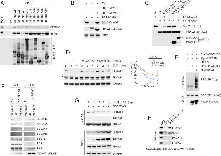
Figure 1. SCFFBXW5 interacts with SEC23B and targets it for ubiquitylation and proteasome-mediated degradation. (A) HEK293T cells were transfected with either an empty vector (EV) or the indicated Streptag-FLAG-tagged (SF) F-box proteins (FBPs). Twenty-four hours after transfection, cells were treated with MLN4924 for 4 hr before harvesting them for affinity-purification (AP) with Streptactin (ST) beads and immunoblotting as indicated. (WCE, whole cell extracts). The white asterisks indicate individual F-box proteins. (B) HEK293T cells were transfected with an EV, FLAG-HA-tagged FBXW5 (FH-FBXW5), or FH-FBXW5(ΔF) together with SF-tagged SEC23B. Twenty-four hours after transfection, cells were harvested for immunoblotting. (C) HEK293T cells were transfected with FH-FBXW5 and SF-SEC23B in combination with either an EV, MYC-tagged DN-CUL1, or MYC-tagged DN-CUL4 as indicated. Twenty-four hours after transfection, cells were either left untreated (UT) or treated with MG132 for 6 hr, and finally harvested for immunoblotting. (D) U-2OS cells were transfected with either a non-targeting siRNA oligo (NT) or two different FBXW5 siRNA oligos (individually). Seventy-two hours after siRNA transfection, cells were treated with cycloheximide (CHX) for the indicated times and harvested for immunoblotting. The asterisk indicates a nonspecific band. The graph shows the quantification of SEC23B levels from three independent experiments. Error bars indicate standard deviation. (E) HEK293T cells were transfected with an EV, HA-tagged FBXW5, or HA-tagged FBXW5(ΔF) together with MYC-tagged SEC23B and FLAG-TR-TUBE cDNA as indicated. WCLs were immunoprecipitated (IP) with anti-FLAG resin and immunoblotted as indicated. The line on the right marks a ladder of bands corresponding to poly-ubiquitylated SEC23B. (F) HEK293T cells were transfected with either an EV or FH-FBXW5. Twenty-four hours after transfection, cells were either left untreated (UT) or treated with MLN4924 for 4 hr before harvesting them for immunoprecipitation (IP) with FLAG beads and immunoblotting as indicated. (G) HEK293T cells were transfected with SF-FBXW5 and increasing amounts of HA-tagged SEC24B as indicated. Twenty-four hours after transfection, cells were harvested for affinity-purification (AP) with Streptactin (ST) beads and immunoblotting as indicated. (H) WCEs from HEK293T cells were incubated with either unconjugated beads or beads coupled to a SEC23B peptide (a.a. 180–194, CEGISKSYVFRGTKD). Beads were washed with lysis buffer and bound proteins were eluted and subjected to SDS-PAGE and immunoblotting.
Figure 1. SCFFBXW5 interacts with SEC23B and targets it for ubiquitylation and proteasome-mediated degradation. (A) HEK293T cells were transfected with either an empty vector (EV) or the indicated Streptag-FLAG-tagged (SF) F-box proteins (FBPs). Twenty-four hours after transfection, cells were treated with MLN4924 for 4 hr before harvesting them for affinity-purification (AP) with Streptactin (ST) beads and immunoblotting as indicated. (WCE, whole cell extracts). The white asterisks indicate individual F-box proteins. (B) HEK293T cells were transfected with an EV, FLAG-HA-tagged FBXW5 (FH-FBXW5), or FH-FBXW5(ΔF) together with SF-tagged SEC23B. Twenty-four hours after transfection, cells were harvested for immunoblotting. (C) HEK293T cells were transfected with FH-FBXW5 and SF-SEC23B in combination with either an EV, MYC-tagged DN-CUL1, or MYC-tagged DN-CUL4 as indicated. Twenty-four hours after transfection, cells were either left untreated (UT) or treated with MG132 for 6 hr, and finally harvested for immunoblotting. (D) U-2OS cells were transfected with either a non-targeting siRNA oligo (NT) or two different FBXW5 siRNA oligos (individually). Seventy-two hours after siRNA transfection, cells were treated with cycloheximide (CHX) for the indicated times and harvested for immunoblotting. The asterisk indicates a nonspecific band. The graph shows the quantification of SEC23B levels from three independent experiments. Error bars indicate standard deviation. (E) HEK293T cells were transfected with an EV, HA-tagged FBXW5, or HA-tagged FBXW5(ΔF) together with MYC-tagged SEC23B and FLAG-TR-TUBE cDNA as indicated. WCLs were immunoprecipitated (IP) with anti-FLAG resin and immunoblotted as indicated. The line on the right marks a ladder of bands corresponding to poly-ubiquitylated SEC23B. (F) HEK293T cells were transfected with either an EV or FH-FBXW5. Twenty-four hours after transfection, cells were either left untreated (UT) or treated with MLN4924 for 4 hr before harvesting them for immunoprecipitation (IP) with FLAG beads and immunoblotting as indicated. (G) HEK293T cells were transfected with SF-FBXW5 and increasing amounts of HA-tagged SEC24B as indicated. Twenty-four hours after transfection, cells were harvested for affinity-purification (AP) with Streptactin (ST) beads and immunoblotting as indicated. (H) WCEs from HEK293T cells were incubated with either unconjugated beads or beads coupled to a SEC23B peptide (a.a. 180–194, CEGISKSYVFRGTKD). Beads were washed with lysis buffer and bound proteins were eluted and subjected to SDS-PAGE and immunoblotting.CC BY · PubMed Central
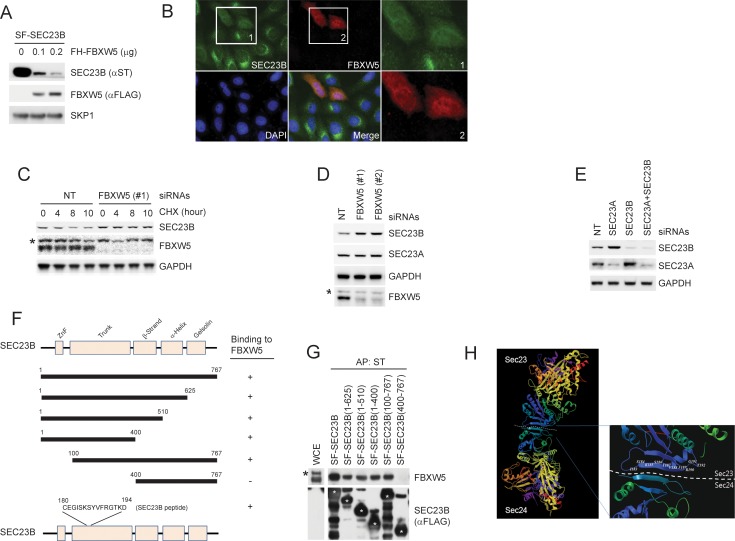
Figure 1—figure supplement 1. FBXW5 interacts with and promotes the degradation of SEC23B. (A) HEK293T cells were transfected with Streptag-FLAG-tagged SEC23B (SF-SEC23B) and increasing amounts of FLAG-HA-tagged FBXW5 (FH-FBXW5). Twenty-four hours after transfection, cells were harvested for immunoblotting as indicated. (B) U-2OS cells stable expressing FLAG-tagged SEC23B were transfected with HA-tagged FBXW5. Twenty-four hours after transfection, cells were fixed with 4% paraformaldehyde in PBS. An anti-FLAG antibody and an anti-HA antibody were used for the detection of SEC23B and FBXW5, respectively. Boxed images were enlarged for clarity. (C) RPE1-hTERT cells were transfected with either a non-targeting siRNA oligo (NT) or a FBXW5 siRNA oligo. Seventy-two hours after siRNA transfection, cells were treated with cycloheximide (CHX) for the indicated times and harvested for immunoblotting as indicated. The asterisk indicates a nonspecific band. (D) RPE1-hTERT cells were transfected with either a non-targeting siRNA oligo (NT) or two different FBXW5 siRNA oligos (each individually). Seventy-two hours after siRNA transfection, cells were harvested for immunoblotting as indicated. The asterisk indicates a nonspecific band recognized by the anti-FBXW5 antibody. (E) RPE1-hTERT cells were transfected with siRNA oligos to the indicated mRNAs. Seventy-two hours after siRNA transfection, cells were harvested for immunoblotting. (F) Schematic view of the SEC23B mutants used in Figure 1—figure supplement 1G and the SEC23B degron peptide. (G) HEK293T cells were transfected with the truncated mutants shown in Figure 1—figure supplement 1F. Twenty-four hours after transfection, cells were harvested for affinity-purification (AP) with Streptactin (ST) beads followed by immunoblotting as indicated. The black asterisk indicates a nonspecific band. The white asterisks indicate bands corresponding to SEC23B or SEC23B mutants. (H) Crystal structure showing the position of Serine 186 of SEC23. Images (PDB: 3EFO; Mancias and Goldberg, 2008) were created by MolBrowser (Molsoft). The dashed line indicates the interface between SEC23 and SEC24 where serine 186 resides.
Figure 1—figure supplement 1. FBXW5 interacts with and promotes the degradation of SEC23B. (A) HEK293T cells were transfected with Streptag-FLAG-tagged SEC23B (SF-SEC23B) and increasing amounts of FLAG-HA-tagged FBXW5 (FH-FBXW5). Twenty-four hours after transfection, cells were harvested for immunoblotting as indicated. (B) U-2OS cells stable expressing FLAG-tagged SEC23B were transfected with HA-tagged FBXW5. Twenty-four hours after transfection, cells were fixed with 4% paraformaldehyde in PBS. An anti-FLAG antibody and an anti-HA antibody were used for the detection of SEC23B and FBXW5, respectively. Boxed images were enlarged for clarity. (C) RPE1-hTERT cells were transfected with either a non-targeting siRNA oligo (NT) or a FBXW5 siRNA oligo. Seventy-two hours after siRNA transfection, cells were treated with cycloheximide (CHX) for the indicated times and harvested for immunoblotting as indicated. The asterisk indicates a nonspecific band. (D) RPE1-hTERT cells were transfected with either a non-targeting siRNA oligo (NT) or two different FBXW5 siRNA oligos (each individually). Seventy-two hours after siRNA transfection, cells were harvested for immunoblotting as indicated. The asterisk indicates a nonspecific band recognized by the anti-FBXW5 antibody. (E) RPE1-hTERT cells were transfected with siRNA oligos to the indicated mRNAs. Seventy-two hours after siRNA transfection, cells were harvested for immunoblotting. (F) Schematic view of the SEC23B mutants used in Figure 1—figure supplement 1G and the SEC23B degron peptide. (G) HEK293T cells were transfected with the truncated mutants shown in Figure 1—figure supplement 1F. Twenty-four hours after transfection, cells were harvested for affinity-purification (AP) with Streptactin (ST) beads followed by immunoblotting as indicated. The black asterisk indicates a nonspecific band. The white asterisks indicate bands corresponding to SEC23B or SEC23B mutants. (H) Crystal structure showing the position of Serine 186 of SEC23. Images (PDB: 3EFO; Mancias and Goldberg, 2008) were created by MolBrowser (Molsoft). The dashed line indicates the interface between SEC23 and SEC24 where serine 186 resides.CC BY · PubMed Central
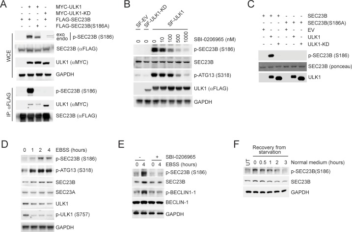
Figure 2. ULK1 phosphorylates SEC23B on Serine 186. (A) HEK293T cells were transfected with either FLAG-tagged SEC23B or FLAG-tagged SEC23B(S186A) in combination with MYC-tagged ULK1 or MYC-tagged ULK1-KD as indicated. Twenty-four hours after transfection, cells were harvested for immunoprecipitation (IP) and immunoblotting. exo and endo indicate the exogenous and endogenous SEC23B, respectively. (B) HEK293T cells were transfected with the SF-ULK1 or SF- ULK1-KD as indicated. Twenty-four hours after transfection, cells were treated with various doses of SBI-0206965 (an ULK1 inhibitor) for 4 hr before harvesting them for immunoblotting. (C) In vitro kinase assays were performed using purified SEC23B (wild-type or the S186A mutant) and ULK1 (wild-type or a kinase-dead mutant) as substrate and kinase, respectively. Purified SEC23B and ULK1 proteins were prepared by immunoprecipitation (followed by elution) from extracts of HEK293T cells transfected with each corresponding plasmid. (D) HEK293T cells were nutrient-starved with EBSS for the indicated times and harvested for immunoblotting. (E) HEK293T cells were nutrient-starved with EBSS for the indicated times (in the presence or absence of SBI-0206965) and harvested for immunoblotting at the indicated times. (F) HEK293T cells were recovered from nutrient-starvation (EBSS for 4 hr) for the indicated times and harvested for immunoblotting.
Figure 2. ULK1 phosphorylates SEC23B on Serine 186. (A) HEK293T cells were transfected with either FLAG-tagged SEC23B or FLAG-tagged SEC23B(S186A) in combination with MYC-tagged ULK1 or MYC-tagged ULK1-KD as indicated. Twenty-four hours after transfection, cells were harvested for immunoprecipitation (IP) and immunoblotting. exo and endo indicate the exogenous and endogenous SEC23B, respectively. (B) HEK293T cells were transfected with the SF-ULK1 or SF- ULK1-KD as indicated. Twenty-four hours after transfection, cells were treated with various doses of SBI-0206965 (an ULK1 inhibitor) for 4 hr before harvesting them for immunoblotting. (C) In vitro kinase assays were performed using purified SEC23B (wild-type or the S186A mutant) and ULK1 (wild-type or a kinase-dead mutant) as substrate and kinase, respectively. Purified SEC23B and ULK1 proteins were prepared by immunoprecipitation (followed by elution) from extracts of HEK293T cells transfected with each corresponding plasmid. (D) HEK293T cells were nutrient-starved with EBSS for the indicated times and harvested for immunoblotting. (E) HEK293T cells were nutrient-starved with EBSS for the indicated times (in the presence or absence of SBI-0206965) and harvested for immunoblotting at the indicated times. (F) HEK293T cells were recovered from nutrient-starvation (EBSS for 4 hr) for the indicated times and harvested for immunoblotting.CC BY · PubMed Central
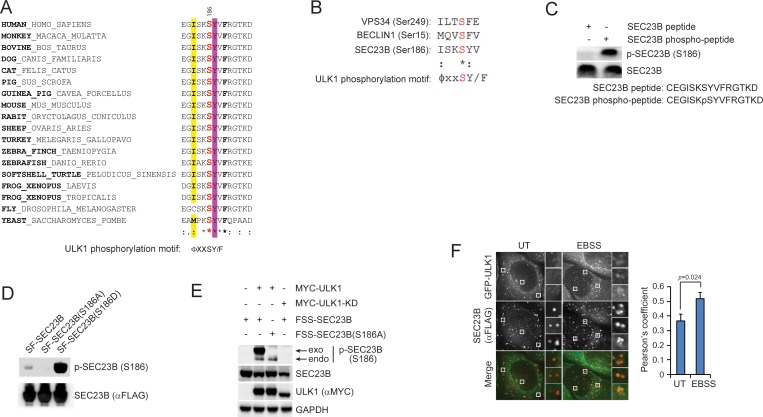
Figure 2—figure supplement 1. Characterization of the phospho-SEC23B (Ser186) antibody and the phospho-mimetic SEC23B mutant. (A) An ULK1 phosphorylation motif (ΦxxSY/F)(Egan et al., 2015) is highly conserved throughout evolution in the FBXW5-binding region of SEC23B. Φ, hydrophobic amino acids. (B) Sequence alignment of the previously characterized ULK1-substrates with SEC23B. (C) A non-phosphorylated SEC23B peptide and an equivalent phospho-peptide (sequences are indicated below the panels) were separated by SDS-PAGE and subjected to immunoblotting either with a phospho-specific SEC23B (Ser186) antibody or with an anti-SEC23B antibody reactive against both phosphorylated and non-phosphorylated species of SEC23B. (D) HEK239T cells were transfected with either SF- tagged SEC23B or the indicated SF-tagged SEC23B mutants. Twenty-four hours after transfection, cells were immunoblotted as indicated. (E) HEK293T cells were transfected with either FLAG-Streptag-Streptag-tagged SEC23B or FLAG-Streptag-Streptag-tagged SEC23B(S186A) in combination with MYC-tagged ULK1 or MYC-tagged ULK1-KD as indicated. Twenty-four hours after transfection, cells were harvested for immunoblotting as indicated. exo and endo indicate the exogenous and endogenous SEC23B, respectively. (F) U-2OS cells stably expressing GFP-ULK1 were transfected with FLAG-HA-tagged SEC23B. Twenty-four hours after transfection, cells were fixed for immunofluorescence as indicated. Images were analysed by ImageJ with at least 100 cells counted per sample. Quantification of SEC23B overlapping with GFP-ULK1 was performed using the Pearson's correlation coefficient. The data are presented as mean ±SD (right panel). Scale bar, 10 μm.
Figure 2—figure supplement 1. Characterization of the phospho-SEC23B (Ser186) antibody and the phospho-mimetic SEC23B mutant. (A) An ULK1 phosphorylation motif (ΦxxSY/F)(Egan et al., 2015) is highly conserved throughout evolution in the FBXW5-binding region of SEC23B. Φ, hydrophobic amino acids. (B) Sequence alignment of the previously characterized ULK1-substrates with SEC23B. (C) A non-phosphorylated SEC23B peptide and an equivalent phospho-peptide (sequences are indicated below the panels) were separated by SDS-PAGE and subjected to immunoblotting either with a phospho-specific SEC23B (Ser186) antibody or with an anti-SEC23B antibody reactive against both phosphorylated and non-phosphorylated species of SEC23B. (D) HEK239T cells were transfected with either SF- tagged SEC23B or the indicated SF-tagged SEC23B mutants. Twenty-four hours after transfection, cells were immunoblotted as indicated. (E) HEK293T cells were transfected with either FLAG-Streptag-Streptag-tagged SEC23B or FLAG-Streptag-Streptag-tagged SEC23B(S186A) in combination with MYC-tagged ULK1 or MYC-tagged ULK1-KD as indicated. Twenty-four hours after transfection, cells were harvested for immunoblotting as indicated. exo and endo indicate the exogenous and endogenous SEC23B, respectively. (F) U-2OS cells stably expressing GFP-ULK1 were transfected with FLAG-HA-tagged SEC23B. Twenty-four hours after transfection, cells were fixed for immunofluorescence as indicated. Images were analysed by ImageJ with at least 100 cells counted per sample. Quantification of SEC23B overlapping with GFP-ULK1 was performed using the Pearson's correlation coefficient. The data are presented as mean ±SD (right panel). Scale bar, 10 μm.CC BY · PubMed Central
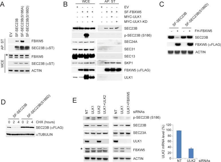
Figure 3. ULK1-mediated phosphorylation of SEC23B on S186 prevents the FBXW5-dependent degradation of SEC23B. (A) HEK293T cells were transfected with either EV, SF-SEC23B, or the indicated SF-SEC23B mutants. Twenty-four hours after transfection, cells were treated with MLN4924 for 4 hr before harvesting them for affinity-purification (AP) with Streptactin (ST) beads and immunoblotting. The asterisk indicates a nonspecific band. (B) HEK293T cells were transfected with SF-FBXW5 in combination with either MYC-tagged ULK1 or MYC-tagged ULK1-KD. Twenty-four hours after transfection, cells were treated with MLN4924 for 4 hr before harvesting them for affinity-purification (AP) with Streptactin (ST) beads and immunoblotting as indicated. (C) HEK293T cells were transfected with FH-FBXW5 in combination with SF-SEC23B or SF-SEC23B(S186D). Twenty-four hours after transfection, cells were harvested for immunoblotting. (D) U-2OS cells stably infected with viruses expressing either SEC23B or SEC23B(S186D) were treated with cycloheximide for the indicated times. The cells were then harvested for immunoblotting. (E) RPE1-hTERT cells were transfected with siRNAs against the indicated mRNAs. Sixty-eight hours after transfection, cells were nutrient-starved with EBSS for 4 hr and harvested for immunoblotting (left panel) and real-time PCR using ULK2 and GAPDH primers (right panel). The asterisk indicates the nonspecific band.
Figure 3. ULK1-mediated phosphorylation of SEC23B on S186 prevents the FBXW5-dependent degradation of SEC23B. (A) HEK293T cells were transfected with either EV, SF-SEC23B, or the indicated SF-SEC23B mutants. Twenty-four hours after transfection, cells were treated with MLN4924 for 4 hr before harvesting them for affinity-purification (AP) with Streptactin (ST) beads and immunoblotting. The asterisk indicates a nonspecific band. (B) HEK293T cells were transfected with SF-FBXW5 in combination with either MYC-tagged ULK1 or MYC-tagged ULK1-KD. Twenty-four hours after transfection, cells were treated with MLN4924 for 4 hr before harvesting them for affinity-purification (AP) with Streptactin (ST) beads and immunoblotting as indicated. (C) HEK293T cells were transfected with FH-FBXW5 in combination with SF-SEC23B or SF-SEC23B(S186D). Twenty-four hours after transfection, cells were harvested for immunoblotting. (D) U-2OS cells stably infected with viruses expressing either SEC23B or SEC23B(S186D) were treated with cycloheximide for the indicated times. The cells were then harvested for immunoblotting. (E) RPE1-hTERT cells were transfected with siRNAs against the indicated mRNAs. Sixty-eight hours after transfection, cells were nutrient-starved with EBSS for 4 hr and harvested for immunoblotting (left panel) and real-time PCR using ULK2 and GAPDH primers (right panel). The asterisk indicates the nonspecific band.CC BY · PubMed Central
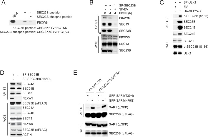
Figure 3—figure supplement 1. ULK1-mediated phosphorylation of SEC23B on S186 prevents the FBXW5-dependent degradation of SEC23B. (A) A non-phosphorylated SEC23B peptide and an equivalent phospho-peptide (sequences are indicated below the panels) were conjugated to beads and used in an in vitro binding assay with FBXW5 that was in vitro transcribed/translated using a rabbit reticulocyte system. Bound FBXW5 was detected by immunoblotting. (B) HEK293T cells were transfected with either a Streptag-FLAG empty vector (SF-EV) or Streptag-FLAG-tagged SEC23B (SF-SEC23B). Twenty-four hours after transfection, cells were nutrient-starved with EBSS for the indicated times and treated with MLN4924 for 4 hr before harvesting them for affinity-purification (AP) with Streptactin (ST) beads and immunoblotting as indicated. (WCE, whole cell extracts). The white asterisk indicates a non-specific band. (C) HEK239T cells were transfected with an empty vector (EV) or HA-tagged SEC24B together with Streptag-FLAG tagged ULK1 (SF-ULK1) as indicated. Twenty-four hours after transfection, cells were harvested for immunoprecipitation (IP) with HA beads and immunoblotting as indicated. (WCE, whole cell extracts). (D) HEK239T cells were transfected with either Streptag-FLAG tagged wild-type SEC23B or the phospho-mimetic SEC23B(S186D) mutant (also Streptag-FLAG tagged). Twenty-four hours after transfection, cells were harvested for affinity-purification (AP) with Streptactin (ST) beads and immunoblotting as indicated. (WCE, whole cell extracts). (E) HEK239T cells were transfected with either Streptag-FLAG tagged SEC23B (SF-SEC23B) or SF-SEC23B(S186D), in combination with the indicated GFP-tagged SAR1 mutants. Twenty-four hours after transfection, cells were harvested for affinity-purification (AP) with Streptactin (ST) beads and immunoblotting as indicated.
Figure 3—figure supplement 1. ULK1-mediated phosphorylation of SEC23B on S186 prevents the FBXW5-dependent degradation of SEC23B. (A) A non-phosphorylated SEC23B peptide and an equivalent phospho-peptide (sequences are indicated below the panels) were conjugated to beads and used in an in vitro binding assay with FBXW5 that was in vitro transcribed/translated using a rabbit reticulocyte system. Bound FBXW5 was detected by immunoblotting. (B) HEK293T cells were transfected with either a Streptag-FLAG empty vector (SF-EV) or Streptag-FLAG-tagged SEC23B (SF-SEC23B). Twenty-four hours after transfection, cells were nutrient-starved with EBSS for the indicated times and treated with MLN4924 for 4 hr before harvesting them for affinity-purification (AP) with Streptactin (ST) beads and immunoblotting as indicated. (WCE, whole cell extracts). The white asterisk indicates a non-specific band. (C) HEK239T cells were transfected with an empty vector (EV) or HA-tagged SEC24B together with Streptag-FLAG tagged ULK1 (SF-ULK1) as indicated. Twenty-four hours after transfection, cells were harvested for immunoprecipitation (IP) with HA beads and immunoblotting as indicated. (WCE, whole cell extracts). (D) HEK239T cells were transfected with either Streptag-FLAG tagged wild-type SEC23B or the phospho-mimetic SEC23B(S186D) mutant (also Streptag-FLAG tagged). Twenty-four hours after transfection, cells were harvested for affinity-purification (AP) with Streptactin (ST) beads and immunoblotting as indicated. (WCE, whole cell extracts). (E) HEK239T cells were transfected with either Streptag-FLAG tagged SEC23B (SF-SEC23B) or SF-SEC23B(S186D), in combination with the indicated GFP-tagged SAR1 mutants. Twenty-four hours after transfection, cells were harvested for affinity-purification (AP) with Streptactin (ST) beads and immunoblotting as indicated.CC BY · PubMed Central
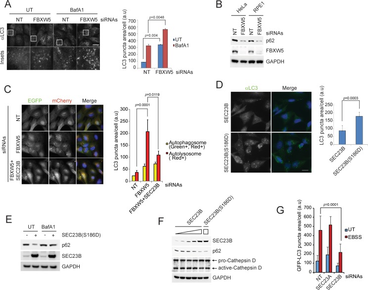
Figure 4. The FBXW5-mediated degradation of SEC23B limits the autophagic flux in the presence of nutrients. (A) RPE1-hTERT cells were transfected with a non-targeting (NT) oligo or a FBXW5-targeting siRNA oligo. Forty-eight hours after transfection, cells were re-plated onto coverglass for immunofluorescence with an anti-LC3 antibody. Where indicated, cells were treated with Bafilomycin A1 (BafA1) for 4 hr before fixation. Images of endogenous LC3 puncta underwent automated processing with at least 300 cells counted per sample. Because in several images LC3·puncta were too close to be distinguished, we adopted LC3 puncta area as a criterion for our analysis. The data are presented as mean ±SD (right panel). Scale bar, 10 μm. (B) HeLa and RPE1-hTERT cells were transfected with the indicated siRNAs. Seventy-two hours after transfection, the cells were harvested for immunoblotting. (C) U-2OS cells stably expressing tandem fluorescent-tagged LC3 (pBabe-mCherry-EGFP-LC3) were transfected with a NT oligo or a FBXW5 siRNA oligo, alone or in combination with a SEC23B-targeting siRNA oligo as indicated. Forty-eight hours after transfection, cells were replated onto coverglass, followed by fixation twenty-four hours after replating. Images of mCherry-EGFP-LC3 puncta underwent automated processing with at least 100 cells counted per sample. The data are presented as mean ±SD (right panel). The yellow and red bars represent green +red double positive LC3 puncta (autophagosome) and red only positive LC3 puncta (autolysosome), respectively. Scale bar, 10 μm. (D) U-2OS cells were infected with lentiviruses expressing either wild-type SEC23B or SEC23B(S186D). Twenty-four hours after infection, cells were fixed for immunofluorescence. Images of endogenous LC3 puncta underwent automated processing with at least 300 cells counted per sample. The data are presented as mean ±SD (right panel). Scale bar, 10 μm. (E) U-2OS cells were infected with lentivirus expressing SEC23B(S186D). Where indicated, forty-eight hours after infection, cells were treated with BafA1 prior to harvest and immunoblotting. (F) U-2OS cells were infected with the increasing amounts of lentivirus expressing SEC23B. Forty-eight hours after infection, cells were harvested for immunoblotting. (G) RPE1-hTERT cells stably expressing GFP-tagged LC3 were transfected with a NT oligo or the indicated siRNA oligos. Forty-eight hours after transfection, cells were replated onto coverglass, followed by treatment with EBSS for 1 hr and fixation. Images of GFP-LC3 puncta underwent automated processing with at least 300 cells counted per sample. The data are presented as mean ±SD.
Figure 4. The FBXW5-mediated degradation of SEC23B limits the autophagic flux in the presence of nutrients. (A) RPE1-hTERT cells were transfected with a non-targeting (NT) oligo or a FBXW5-targeting siRNA oligo. Forty-eight hours after transfection, cells were re-plated onto coverglass for immunofluorescence with an anti-LC3 antibody. Where indicated, cells were treated with Bafilomycin A1 (BafA1) for 4 hr before fixation. Images of endogenous LC3 puncta underwent automated processing with at least 300 cells counted per sample. Because in several images LC3·puncta were too close to be distinguished, we adopted LC3 puncta area as a criterion for our analysis. The data are presented as mean ±SD (right panel). Scale bar, 10 μm. (B) HeLa and RPE1-hTERT cells were transfected with the indicated siRNAs. Seventy-two hours after transfection, the cells were harvested for immunoblotting. (C) U-2OS cells stably expressing tandem fluorescent-tagged LC3 (pBabe-mCherry-EGFP-LC3) were transfected with a NT oligo or a FBXW5 siRNA oligo, alone or in combination with a SEC23B-targeting siRNA oligo as indicated. Forty-eight hours after transfection, cells were replated onto coverglass, followed by fixation twenty-four hours after replating. Images of mCherry-EGFP-LC3 puncta underwent automated processing with at least 100 cells counted per sample. The data are presented as mean ±SD (right panel). The yellow and red bars represent green +red double positive LC3 puncta (autophagosome) and red only positive LC3 puncta (autolysosome), respectively. Scale bar, 10 μm. (D) U-2OS cells were infected with lentiviruses expressing either wild-type SEC23B or SEC23B(S186D). Twenty-four hours after infection, cells were fixed for immunofluorescence. Images of endogenous LC3 puncta underwent automated processing with at least 300 cells counted per sample. The data are presented as mean ±SD (right panel). Scale bar, 10 μm. (E) U-2OS cells were infected with lentivirus expressing SEC23B(S186D). Where indicated, forty-eight hours after infection, cells were treated with BafA1 prior to harvest and immunoblotting. (F) U-2OS cells were infected with the increasing amounts of lentivirus expressing SEC23B. Forty-eight hours after infection, cells were harvested for immunoblotting. (G) RPE1-hTERT cells stably expressing GFP-tagged LC3 were transfected with a NT oligo or the indicated siRNA oligos. Forty-eight hours after transfection, cells were replated onto coverglass, followed by treatment with EBSS for 1 hr and fixation. Images of GFP-LC3 puncta underwent automated processing with at least 300 cells counted per sample. The data are presented as mean ±SD.CC BY · PubMed Central
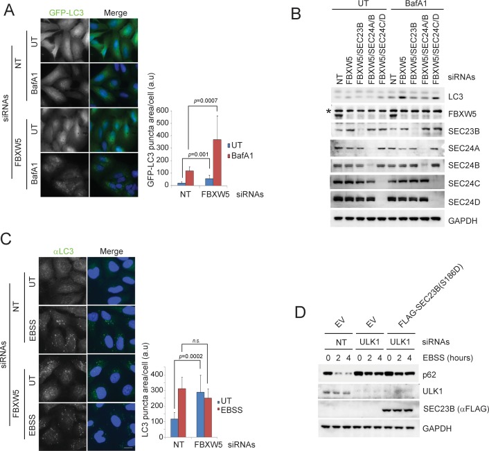
Figure 4—figure supplement 1. The FBXW5-mediated degradation of SEC23B limits the autophagic flux in the presence of nutrients. (A) RPE1-hTERT cells stably expressing GFP-LC3 were transfected with either a non-targeting siRNA oligo (NT) or an siRNA oligo targeting FBXW5 RNA. Forty-eight hours after transfection, cells were replated onto coverglass. Where indicated, cells were treated with Bafilomycin A1 (BafA1) for 4 hr before fixation with cold methanol. Images of the GFP-LC3 puncta underwent automated processing with at least 300 cells counted per sample. The data are presented as mean ±SD (right panel). Scale bar, 10 μm. (B) RPE1-hTERT cells were transfected with the indicated siRNAs. Seventy-two hours after transfection, where indicated, cells were treated with Bafilomycin A1 (BafA1) for one hour prior to harvest for immunoblotting. (C) RPE1-hTERT cells stably expressing GFP-LC3 were transfected with either a non-targeting siRNA oligo (NT) or an siRNA oligo targeting FBXW5 RNA. Forty-eight hours after transfection, cells were replated onto coverglass for immunofluorescence with an anti-LC3 antibody. Cells were nutrient-starved with EBSS for 4 hr before fixation with cold methanol. Images of the LC3 puncta underwent automated processing with at least 300 cells counted per sample. The data are presented as mean ±SD (right panel). Scale bar, 10 μm. (D) U-2OS cells infected with either an empty virus (EV) or lentivirus expressing SEC23B(S186D) were transfected with the indicated siRNAs. Seventy-two hours after transfection, cells were treated with EBSS prior to harvest for immunoblotting.
Figure 4—figure supplement 1. The FBXW5-mediated degradation of SEC23B limits the autophagic flux in the presence of nutrients. (A) RPE1-hTERT cells stably expressing GFP-LC3 were transfected with either a non-targeting siRNA oligo (NT) or an siRNA oligo targeting FBXW5 RNA. Forty-eight hours after transfection, cells were replated onto coverglass. Where indicated, cells were treated with Bafilomycin A1 (BafA1) for 4 hr before fixation with cold methanol. Images of the GFP-LC3 puncta underwent automated processing with at least 300 cells counted per sample. The data are presented as mean ±SD (right panel). Scale bar, 10 μm. (B) RPE1-hTERT cells were transfected with the indicated siRNAs. Seventy-two hours after transfection, where indicated, cells were treated with Bafilomycin A1 (BafA1) for one hour prior to harvest for immunoblotting. (C) RPE1-hTERT cells stably expressing GFP-LC3 were transfected with either a non-targeting siRNA oligo (NT) or an siRNA oligo targeting FBXW5 RNA. Forty-eight hours after transfection, cells were replated onto coverglass for immunofluorescence with an anti-LC3 antibody. Cells were nutrient-starved with EBSS for 4 hr before fixation with cold methanol. Images of the LC3 puncta underwent automated processing with at least 300 cells counted per sample. The data are presented as mean ±SD (right panel). Scale bar, 10 μm. (D) U-2OS cells infected with either an empty virus (EV) or lentivirus expressing SEC23B(S186D) were transfected with the indicated siRNAs. Seventy-two hours after transfection, cells were treated with EBSS prior to harvest for immunoblotting.CC BY · PubMed Central
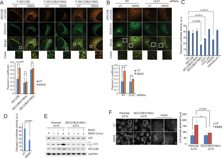
Figure 5. Ser186 in SEC23B is necessary for its localization to the ERGIC and an efficient autophagic response upon nutrient deprivation. (A) U-2OS cells were transfected with either FLAG-HA-tagged wild-type SEC23B, SEC23B(S186A), or SEC23B(S186D). Twenty-four hours after transfection, cells were fixed for immunofluorescence as indicated. Images were analysed by ImageJ with at least 100 cells counted per sample. Quantification of SEC23B overlapping with ERGIC53 was performed using the Pearson's correlation coefficient. The data are presented as mean ±SD (bottom panel). Scale bar, 10 μm. (B) U-2OS cells stably expressing FLAG-HA-tagged SEC23B were transfected with ULK1 siRNAs. Seventy-two hours after transfection, cells were fixed for immunofluorescence as indicated. Images were analysed by ImageJ with at least 100 cells counted per sample. Quantification of SEC23B overlapping with ERGIC53 was performed using the Pearson's correlation coefficient. The data are presented as mean ±SD (bottom panel). Scale bar, 10 μm. (C) HEK239T cells were transfected with a plasmid expressing Gaussia luciferase in combination with the indicated constructs. Twenty-four hours after transfection, cells were replated onto 96-well plates. After another forty-eight hours, fresh media was added to the cells, and four hours after, the culture media were collected to measure Gaussia luciferase activity. The data are presented as mean ±SD of the Gaussia luciferase activity of triplicate samples. Expression of FBXW5 was used as a positive control since it results in the downregulation of SEC23B and, therefore, it is expected to inhibit trafficking. (D) A375 cells stably expressing Gaussia luciferase were plated onto 96-well plates. After forty-eight hours, either fresh media or EBSS was added to the cells and four hours after, the culture media were collected to measure Gaussia luciferase activity. The data are presented as mean ±SD of the Gaussia luciferase activity of triplicate samples. (E) A375 parental cells or SEC23B(S186A)-A357 knock-in cells were starved with EBSS for the indicated times (±BafA1) and harvested for immunoblotting as indicated. (F) A375 parental cells or SEC23B(S186A)-A357 knock-in cells were starved with EBSS and fixed for immunofluorescence. Images of endogenous LC3 puncta underwent automated processing with at least 100 cells counted per sample. The data are presented as mean ±SD (right panel). Scale bar, 10 μm.
Figure 5. Ser186 in SEC23B is necessary for its localization to the ERGIC and an efficient autophagic response upon nutrient deprivation. (A) U-2OS cells were transfected with either FLAG-HA-tagged wild-type SEC23B, SEC23B(S186A), or SEC23B(S186D). Twenty-four hours after transfection, cells were fixed for immunofluorescence as indicated. Images were analysed by ImageJ with at least 100 cells counted per sample. Quantification of SEC23B overlapping with ERGIC53 was performed using the Pearson's correlation coefficient. The data are presented as mean ±SD (bottom panel). Scale bar, 10 μm. (B) U-2OS cells stably expressing FLAG-HA-tagged SEC23B were transfected with ULK1 siRNAs. Seventy-two hours after transfection, cells were fixed for immunofluorescence as indicated. Images were analysed by ImageJ with at least 100 cells counted per sample. Quantification of SEC23B overlapping with ERGIC53 was performed using the Pearson's correlation coefficient. The data are presented as mean ±SD (bottom panel). Scale bar, 10 μm. (C) HEK239T cells were transfected with a plasmid expressing Gaussia luciferase in combination with the indicated constructs. Twenty-four hours after transfection, cells were replated onto 96-well plates. After another forty-eight hours, fresh media was added to the cells, and four hours after, the culture media were collected to measure Gaussia luciferase activity. The data are presented as mean ±SD of the Gaussia luciferase activity of triplicate samples. Expression of FBXW5 was used as a positive control since it results in the downregulation of SEC23B and, therefore, it is expected to inhibit trafficking. (D) A375 cells stably expressing Gaussia luciferase were plated onto 96-well plates. After forty-eight hours, either fresh media or EBSS was added to the cells and four hours after, the culture media were collected to measure Gaussia luciferase activity. The data are presented as mean ±SD of the Gaussia luciferase activity of triplicate samples. (E) A375 parental cells or SEC23B(S186A)-A357 knock-in cells were starved with EBSS for the indicated times (±BafA1) and harvested for immunoblotting as indicated. (F) A375 parental cells or SEC23B(S186A)-A357 knock-in cells were starved with EBSS and fixed for immunofluorescence. Images of endogenous LC3 puncta underwent automated processing with at least 100 cells counted per sample. The data are presented as mean ±SD (right panel). Scale bar, 10 μm.CC BY · PubMed Central
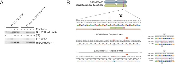
Figure 5—figure supplement 1. Ser186 in SEC23B is necessary for its localization to the ERGIC and an efficient autophagic response upon nutrient deprivation. (A) HeLa cells were permeabilized, incubated with either purified recombinant FLAG-tagged SEC23B or purified recombinant FLAG-tagged SEC23B(S186D), and subjected to subcellular fractionation. Each fraction was subjected to immunoblotting with the indicated antibodies. ERGIC53 was used as a marker of the ERGIC, and RIBOPHORIN-1 as a marker of the ER. SEC23B bands were subjected to densitometry analysis and quantified as percentage of total SEC23B or SEC23B(S186D). (B) Schematic representation of the SEC23B genomic locus and gRNA target location. Exon five refers to human SEC23B gene (Gene ID: 10483). Wild-type genomic DNA template and knock-in mutant sequences are shown for both SEC23B(S186A) and SEC23B(S186N).
Figure 5—figure supplement 1. Ser186 in SEC23B is necessary for its localization to the ERGIC and an efficient autophagic response upon nutrient deprivation. (A) HeLa cells were permeabilized, incubated with either purified recombinant FLAG-tagged SEC23B or purified recombinant FLAG-tagged SEC23B(S186D), and subjected to subcellular fractionation. Each fraction was subjected to immunoblotting with the indicated antibodies. ERGIC53 was used as a marker of the ERGIC, and RIBOPHORIN-1 as a marker of the ER. SEC23B bands were subjected to densitometry analysis and quantified as percentage of total SEC23B or SEC23B(S186D). (B) Schematic representation of the SEC23B genomic locus and gRNA target location. Exon five refers to human SEC23B gene (Gene ID: 10483). Wild-type genomic DNA template and knock-in mutant sequences are shown for both SEC23B(S186A) and SEC23B(S186N).CC BY · PubMed Central
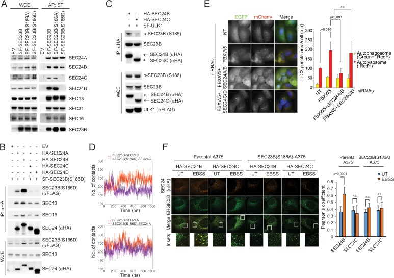
Figure 6. SEC24A/B, but not SEC24C/D, specifically associate with phosphorylated SEC23B and contribute to autophagy. (A) HEK293T cells were transfected with an EV, SF-SEC23B, or SF-SEC23B mutants. Twenty-four hours after transfection, cells were treated with MLN4924 for 4 hr before harvesting them for affinity-purification (AP) with Streptactin (ST) beads and immunoblotting. (B) HEK293T cells were transfected with an EV or SF) SEC23B(S186D) in combination with either HA-tagged SEC24 paralogs as indicated. Twenty-four hours after transfection, cells were treated with MLN4924 for 4 hr before harvesting them for immunoprecipitation (IP) with anti-HA beads and immunoblotting. (C) HEK293T cells were transfected with either HA-tagged SEC24B or HA-tagged SEC24C together with SF-ULK1. Twenty-four hours after transfection, cells were treated with MLN4924 for 4 hr before harvesting them for immunoprecipitation (IP) with anti-HA beads and immunoblotting. (D) Evolution of the inter-molecular contacts between monomers in the four studied systems during 1 μs molecular dynamics simulations. Upper panel: SEC23B-SEC24C (red), SEC23B(S186D)-SEC24C (violet); bottom panel: SEC23B-SEC24A (red), SEC23B(S186D)-SEC24A (violet). Contacts were calculated as the number of heavy atom interacting pairs within a distance of 4.4 Å. (E) U-2OS cells stably expressing tandem fluorescent-tagged LC3 (pBabe-mCherry-EGFP-LC3) were transfected with a NT oligo or a FBXW5 siRNA oligo in combination with the indicated siRNA oligos. Forty-eight hours after transfection, cells were replated onto coverglass followed by fixation twenty-four hours after replating. Images of mCherry-EGFP-LC3 puncta underwent automated processing with at least 100 cells counted per sample. The data are presented as mean ±SD (right panel). Scale bar, 10 μm (F) A375 parental cells or SEC23B(S186D)-A357 knock-in cells were transfected with either HA-tagged SEC24B or HA-tagged SEC24C. Twenty-four hours after transfection, cells were either left untreated (UT) or starved with EBSS for two hours. Next, cells were fixed for immunofluorescence as indicated. Images were analysed by ImageJ with at least 100 cells counted per sample. Quantification of SEC24 overlapped with ERGIC53 was performed using the Pearson's correlation coefficient. The data are presented as mean ±SD (right panel). Scale bar, 10 μM.
Figure 6. SEC24A/B, but not SEC24C/D, specifically associate with phosphorylated SEC23B and contribute to autophagy. (A) HEK293T cells were transfected with an EV, SF-SEC23B, or SF-SEC23B mutants. Twenty-four hours after transfection, cells were treated with MLN4924 for 4 hr before harvesting them for affinity-purification (AP) with Streptactin (ST) beads and immunoblotting. (B) HEK293T cells were transfected with an EV or SF) SEC23B(S186D) in combination with either HA-tagged SEC24 paralogs as indicated. Twenty-four hours after transfection, cells were treated with MLN4924 for 4 hr before harvesting them for immunoprecipitation (IP) with anti-HA beads and immunoblotting. (C) HEK293T cells were transfected with either HA-tagged SEC24B or HA-tagged SEC24C together with SF-ULK1. Twenty-four hours after transfection, cells were treated with MLN4924 for 4 hr before harvesting them for immunoprecipitation (IP) with anti-HA beads and immunoblotting. (D) Evolution of the inter-molecular contacts between monomers in the four studied systems during 1 μs molecular dynamics simulations. Upper panel: SEC23B-SEC24C (red), SEC23B(S186D)-SEC24C (violet); bottom panel: SEC23B-SEC24A (red), SEC23B(S186D)-SEC24A (violet). Contacts were calculated as the number of heavy atom interacting pairs within a distance of 4.4 Å. (E) U-2OS cells stably expressing tandem fluorescent-tagged LC3 (pBabe-mCherry-EGFP-LC3) were transfected with a NT oligo or a FBXW5 siRNA oligo in combination with the indicated siRNA oligos. Forty-eight hours after transfection, cells were replated onto coverglass followed by fixation twenty-four hours after replating. Images of mCherry-EGFP-LC3 puncta underwent automated processing with at least 100 cells counted per sample. The data are presented as mean ±SD (right panel). Scale bar, 10 μm (F) A375 parental cells or SEC23B(S186D)-A357 knock-in cells were transfected with either HA-tagged SEC24B or HA-tagged SEC24C. Twenty-four hours after transfection, cells were either left untreated (UT) or starved with EBSS for two hours. Next, cells were fixed for immunofluorescence as indicated. Images were analysed by ImageJ with at least 100 cells counted per sample. Quantification of SEC24 overlapped with ERGIC53 was performed using the Pearson's correlation coefficient. The data are presented as mean ±SD (right panel). Scale bar, 10 μM.CC BY · PubMed Central
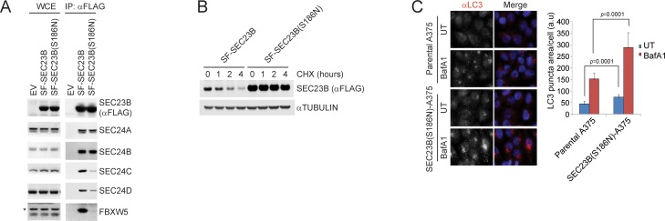
Figure 7. A human melanoma-associated mutation in SEC23B results in its stabilization and increased autophagy flux. (A) HEK239T cells were transfected with either SF-SEC23B or SF-SEC23B(S186N). Twenty-four hours after transfection, cells were harvested for immunoprecipitation (IP) with FLAG-M2 beads and immunoblotted as indicated. (B) HEK293T cells were transfected with either Streptag-FLAG-tagged (SF) wild-type SEC23B (SF-SEC23B) or Streptag-FLAG-tagged (SF) SEC23B(S186N). Twenty-four hours after transfection, cells were treated with cycloheximide (CHX) for the indicated time and subjected to immunoblot analysis. (C) A375 parental cells or SEC23B(S186N)-A357 knock-in cells were treated with BafA1 and fixed for immunofluorescence. Images of endogenous LC3 puncta underwent automated processing with at least 100 cells counted per sample. The data are presented as mean ±SD (right panel). Scale bar, 10 μm.
Figure 7. A human melanoma-associated mutation in SEC23B results in its stabilization and increased autophagy flux. (A) HEK239T cells were transfected with either SF-SEC23B or SF-SEC23B(S186N). Twenty-four hours after transfection, cells were harvested for immunoprecipitation (IP) with FLAG-M2 beads and immunoblotted as indicated. (B) HEK293T cells were transfected with either Streptag-FLAG-tagged (SF) wild-type SEC23B (SF-SEC23B) or Streptag-FLAG-tagged (SF) SEC23B(S186N). Twenty-four hours after transfection, cells were treated with cycloheximide (CHX) for the indicated time and subjected to immunoblot analysis. (C) A375 parental cells or SEC23B(S186N)-A357 knock-in cells were treated with BafA1 and fixed for immunofluorescence. Images of endogenous LC3 puncta underwent automated processing with at least 100 cells counted per sample. The data are presented as mean ±SD (right panel). Scale bar, 10 μm.CC BY · PubMed Central

Verification

Claims the paper makes

The claims and statistics of this paper haven’t been checked yet.

Run the check — free, no account needed. It reads the open-access text and checks the sources it cites.

A few free checks a day · papers already verified open instantly · results are public

Claims other papers make about this one

Each is checked against this paper’s own text: does it say what the citing paper says it does?

Self-check

Percentages, sums and reported differences re-computed from the paper’s own numbers.

The self-check needs the paper’s full text.

Citations

Cites

Show all 4 references

Research works printed but not linked: 52

  1. Abraham, 2015, GROMACS: High performance molecular simulations through multi-level parallelism from laptops to supercomputers, SoftwareX, 1-2, 19, 10.1016/j.softx.2015.06.001 · DOI · Paper page
  2. Anisimov, 2011, Quantum mechanical binding free energy calculation for phosphopeptide inhibitors of the Lck SH2 domain, Journal of Computational Chemistry, 32, 2254, 10.1002/jcc.21808 · DOI
  3. Badr, 2007, A highly sensitive assay for monitoring the secretory pathway and ER stress, PLoS One, 2, e571, 10.1371/journal.pone.0000571 · DOI
  4. Boyadjiev, 2006, Cranio-lenticulo-sutural dysplasia is caused by a SEC23A mutation leading to abnormal endoplasmic-reticulum-to-Golgi trafficking, Nature Genetics, 38, 1192, 10.1038/ng1876 · DOI
  5. Case, 2017, AMBER 2017 Reference Manual
  6. Dankert, 2016, Cyclin F-mediated degradation of SLBP limits H2A.X accumulation and apoptosis upon genotoxic stress in G2, Molecular Cell, 64, 507, 10.1016/j.molcel.2016.09.010 · DOI
  7. Davis, 2016, Sec24 phosphorylation regulates autophagosome abundance during nutrient deprivation, eLife, 5, e21167, 10.7554/eLife.21167 · DOI
  8. Davis, 2017, Crosstalk between the secretory and autophagy pathways regulates autophagosome formation, Developmental Cell, 41, 23, 10.1016/j.devcel.2017.03.015 · DOI
  9. Egan, 2015, Small molecule inhibition of the autophagy Kinase ULK1 and Identification of ULK1 Substrates, Molecular Cell, 59, 285, 10.1016/j.molcel.2015.05.031 · DOI · Paper page
  10. Essmann, 1995, A smooth particle mesh ewald method, The Journal of Chemical Physics, 103, 8577, 10.1063/1.470117 · DOI
  11. Florens, 2006, Proteomic analysis by multidimensional protein identification technology, Methods in Molecular Biology, 328, 159, 10.1385/1-59745-026-X:159 · DOI
  12. Frescas, 2008, Deregulated proteolysis by the F-box proteins SKP2 and beta-TrCP: tipping the scales of cancer, Nature Reviews Cancer, 8, 438, 10.1038/nrc2396 · DOI
  13. Fromme, 2008, Coordination of COPII vesicle trafficking by Sec23, Trends in Cell Biology, 18, 330, 10.1016/j.tcb.2008.04.006 · DOI
  14. Gan, 2017, ULK1 phosphorylates Sec23A and mediates autophagy-induced inhibition of ER-to-Golgi traffic, BMC Cell Biology, 18, 22, 10.1186/s12860-017-0138-8 · DOI · Paper page
  15. Ge, 2013, The ER-Golgi intermediate compartment is a key membrane source for the LC3 lipidation step of autophagosome biogenesis, eLife, 2, e00947, 10.7554/eLife.00947 · DOI
  16. Ge, 2014, Phosphatidylinositol 3-kinase and COPII generate LC3 lipidation vesicles from the ER-Golgi intermediate compartment, eLife, 3, e04135, 10.7554/eLife.04135 · DOI
  17. Ge, 2015, Biogenesis of autophagosomal precursors for LC3 lipidation from the ER-Golgi intermediate compartment, Autophagy, 11, 2372, 10.1080/15548627.2015.1105422 · DOI
  18. Ge, 2017, Remodeling of ER-exit sites initiates a membrane supply pathway for autophagosome biogenesis, EMBO Reports, 18, 1586, 10.15252/embr.201744559 · DOI
  19. Genheden, 2015, The MM/PBSA and MM/GBSA methods to estimate ligand-binding affinities, Expert Opinion on Drug Discovery, 10, 449, 10.1517/17460441.2015.1032936 · DOI
  20. Hornak, 2006, Comparison of multiple Amber force fields and development of improved protein backbone parameters, Proteins: Structure, Function, and Bioinformatics, 65, 712, 10.1002/prot.21123 · DOI
  21. Hu, 2008, WD40 protein FBW5 promotes ubiquitination of tumor suppressor TSC2 by DDB1-CUL4-ROC1 ligase, Genes & Development, 22, 866, 10.1101/gad.1624008 · DOI
  22. Hurley, 2017, Mechanisms of autophagy initiation, Annual Review of Biochemistry, 86, 225, 10.1146/annurev-biochem-061516-044820 · DOI
  23. Ishihara, 2001, Autophagosome requires specific early Sec proteins for its formation and NSF/SNARE for vacuolar fusion, Molecular Biology of the Cell, 12, 3690, 10.1091/mbc.12.11.3690 · DOI
  24. Jeong, 2013, FBH1 promotes DNA double-strand breakage and apoptosis in response to DNA replication stress, The Journal of Cell Biology, 200, 141, 10.1083/jcb.201209002 · DOI
  25. Jiang, 2014, Autophagy and human diseases, Cell Research, 24, 69, 10.1038/cr.2013.161 · DOI
  26. Joo, 2016, The noncanonical role of ULK/ATG1 in ER-to-Golgi trafficking is essential for cellular homeostasis, Molecular Cell, 62, 982, 10.1016/j.molcel.2016.05.030 · DOI
  27. Karanasios, 2016, Autophagy initiation by ULK complex assembly on ER tubulovesicular regions marked by ATG9 vesicles, Nature Communications, 7, 12420, 10.1038/ncomms12420 · DOI · Paper page
  28. Kim, 2005, Uncoupled packaging of amyloid precursor protein and presenilin 1 into coat protein complex II vesicles, Journal of Biological Chemistry, 280, 7758, 10.1074/jbc.M411091200 · DOI
  29. Kim, 2013, CRL4A-FBXW5-mediated degradation of DLC1 Rho GTPase-activating protein tumor suppressor promotes non-small cell lung cancer cell growth, PNAS, 110, 16868, 10.1073/pnas.1306358110 · DOI
  30. Kuchay, 2013, FBXL2- and PTPL1-mediated degradation of p110-free p85β regulatory subunit controls the PI(3)K signalling cascade, Nature Cell Biology, 15, 472, 10.1038/ncb2731 · DOI
  31. Kuchay, 2017, PTEN counteracts FBXL2 to promote IP3R3- and Ca2+-mediated apoptosis limiting tumour growth, Nature, 546, 554, 10.1038/nature22965 · DOI · Paper page
  32. Lamb, 2013, The autophagosome: origins unknown, biogenesis complex, Nature Reviews Molecular Cell Biology, 14, 759, 10.1038/nrm3696 · DOI
  33. Lang, 2006, Secretory COPII coat component Sec23a is essential for craniofacial chondrocyte maturation, Nature Genetics, 38, 1198, 10.1038/ng1880 · DOI
  34. Lemus, 2016, An ER-Localized SNARE Protein Is Exported in Specific COPII Vesicles for Autophagosome Biogenesis, Cell Reports, 14, 1710, 10.1016/j.celrep.2016.01.047 · DOI
  35. Mancias, 2008, Structural basis of cargo membrane protein discrimination by the human COPII coat machinery, The EMBO Journal, 27, 2918, 10.1038/emboj.2008.208 · DOI
  36. Petroski, 2005, Function and regulation of cullin-RING ubiquitin ligases, Nature Reviews Molecular Cell Biology, 6, 9, 10.1038/nrm1547 · DOI · Paper page
  37. Reggiori, 2004, Early stages of the secretory pathway, but not endosomes, are required for Cvt vesicle and autophagosome assembly in Saccharomyces cerevisiae, Molecular Biology of the Cell, 15, 2189, 10.1091/mbc.e03-07-0479 · DOI
  38. Rybstein, 2018, The autophagic network and cancer, Nature Cell Biology, 20, 243, 10.1038/s41556-018-0042-2 · DOI
  39. Sanchez-Wandelmer, 2015, ERES: sites for autophagosome biogenesis and maturation?, Journal of Cell Science, 128, 185, 10.1242/jcs.158758 · DOI
  40. Schwarz, 2009, Mutations affecting the secretory COPII coat component SEC23B cause congenital dyserythropoietic anemia type II, Nature Genetics, 41, 936, 10.1038/ng.405 · DOI
  41. Skaar, 2013, Mechanisms and function of substrate recruitment by F-box proteins, Nature Reviews Molecular Cell Biology, 14, 369, 10.1038/nrm3582 · DOI
  42. Skaar, 2014, SCF ubiquitin ligase-targeted therapies, Nature Reviews Drug Discovery, 13, 889, 10.1038/nrd4432 · DOI
  43. Stadel, 2015, TECPR2 Cooperates with LC3C to Regulate COPII-Dependent ER Export, Molecular Cell, 60, 89, 10.1016/j.molcel.2015.09.010 · DOI · Paper page
  44. Tan, 2013, The EM structure of the TRAPPIII complex leads to the identification of a requirement for COPII vesicles on the macroautophagy pathway, PNAS, 110, 19432, 10.1073/pnas.1316356110 · DOI
  45. van der Walt, 2014, scikit-image: image processing in Python, PeerJ, 2, e453, 10.7717/peerj.453 · DOI
  46. Venditti, 2012, Sedlin controls the ER export of procollagen by regulating the Sar1 cycle, Science, 337, 1668, 10.1126/science.1224947 · DOI
  47. Wang, 2014, A requirement for ER-derived COPII vesicles in phagophore initiation, Autophagy, 10, 708, 10.4161/auto.28103 · DOI
  48. Yoshida, 2015, A comprehensive method for detecting ubiquitinated substrates using TR-TUBE, PNAS, 112, 4630, 10.1073/pnas.1422313112 · DOI
  49. Zacharogianni, 2011, ERK7 is a negative regulator of protein secretion in response to amino-acid starvation by modulating Sec16 membrane association, The EMBO Journal, 30, 3684, 10.1038/emboj.2011.253 · DOI
  50. Zacharogianni, 2014, A stress assembly that confers cell viability by preserving ERES components during amino-acid starvation, eLife, 3, e04132, 10.7554/eLife.04132 · DOI
  51. Zanetti, 2011, COPII and the regulation of protein sorting in mammals, Nature Cell Biology, 14, 20, 10.1038/ncb2390 · DOI
  52. Zoppino, 2010, Autophagosome formation depends on the small GTPase Rab1 and functional ER exit sites, Traffic, 11, 1246, 10.1111/j.1600-0854.2010.01086.x · DOI

Cited by

Sorted by citations

Metadata

Authors
Jeong, Y.-T., Simoneschi, D., Keegan, S., Melville, D., Adler, N. S., Saraf, A., Florens, L., Washburn, M. P., Cavasotto, C. N., Fenyö, D., Cuervo, A. M., Rossi, M., & Pagano, M.
Venue
eLife
Published
2018-12-31
Volume
7
ISSN
2050-084X
Cited by
79
References
4
Language
en
Funders
  • National Institutes of Health (R01‐CA076584, R01‐GM057587)
  • Agencia Nacional de Promoción Científica y Tecnológica (PICT‐2014‐0458, PICT2016‐2620, PICT‐ 2014‐3599)

About these checks

Registry, retraction, Cites and statistics results are computed, not judged, with one exception: a printed reference that no index lists is read once by a model before it counts as a citation error. Claims are extracted by a model and kept only when their quote appears verbatim in the text; each cited source is then checked, with a model’s help, for whether it says what the claim relies on. Those verdicts can be wrong — read the claim’s quote and the cited source before relying on one.

Statistics and arithmetic are recomputed, not judged.

Confidence bands are shown only for fields calibrated on our annotated test set. None is calibrated yet, so no band is shown.

What counts as a claim ›How we measure confidence ›How we check this ›

Are you an author, or do you see a problem with this page? Request a review