

The DOI matches its Crossref record; 123 of the 123 references it cites were found in a registry or an index.
The right time for senescence
Open accessAbstract
Cellular senescence is a highly complex and programmed cellular state with diverse and, at times, conflicting physiological and pathological roles across the lifespan of an organism. Initially considered a cell culture artifact, senescence evolved from an age-related circumstance to an intricate cellular defense mechanism in response to stress, implicated in a wide spectrum of biological processes like tissue remodelling, injury and cancer. The development of new tools to study senescence in vivo paved the way to uncover its functional roles in various frameworks, which are sometimes hard to reconcile. Here, we review the functional impact of senescent cells on different organismal contexts. We provide updated insights on the role of senescent cells in tissue repair and regeneration, in which they essentially modulate the levels of fibrosis and inflammation, discussing how "time" seems to be the key maestro of their effects. Finally, we overview the current clinical research landscape to target senescent cells and contemplate its repercussions on this fast-evolving field.
Overview
The claims and statistics of this paper haven’t been checked yet.
Run the check — free, no account needed. It reads the open-access text and checks the sources it cites.
Integrity signals
Registry
Cites
Statistics
Figures
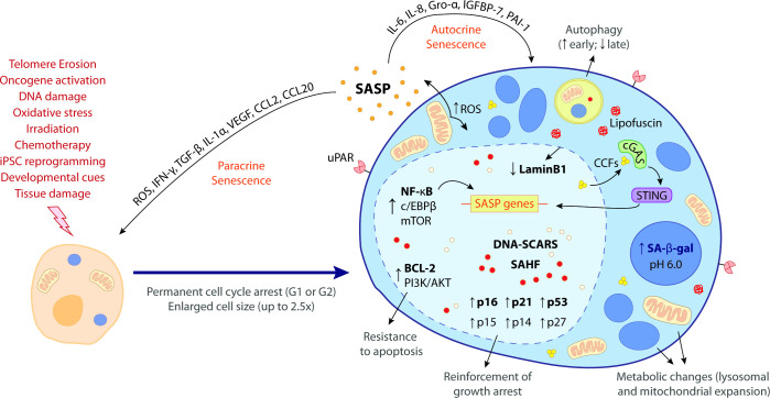
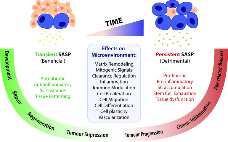
Verification
Claims the paper makes
The claims and statistics of this paper haven’t been checked yet.
Run the check — free, no account needed. It reads the open-access text and checks the sources it cites.
Claims other papers make about this one
Each is checked against this paper’s own text: does it say what the citing paper says it does?
In the citing paper
“Conversely, the progressive accumulation of senescent cells (facilitated by defective immunosurveillance) contributes to the phenomena of tissue fibrosis and lowgrade inflammation that define the aging process and exacerbate the risk of neoplastic transformation.176”
From Meta-hallmarks of aging and cancer (2023) · cites this paper as [176]
Self-check
Percentages, sums and reported differences re-computed from the paper’s own numbers.
The self-check needs the paper’s full text.
Citations
Cites
Takahashi, K., & Yamanaka, S. (2006) · Cell · doi:10.1016/j.cell.2006.07.024 · Cited by 26,673
Induction of Pluripotent Stem Cells from Adult Human Fibroblasts by Defined Factors
Takahashi, K., Tanabe, K., Ohnuki, M., Narita, M., Ichisaka, T., Tomoda, K., & Yamanaka, S. (2007) · Cell · doi:10.1016/j.cell.2007.11.019 · Cited by 20,221
The serial cultivation of human diploid cell strains
Hayflick, L., & Moorhead, P. S. (1961) · Experimental Cell Research · doi:10.1016/0014-4827(61)90192-6 · Cited by 8,508
A biomarker that identifies senescent human cells in culture and in aging skin in vivo.
Dimri, G. P., Lee, X., Basile, G., Acosta, M., Scott, G., Roskelley, C., Medrano, E. E., Linskens, M., Rubelj, I., & Pereira-Smith, O. (1995) · Proceedings of the National Academy of Sciences · doi:10.1073/pnas.92.20.9363 · Cited by 7,477
The limited in vitro lifetime of human diploid cell strains
Hayflick, L. (1965) · Experimental Cell Research · doi:10.1016/0014-4827(65)90211-9 · Cited by 5,876
Telomeres shorten during ageing of human fibroblasts
Harley, C. B., Futcher, A. B., & Greider, C. W. (1990) · Nature · doi:10.1038/345458a0 · Cited by 5,750
Oncogenic ras provokes premature cell senescence associated with accumulation of p53 and p16INK4a
Serrano, M., Lin, A. W., McCurrach, M. E., Beach, D., & Lowe, S. W. (1997) · Cell · doi:10.1016/s0092-8674(00)81902-9 · Cited by 5,218 · cited 2×
“The first experimental evidence of OIS came from the overexpression of oncogenic Ras in human fibroblasts, resulting in upregulation of p53, p16, pRB, and permanent cell cycle arrest (Serrano et al., 1997).”
BackgroundThe senescence-associated secretory phenotype: the dark side of tumor suppression
Coppé, J.-P., Desprez, P.-Y., Krtolica, A., & Campisi, J. (2010) · Annual Review of Pathology: Mechanisms of Disease · doi:10.1146/annurev-pathol-121808-102144 · Cited by 5,206
Chronic inflammation in the etiology of disease across the life span
Furman, D., Campisi, J., Verdin, E., Carrera-Bastos, P., Targ, S., Franceschi, C., Ferrucci, L., Gilroy, D. W., Fasano, A., Miller, G. W., et al. (2019) · Nature Medicine · doi:10.1038/s41591-019-0675-0 · Cited by 4,915
Clearance of p16Ink4a-positive senescent cells delays ageing-associated disorders
Baker, D. J., Wijshake, T., Tchkonia, T., LeBrasseur, N. K., Childs, B. G., van de Sluis, B., Kirkland, J. L., & van Deursen, J. M. (2011) · Nature · doi:10.1038/nature10600 · Cited by 3,670 · cited 1×
“Remarkably, the use of progeroid mouse models has provided evidence that ablation of p16 cells improves many of these disease symptoms, reinforcing a detrimental role of SCs in aged tissues (Baker et al., 2011; Rhinn et al., 2019).”
Background
Cited by
Sorted by citations
Cellular senescence: a key therapeutic target in aging and diseases
Zhang, L., Pitcher, L. E., Yousefzadeh, M. J., Niedernhofer, L. J., Robbins, P. D., & Zhu, Y. (2022) · Journal of Clinical Investigation · doi:10.1172/jci158450 · Cited by 631
Meta-hallmarks of aging and cancer
López-Otín, C., Pietrocola, F., Roiz-Valle, D., Galluzzi, L., & Kroemer, G. (2023) · Cell Metabolism · doi:10.1016/j.cmet.2022.11.001 · Cited by 529
Local immune recognition of trophoblast in early human pregnancy: controversies and questions
Moffett, A., & Shreeve, N. (2022) · Nature reviews. Immunology · doi:10.1038/s41577-022-00777-2 · Cited by 152
Emerging role of senescent microglia in brain aging-related neurodegenerative diseases
Rim, C., You, M., Nahm, M., & Kwon, M. (2024) · Translational Neurodegeneration · doi:10.1186/s40035-024-00402-3 · Cited by 158
Role of cellular senescence in inflammation and regeneration
Saito, Y., Yamamoto, S., & Chikenji, T. (2024) · Inflammation and Regeneration · doi:10.1186/s41232-024-00342-5 · Cited by 127
The aged extracellular matrix and the profibrotic role of senescence-associated secretory phenotype
Mebratu, Y. A., Soni, S., Rosas, L., Rojas, M. M., Horowitz, J. C., & Nho, R. S. (2023) · American Journal of Physiology-Cell Physiology · doi:10.1152/ajpcell.00124.2023 · Cited by 95
TFEB‐dependent lysosome biogenesis is required for senescence
Curnock, R., Yalci, K., Palmfeldt, J., Jäättelä, M., Liu, B., & Carroll, B. (2023) · The EMBO Journal · doi:10.15252/embj.2022111241 · Cited by 91
Cellular senescence and the host immune system in aging and age-related disorders
Giannoula, Y., Kroemer, G., & Pietrocola, F. (2023) · Biomedical Journal · doi:10.1016/j.bj.2023.02.001 · Cited by 81
Jang, D., Shin, J., Shim, E., Ohtani, N., & Jeon, O. H. (2024) · Aging Cell · doi:10.1111/acel.14315 · Cited by 84
A coalition to heal—the impact of the cardiac microenvironment
Tzahor, E., & Dimmeler, S. (2022) · Science · doi:10.1126/science.abm4443 · Cited by 72
Stress-induced β cell early senescence confers protection against type 1 diabetes
Lee, H., Sahin, G. S., Chen, C.-W., Sonthalia, S., Cañas, S. M., Oktay, H. Z., Duckworth, A. T., Brawerman, G. M., Thompson, P. J., Hatzoglou, M., et al. (2023) · Cell Metabolism · doi:10.1016/j.cmet.2023.10.014 · Cited by 66
Roadmap for alleviating the manifestations of ageing in the cardiovascular system
Liberale, L., Tual-Chalot, S., Sedej, S., Ministrini, S., Georgiopoulos, G., Grunewald, M., Bäck, M., Bochaton-Piallat, M.-L., Boon, R. A., Ramos, G. C., et al. (2025) · Nature Reviews Cardiology · doi:10.1038/s41569-025-01130-5 · Cited by 59
Cognitive interventions for memory and psychological well-being in aging and dementias
Dinius, C. J., Pocknell, C. E., Caffrey, M. P., & Roche, R. A. P. (2023) · Frontiers in Psychology · doi:10.3389/fpsyg.2023.1070012 · Cited by 62
Kaur, G., Muthumalage, T., & Rahman, I. (2023) · Aging Cell · doi:10.1111/acel.13850 · Cited by 53
Gut-specific telomerase expression counteracts systemic aging in telomerase-deficient zebrafish
Maï, M. E., Bird, M., Allouche, A., Targen, S., Şerifoğlu, N., Lopes-Bastos, B., Guigonis, J., Kang, D., Pourcher, T., Yue, J., et al. (2023) · Nature Aging · doi:10.1038/s43587-023-00401-5 · Cited by 59
Subcellular structure, heterogeneity, and plasticity of senescent cells
Bitencourt, T. C., Vargas, J. E., Silva, A. O., Fraga, L. R., & Filippi‐Chiela, E. C. (2024) · Aging Cell · doi:10.1111/acel.14154 · Cited by 46
Cell senescence in cardiometabolic diseases
Grootaert, M. O. J. (2024) · npj Aging · doi:10.1038/s41514-024-00170-4 · Cited by 56
Macrophage Involvement in Aging-Associated Skeletal Muscle Regeneration
Cui, C., Ferrucci, L., & Gorospe, M. (2023) · Cells · doi:10.3390/cells12091214 · Cited by 49
Senescence-induced cellular reprogramming drives cnidarian whole-body regeneration
Salinas-Saavedra, M., Febrimarsa, Krasovec, G., Horkan, H. R., Baxevanis, A. D., & Frank, U. (2023) · Cell Reports · doi:10.1016/j.celrep.2023.112687 · Cited by 45
Cell Cycle and Senescence Regulation by Podocyte Histone Deacetylase 1 and 2
Rangel, P. X. M., Cross, E., Liu, C., Pedigo, C. E., Tian, X., Gutiérrez-Calabrés, E., Nagata, S., Priyadarshini, A., Lerner, G., Bunda, P., et al. (2022) · Journal of the American Society of Nephrology · doi:10.1681/asn.2022050598 · Cited by 46
Delenko, J., Xue, X., Chatterjee, P. K., Hyman, N., Shih, A., Adelson, R. P., Tepeš, P. Š., Gregersen, P. K., & Metz, C. N. (2024) · Reproductive Biology and Endocrinology · doi:10.1186/s12958-024-01265-z · Cited by 50
Human iPSC-derived renal collecting duct organoid model cystogenesis in ADPKD
Mae, S.-I., Hattanda, F., Morita, H., Nozaki, A., Katagiri, N., Ogawa, H., Teranaka, K., Nishimura, Y., Kudoh, A., Yamanaka, S., et al. (2023) · Cell Reports · doi:10.1016/j.celrep.2023.113431 · Cited by 42
Wong, G. C., & Chow, H. (2022) · Journal of Alzheimer s Disease · doi:10.3233/jad-220203 · Cited by 41
Senescent cells at the crossroads of aging, disease, and tissue homeostasis
Kuehnemann, C., & Wiley, C. D. (2023) · Aging Cell · doi:10.1111/acel.13988 · Cited by 44
Egr1 regulates regenerative senescence and cardiac repair
Zhang, L., Elkahal, J., Wang, T., Rimmer, R., Genzelinakh, A., Bassat, E., Wang, J., Perez, D. E., Kain, D., Lendengolts, D., et al. (2024) · Nature Cardiovascular Research · doi:10.1038/s44161-024-00493-1 · Cited by 37
Oda, T., Gotoh, N., Kasamatsu, T., Handa, H., Saitoh, T., & Sasaki, N. (2023) · Cell Proliferation · doi:10.1111/cpr.13398 · Cited by 33
Is “cellular senescence” a misnomer?
Gems, D., & Kern, C. C. (2022) · GeroScience · doi:10.1007/s11357-022-00652-x · Cited by 33
Senolytic treatment for low back pain
Mannarino, M., Cherif, H., Darband, S. G., Martinez, O. W., Sheng, K., Cousineau, E., Lee, S., Millecamps, M., Gao, C., Gilbert, A., et al. (2025) · Science Advances · doi:10.1126/sciadv.adr1719 · Cited by 33
Tieing together loose ends: telomere instability in cancer and aging
Borges, G. D. O., Criqui, M., & Harrington, L. A. (2022) · Molecular Oncology · doi:10.1002/1878-0261.13299 · Cited by 34
Endothelial Senescence and Its Impact on Angiogenesis in Alzheimer’s Disease
Georgieva, I., Tchekalarova, J. D., Iliev, D. B., & Tzoneva, R. (2023) · International Journal of Molecular Sciences · doi:10.3390/ijms241411344 · Cited by 29
Understanding and targeting senescence in kidney disease
Chanvillard, L., Mason, T., & Ferenbach, D. A. (2025) · Clinical Kidney Journal · doi:10.1093/ckj/sfaf190 · Cited by 28
Yang, F., Shu, R., Dai, W., Li, B., Liu, C., Yang, H., Johnson, H. M., Yu, S., Bai, D., Yang, W., et al. (2024) · Biomaterials · doi:10.1016/j.biomaterials.2024.122659 · Cited by 25
Adenosine A2A receptor activation reduces chondrocyte senescence
Friedman, B., Larranaga‐Vera, A., Castro, C. M., Corciulo, C., Rabbani, P. S., & Cronstein, B. N. (2023) · The FASEB Journal · doi:10.1096/fj.202201212rr · Cited by 23
The context-dependent effect of cellular senescence: From embryogenesis and wound healing to aging
Lavarti, R., Alvarez-Diaz, T., Marti, K., Kar, P., & Raju, R. P. (2025) · Ageing Research Reviews · doi:10.1016/j.arr.2025.102760 · Cited by 21
Qamsari, E. S., & Stewart, D. J. (2024) · Frontiers in Immunology · doi:10.3389/fimmu.2024.1403669 · Cited by 22
Senescence: a double-edged sword in beta-cell health and failure?
Varghese, S. S., & Dhawan, S. (2023) · Frontiers in Endocrinology · doi:10.3389/fendo.2023.1196460 · Cited by 23
Lack of apoptosis leads to cellular senescence and tumorigenesis in Drosophila epithelial cells
Garcia-Arias, J. M., Pinal, N., Cristobal-Vargas, S., Estella, C., & Morata, G. (2023) · Cell Death Discovery · doi:10.1038/s41420-023-01583-y · Cited by 19
Transposon control as a checkpoint for tissue regeneration
Angileri, K. M., Bagia, N. A., & Feschotte, C. (2022) · Development · doi:10.1242/dev.191957 · Cited by 15
Targeting senescent cells in atherosclerosis: Pathways to novel therapies
Tian, Y., Shao, S., Feng, H., Zeng, R., Li, S., & Zhang, Q. (2024) · Ageing Research Reviews · doi:10.1016/j.arr.2024.102502 · Cited by 15
The Potent and Paradoxical Biology of Cellular Senescence in Cancer
Romesser, P. B., & Lowe, S. W. (2023) · Annual Review of Cancer Biology · doi:10.1146/annurev-cancerbio-061421-124434 · Cited by 15
Fujita, M., Sasada, M., Iyoda, T., & Fukai, F. (2024) · International Journal of Molecular Sciences · doi:10.3390/ijms25126591 · Cited by 14
Morelli, N. R., Préfontaine, C., Pipella, J., & Thompson, P. J. (2024) · American Journal of Physiology-Endocrinology and Metabolism · doi:10.1152/ajpendo.00257.2024 · Cited by 12
Specific types of male infertility are correlated with T cell exhaustion or senescence signatures
Amodio, G., Giacomini, G., Boeri, L., Raffo, M., Cilio, S., Pozzi, E., Belladelli, F., Negri, F., Ferrara, A. M., d’Arma, A., et al. (2025) · Nature Communications · doi:10.1038/s41467-025-56193-2 · Cited by 16
Increased chromatin accessibility underpins senescence
Lopes‐Paciencia, S., & Ferbeyre, G. (2025) · FEBS Journal · doi:10.1111/febs.70136 · Cited by 18
Jiang, R., Fang, Z., Lai, Y., Li, L., Tan, J., Yu, C., Fan, M., Tao, L., Shen, W., Xu, C., et al. (2024) · European Journal of Pharmacology · doi:10.1016/j.ejphar.2024.176318 · Cited by 13
Mapping the core senescence phenotype of primary human colon fibroblasts
Hattangady, N. G., Carter, K. T., Maroni-Rana, B., Wang, T., Ayers, J., Yu, M., & Grady, W. M. (2024) · Aging · doi:10.18632/aging.205577 · Cited by 12
Targeted delivery strategy: A beneficial partner for emerging senotherapy
Zhang, Q., Li, S., Chen, F., Zeng, R., & Tong, R. (2022) · Biomedicine & Pharmacotherapy · doi:10.1016/j.biopha.2022.113737 · Cited by 12
Venz, R., Goyala, A., Soto-Gamez, A., Yenice, T., Demaria, M., & Ewald, C. Y. (2023) · GeroScience · doi:10.1007/s11357-023-00909-z · Cited by 13
Kang, T., Moore, E. C., Kopania, E. E. K., King, C. D., Schilling, B., Campisi, J., Good, J. M., & Brem, R. B. (2023) · G3: Genes, Genomes, Genetics · doi:10.1093/g3journal/jkad091 · Cited by 10
Hultberg, J., Blixt, E., Göransson, R., Adolfsson, J., Govender, M., Larsson, M., Nilsdotter‐Augustinsson, Å., Ernerudh, J., & Nyström, S. N. (2023) · Clinical Immunology · doi:10.1016/j.clim.2023.109816 · Cited by 10
Tejerina-Miranda, S., Pérez‐Ginés, V., Torrente-Rodríguez, R. M., Pedrero, M., Montero‐Calle, A., Pingarrón, J. M., Barderas, R., & Campuzano, S. (2024) · Sensors & Diagnostics · doi:10.1039/d3sd00311f · Cited by 10
Datta, I., & Bangi, E. (2024) · Developmental Cell · doi:10.1016/j.devcel.2024.01.009 · Cited by 9
Harada, M., Su-Harada, K., Kimura, T., Ono, K., & Ashida, N. (2024) · Cell Cycle · doi:10.1080/15384101.2024.2325802 · Cited by 10
Liu, C., Pan, J., & Bao, Q. (2025) · Molecular Biology Reports · doi:10.1007/s11033-025-10338-0 · Cited by 13
Ahmed, S., Gozal, D., & Khalyfa, A. (2025) · Sleep Medicine Reviews · doi:10.1016/j.smrv.2025.102170 · Cited by 12
Is Senolytic Therapy in Cardiovascular Diseases Ready for Translation to Clinics?
Yang, Z., Potenza, D. M., & Ming, X. (2025) · Biomolecules · doi:10.3390/biom15040545 · Cited by 10
Polzer, O., Kinloch, E., & Fitzsimons, C. P. (2025) · Aging Brain · doi:10.1016/j.nbas.2025.100141 · Cited by 10
Cellular senescence contributes to spontaneous repair of the rat meniscus
Aimono, Y., Endo, K., & Sekiya, I. (2024) · Aging Cell · doi:10.1111/acel.14385 · Cited by 11
ACSL5 regulated acetyl-CoA to promote bladder cancer cellular senescence via 53BP1 acetylation
Luo, Y., Zeng, Y., Liu, Y., Liu, S., Dai, Y., Pan, W., Zhang, L., Zhu, R., Feng, D., Du, K., et al. (2025) · Oncogene · doi:10.1038/s41388-025-03474-1 · Cited by 10
Promise and Perspective of Nanomaterials in Antisenescence Tissue Engineering Applications
Lu, Z., Singh, G., Lesani, P., & Zreiqat, H. (2022) · ACS Biomaterials Science & Engineering · doi:10.1021/acsbiomaterials.1c01298 · Cited by 8
Cellular senescence in the cancer microenvironment
Meguro, S., & Nakanishi, M. (2025) · The Journal of Biochemistry · doi:10.1093/jb/mvaf001 · Cited by 7
The IGF-1 senescence switch: a biphasic model for SASP-driven aging and precision senomodulation
Manni, E., Al‐kuraishy, H. M., Hussain, N. R., Abdelaziz, A. M., & Batiha, G. E. (2026) · Cytokine · doi:10.1016/j.cyto.2026.157143 · Cited by 8
Cell Senescence and the Genetics of Melanoma Development
Constantinou, S. M., & Bennett, D. C. (2024) · Genes Chromosomes and Cancer · doi:10.1002/gcc.23273 · Cited by 7
Juvenile zebrafish (Danio rerio) are able to recover from lordosis
Printzi, A., Mazurais, D., Witten, P. E., Madec, L., Gonzalez, A., Mialhe, X., Zambonino‐Infante, J., & Koumoundouros, G. (2022) · Scientific Reports · doi:10.1038/s41598-022-26112-2 · Cited by 7
Overview of molecular signatures of senescence and associated resources: pros and cons
Ntintas, O. A., Vagena, S., Pantelis, P., Theocharous, G., Petty, R., Evangelou, K., & Gorgoulis, V. G. (2026) · FEBS Open Bio · doi:10.1002/2211-5463.70134 · Cited by 6
Chromatin architectural factor CTCF is essential for progesterone‐dependent uterine maturation
Hewitt, S. C., Gruzdev, A., Willson, C. J., Wu, S., Lydon, J. P., Galjart, N., & DeMayo, F. J. (2023) · The FASEB Journal · doi:10.1096/fj.202300862r · Cited by 6
Cellular Senescence in Craniofacial Tissue Regeneration: Inducers, Biomarkers, and Interventions
Tang, W., Huo, F., Long, J., Zhang, S., & Tian, W. (2023) · Tissue Engineering Part B Reviews · doi:10.1089/ten.teb.2023.0136 · Cited by 6
Chandrasegaran, S., Sluka, J. P., & Shanley, D. P. (2025) · PLoS Computational Biology · doi:10.1371/journal.pcbi.1012298 · Cited by 8
Júnior, J. X. D. N., Gomes, J. D. C., Filho, R. I., Valença, H. D. M., Branco, J. R., Araújo, A. B., Moreira, A. D. O. E., Crepaldi, L. D., Paixão, L. P., Ochioni, A. C., et al. (2025) · Communications Biology · doi:10.1038/s42003-025-07451-y · Cited by 5
Akagi, K., Koizumi, K., Kadowaki, M., Kitajima, I., & Saito, S. (2023) · Cells · doi:10.3390/cells12182297 · Cited by 5
Impact of Aging and Cellular Senescence in the Pathophysiology of Preeclampsia
Suvakov, S. R., Kattah, A. G., Gojković, T., Enninga, E. A. L., Pruett, J. E., Jayachandran, M., Sousa, C., Santos, J., Hassan, C. A., Suarez, M. L. G., et al. (2023) · Comprehensive physiology · doi:10.1002/cphy.c230003 · Cited by 6
Cellular senescence in reproduction: a two-edged sword
Pretto, L., Nabinger, E., Filippi‐Chiela, E. C., & Fraga, L. R. (2024) · Biology of Reproduction · doi:10.1093/biolre/ioae025 · Cited by 5
de Mendonça, L. B., Lenz, G., & Filippi‐Chiela, E. C. (2025) · Biochemical Society Transactions · doi:10.1042/bst20253054 · Cited by 5
Immunological consequences of senescence in physiology and pathology
Zubova, A., Pietrocola, F., & Morsli, S. (2026) · Journal of Translational Medicine · doi:10.1186/s12967-026-08335-3 · Cited by 6
Damage-Induced Senescent Immune Cells Regulate Regeneration of the Zebrafish Retina
Patton, J. G., Konar, G. J., Flickinger, Z., Sharma, S., Vallone, K. T., Lyon, C. E., Doshier, C., Lingan, A. L., & Lyon, W. (2024) · doi:10.59368/agingbio.20240021 · Cited by 4
Natural Product-Derived Senotherapeutics: Extraction and Biological Evaluation Techniques
Pratsinis, H., Mavrogonatou, E., Zervou, S., Triantis, T. M., Hiskia, A. E., & Kletsas, D. (2025) · Methods in molecular biology · doi:10.1007/978-1-0716-4426-3_19 · Cited by 4
Zhang, F., Wang, Y., Zhang, L., Wang, C., Chen, D., Liu, H., Xu, R., Haynes, C. M., Shim, J., & Ge, X. (2025) · eLife · doi:10.7554/elife.101984.3 · Cited by 3
Cellular Senescence in Cancer: Mechanisms, Roles in Tumor Progression, and Therapeutic Implications
Yan, J., Zhang, Y., Mao, G., Yu, J., Zhou, T., & Hao, J. (2025) · MedComm – Oncology · doi:10.1002/mog2.70029 · Cited by 3
Martic, I., Guerrero‐Navarro, L., Cappuccio, E., Hassan, A., Jenewein, B., Arcalís, E., Hrapovic, N., Visdal‐Johnsen, L., Declercq, L., Jansen‐Dürr, P., et al. (2025) · Scientific Reports · doi:10.1038/s41598-025-28590-6 · Cited by 5
Chromatin Structure from Development to Ageing
Ayala-Guerrero, L., Claudio-Galeana, S., Furlan-Magaril, M., & Castro‐Obregón, S. (2023) · Sub-cellular biochemistry/Subcellular biochemistry · doi:10.1007/978-3-031-21410-3_2 · Cited by 2
Survey on large language model annotation of cellular senescence from figures in review articles
Yamagata, Y., & Yamada, R. (2024) · Genomics & Informatics · doi:10.1186/s44342-024-00011-6 · Cited by 2
A Potential Contribution of S100A11 to Skin Fibrosis and Pulmonary Involvement in Systemic Sclerosis
Takahashi, T., Takahashi, T., Segawa, Y., Ikawa, T., Terui, H., Takahashi, T., Sumida, H., Yoshizaki, A., Sato, S., & Asano, Y. (2024) · Experimental Dermatology · doi:10.1111/exd.70026 · Cited by 2
Two roads to fibrosis: Contrasting initiating mechanisms of Bleomycin and TGFβ-1 in lung fibroblasts
D’Alessandro, N., Möbus, L., del Giudice, G., Perho, N., Serra, A., & Greco, D. (2025) · Toxicology · doi:10.1016/j.tox.2025.154233 · Cited by 2
DRUGS WITH SENOLYTIC ACTIVITY: PROSPECTS AND POSSIBLE LIMITATIONS
Васильевна, М. Г., & Khokhlov, A. N. (2024) · Vestnik Moskovskogo universiteta Seria 16 Biologia · doi:10.55959/msu0137-0952-16-78-4-3 · Cited by 2
Drugs with Senolytic Activity: Prospects and Possible Limitations
Васильевна, М. Г., & Khokhlov, A. N. (2023) · Moscow University Biological Sciences Bulletin · doi:10.3103/s0096392524600455 · Cited by 2
Zhang, F., Wang, Y., Zhang, L., Wang, C., Chen, D., Liu, H., Xu, R., Haynes, C. M., Shim, J., & Ge, X. (2024) · eLife · doi:10.7554/elife.101984 · Cited by 2
Cellular senescence perspective of mTOR in the delay of aging process
Armadivin, F. H., Achadiyani, A., & Atik, N. (2024) · Journal Of Advanced Pharmacy Education And Research · doi:10.51847/ovga9z0wdb · Cited by 1
Cheng, H., Zhao, Z.-W., Liu, D., Wang, Y., & Zhang, M. (2024) · Journal of Zhejiang University SCIENCE B · doi:10.1631/jzus.b2400013 · Cited by 1
Fibrocartilage repair involves chronic cellular senescence in a rat model of bone marrow stimulation
Childress, L., Gatrell, L. B., Wu, H., Ferreira, E., Nookaew, I., & Porter, R. M. (2025) · Osteoarthritis and Cartilage Open · doi:10.1016/j.ocarto.2025.100620 · Cited by 2
Calma, M. L., Navasumrit, P., Parnlob, V., Rattanayut, P., & Ruchirawat, M. (2026) · Ecotoxicology and Environmental Safety · doi:10.1016/j.ecoenv.2025.119670 · Cited by 1
Valieva, Y., Igrunkova, A., Fayzullin, A. L., Serejnikova, N. B., Kurkov, A. V., Fayzullina, N., Valishina, D., Bakulina, A., Timashev, P. S., & Shekhter, A. B. (2023) · Biology · doi:10.3390/biology12040565 · Cited by 2
Zhang, F., Wang, Y., Zhang, L., Wang, C., Chen, D., Liu, H., Xu, R., Haynes, C. M., Shim, J., & Ge, X. (2020) · bioRxiv (Cold Spring Harbor Laboratory) · doi:10.1101/2020.08.03.233874
An Overview of the Role of Senescence in Cancer
Al-Janabi, I. I. (2022) · Al-Rafidain Journal of Medical Sciences · doi:10.54133/ajms.v3i.77
Albagli, O., & Pelczar, H. (2023) · médecine/sciences · doi:10.1051/medsci/2022186
Kopsidas, C. A., Lowe, C. C., Zhang, J., Kang, W., Zhou, X., & Feng, Y. (2023) · bioRxiv (Cold Spring Harbor Laboratory) · doi:10.1101/2023.01.26.525738
Lack of apoptosis leads to cellular senescence and tumorigenesis in Drosophila epithelial cellsPreprint
García-Arias, J. M., Pinal, N., Cristobal‐Vargas, S., Estella, C., & Morata, G. (2023) · bioRxiv (Cold Spring Harbor Laboratory) · doi:10.1101/2023.05.08.539867
Senescent Subtypes Transition ModelPreprint
Singh, G. (2023) · bioRxiv (Cold Spring Harbor Laboratory) · doi:10.1101/2023.06.10.544466
Cellular senescence is required for cardiac regeneration
(2024) · Nature Cardiovascular Research · doi:10.1038/s44161-024-00494-0
Kaur, G., Wang, Q., Tjitropranoto, A., Unwalla, H. J., & Rahman, I. (2024) · Experimental Lung Research · doi:10.1080/01902148.2024.2414974 · Cited by 1
Zhang, F., Wang, Y., Zhang, L., Wang, C., Chen, D., Liu, H., Xu, R., Haynes, C. M., Shim, J., & Ge, X. (2024) · eLife · doi:10.7554/elife.101984.1
Metadata
- Authors
- Paramos-de-Carvalho, D., Jacinto, A., & Saúde, L.
- Affiliations
- University of Lisbon (PT)
- CEDOC, NOVA Medical School, Faculdade de Ciências Médicas da Universidade Nova de Lisboa
- Corresponding author
- Paramos-de-Carvalho, Diogo
- Saúde, Leonor
- Venue
- eLife
- Publisher
- eLife Sciences Publications, Ltd
- Published
- 2021-11-10
- Volume
- 10
- ISSN
- 2050-084X (ISSN-L)
- PMC
- PMC8580479
- OpenAlex
- W3211748729
- Cited by
- 111
- References
- 123
- Citations by year
- 2026: 8 · 2025: 24 · 2024: 30 · 2023: 34 · 2022: 14
- FWCI
- 2.60
- Type
- journal article
- Language
- en
- Open access
- gold
- doi.org
- Licence
- Locations
- eLife (publishedVersion, cc-by)
- PubMed Central (publishedVersion, cc-by)
- PubMed (publishedVersion)
- Portuguese National Funding Agency for Science, Research and Technology (RCAAP Project by FCT) (submittedVersion, other-oa)
- DOAJ (DOAJ: Directory of Open Access Journals) (submittedVersion, cc-by-sa)
- Funders
- Fundação para a Ciência e a Tecnologia (PD/BD/105770/2014, PTDC/MED-NEU/30428/2017, HR18-00187)
- "la Caixa" Banking Foundation (HR18-00187)
- Santa Casa da Misericórdia de Lisboa (MC36-2020 (Melo e Castro Award))
- Topics
- Life Sciences › Biochemistry, Genetics and Molecular Biology › Cell Biology › Telomeres, Telomerase, and Senescence
- Life Sciences › Biochemistry, Genetics and Molecular Biology › Aging › Genetics, Aging, and Longevity in Model Organisms
- Health Sciences › Medicine › Geriatrics and Gerontology › Sirtuins and Resveratrol in Medicine
- Keywords
- cellular senescence, senolytics, tissue repair, tissue regeneration, fibrosis, inflammation, aging, stress response, cancer
- MeSH terms
- Aging, Animals, Fibrosis, Humans, Inflammation, Regeneration, Cellular Senescence
About these checks
Registry, retraction, Cites and statistics results are computed, not judged, with one exception: a printed reference that no index lists is read once by a model before it counts as a citation error. Claims are extracted by a model and kept only when their quote appears verbatim in the text; each cited source is then checked, with a model’s help, for whether it says what the claim relies on. Those verdicts can be wrong — read the claim’s quote and the cited source before relying on one.
Statistics and arithmetic are recomputed, not judged.
Confidence bands are shown only for fields calibrated on our annotated test set. None is calibrated yet, so no band is shown.
What counts as a claim ›How we measure confidence ›How we check this ›
Are you an author, or do you see a problem with this page? Request a review
