

The DOI matches its Crossref record.
Dysphagia as the Presenting Symptom of Laryngeal Tuberculosis
Open accessAbstract
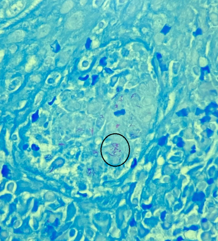
Laryngeal tuberculosis (TB) is a rare form of tuberculosis that most commonly presents with dysphagia and weight loss. We report a case of a 75-year-old Vietnamese male who presented with dysphagia, odynophagia, and a 30-pound weight loss over the two months prior to presentation. Nasopharyngeal evaluation with microdirect laryngoscopy was performed as part of the workup and revealed lesions of the epiglottis and left vocal cord. A tissue biopsy and quantiferon testing confirmed the diagnosis of tuberculosis. The patient was started on quadruple therapy and is currently receiving treatment. This case highlights the need for consideration of the rare, yet important, differential of laryngeal TB in patients presenting with non-specific complaints such as dysphagia and weight loss.
Overview
The claims and statistics of this paper haven’t been checked yet.
Run the check — free, no account needed. It reads the open-access text and checks the sources it cites.
Integrity signals
Registry
Cites
Statistics
Figures
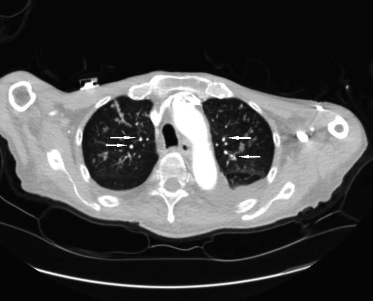
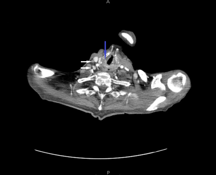
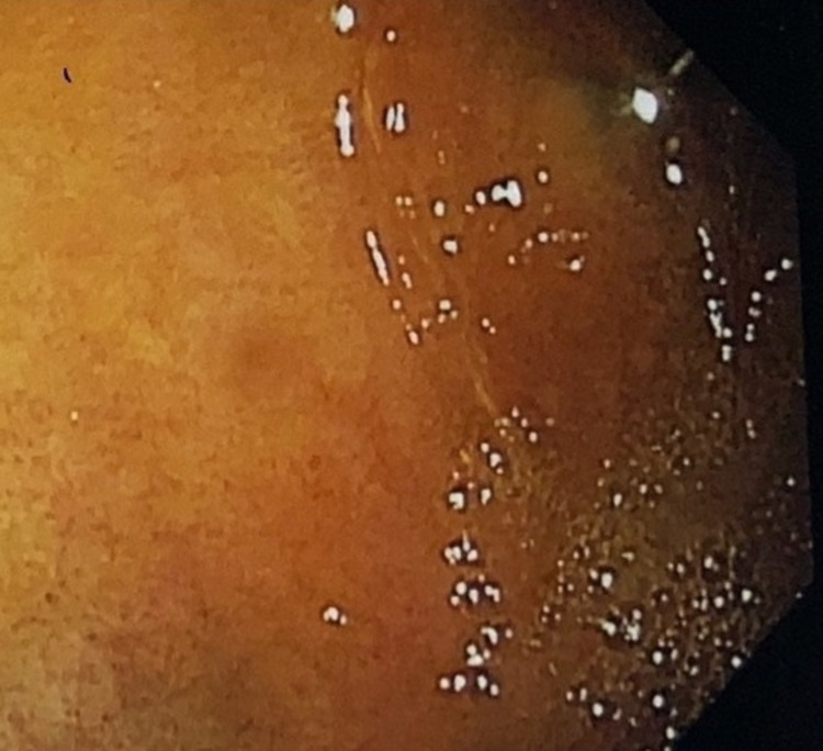
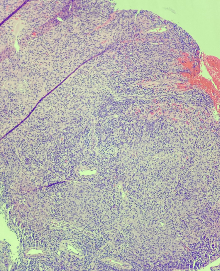
Verification
Claims the paper makes
The claims and statistics of this paper haven’t been checked yet.
Run the check — free, no account needed. It reads the open-access text and checks the sources it cites.
Claims other papers make about this one
Each is checked against this paper’s own text: does it say what the citing paper says it does?
The source backs part of the claim, not all of it — or backs it for a different population, condition or comparison.
In the citing paper
“Laryngeal TB is another notable manifestation, though less common than enlarged cervical lymph nodes. It can present with symptoms such as dysphonia and dysphagia [22,36].”
From Head and Neck Tuberculosis: A Rare Diagnosis and the Role of Surgical Biopsy and Histopathological Evaluation in Extrapulmonary Disease (2025) · cites this paper as [36]
Experimental — accuracy not yet measured
Self-check
Percentages, sums and reported differences re-computed from the paper’s own numbers.
The self-check needs the paper’s full text.
Citations
Cites
No references could be resolved yet.
Research works printed but not linked: 8
- Dheda K, Barry CE 3rd, Maartens G. Tuberculosis. Lancet. 2016, 19:1211-26. 10.1016/S0140-6736(15)00151-8 · DOI
- Furin J, Cox H, Pai M. Tuberculosis. Lancet. 2019, 20:1642-1656. 10.1016/S0140-6736(19)30308-3 · DOI
- Wang CC, Lin CC, Wang CP, Liu SA, Jiang RS. Laryngeal tuberculosis: a review of 26 cases. Otolaryngol Head Neck Surg. 2007, 137:582-8. 10.1016/j.otohns.2007.04.002 · DOI
- Benwill JL, Sarria JC. Laryngeal tuberculosis in the United States of America: a forgotten disease. Scand J Infect Dis. 2014, 46:241-9. 10.3109/00365548.2013.877157 · DOI
- Kiakojuri K, Hasanjani Roushan MR. Laryngeal tuberculosis without pulmonary involvement. Caspian J Intern Med. 2012, 3:397-9.
- Sakthivel P, Singh CA, Sharma SC, Kanodia A, Jat B, Rajeshwari M. Primary laryngeal tuberculosis-“the great masquerader”. Clin Case Rep Rev. 2017, 3:1-3. 10.15761/CCRR.1000335 · DOI
- Kurokawa M, Nibu K, Ichimura K, Nishino H. Laryngeal tuberculosis: a report of 17 cases. Auris Nasus Larynx. 2015, 42:305-10. 10.1016/j.anl.2015.02.012 · DOI
- El Kettani NE, El Hassani M, Chakir N, Jiddane M. Primary laryngeal tuberculosis mimicking laryngeal carcinoma: CT scan features. Indian J Radiol Imaging. 2010, 20:11-2. · DOI
Cited by
Sorted by citations
Mogoantă, C. A., Osman, A., Georgescu, A.-M., Mitroi, A. M., Busuioc, C. I., Tănase, I., Cioboată, R., Liliac, I. M., Cimpeanu, O. L., & Ciolofan, M. S. (2025) · Pathogens · doi:10.3390/pathogens14050479 · Cited by 2
“It can present with symptoms such as dysphonia and dysphagia [22,36].”
Metadata
- Authors
- Kandah, E., Konda, R., Malik, B., Madadha, A., & Kunadi, A.
- Affiliations
- University of Jordan (JO)
- Corresponding author
- Kandah, Emad
- Venue
- Cureus
- Published
- 2021-04-15
- Volume
- 13
- Issue
- 4
- Pages
- e14495-e14495
- ISSN
- 2168-8184 (ISSN-L)
- PMC
- PMC8122223
- OpenAlex
- W3156873650
- Cited by
- 4
- References
- 0
- Citations by year
- 2025: 2 · 2023: 1 · 2021: 1
- FWCI
- 0.47
- Type
- journal article
- Language
- en
- Open access
- diamond
- doi.org
- Licence
- cc-by
- Locations
- Cureus (publishedVersion, cc-by)
- PubMed Central (publishedVersion, cc-by)
- PubMed (publishedVersion)
- Topics
- Health Sciences › Medicine › Infectious Diseases › Diagnosis and treatment of tuberculosis
- Health Sciences › Medicine › Infectious Diseases › Tuberculosis Research and Epidemiology
- Health Sciences › Medicine › Infectious Diseases › Infectious Diseases and Tuberculosis
- Keywords
- laryngeal tuberculosis, dysphagia, odynophagia, weight loss, vocal cord lesions, Quantiferon test, quadruple therapy, differential diagnosis
About these checks
Registry, retraction, Cites and statistics results are computed, not judged, with one exception: a printed reference that no index lists is read once by a model before it counts as a citation error. Claims are extracted by a model and kept only when their quote appears verbatim in the text; each cited source is then checked, with a model’s help, for whether it says what the claim relies on. Those verdicts can be wrong — read the claim’s quote and the cited source before relying on one.
Statistics and arithmetic are recomputed, not judged.
Confidence bands are shown only for fields calibrated on our annotated test set. None is calibrated yet, so no band is shown.
What counts as a claim ›How we measure confidence ›How we check this ›
Are you an author, or do you see a problem with this page? Request a review
How to cite
Cite CoChat for the checks on this page, and the paper for its own findings.
- This page
- CoChat. (2026). Citation check: Dysphagia as the Presenting Symptom of Laryngeal Tuberculosis. https://app.cochat.ai/papers/10.7759/cureus.14495
- The paper
- Kandah, E., Konda, R., Malik, B., Madadha, A., & Kunadi, A. (2021). Dysphagia as the Presenting Symptom of Laryngeal Tuberculosis. Cureus. https://doi.org/10.7759/cureus.14495
