

The DOI matches its Crossref record; 32 of the 32 references it cites were found in a registry or an index.
Evaluation of Combined Artificial Intelligence and Radiologist Assessment to Interpret Screening Mammograms
Open accessAbstract
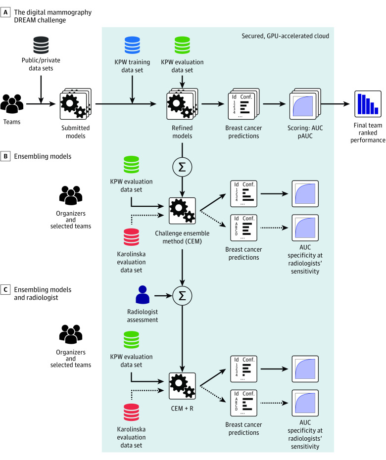
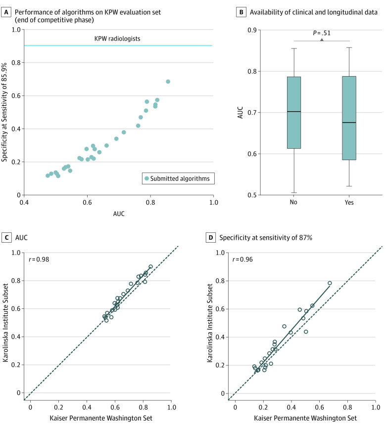
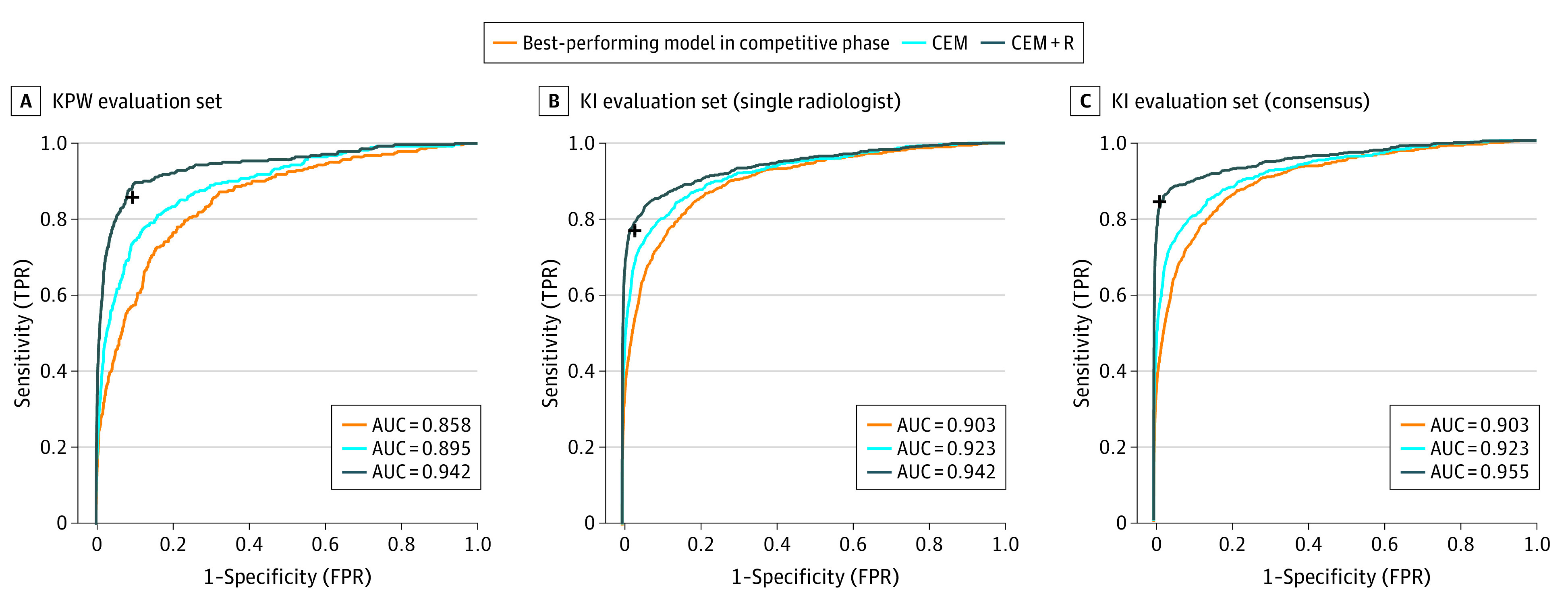
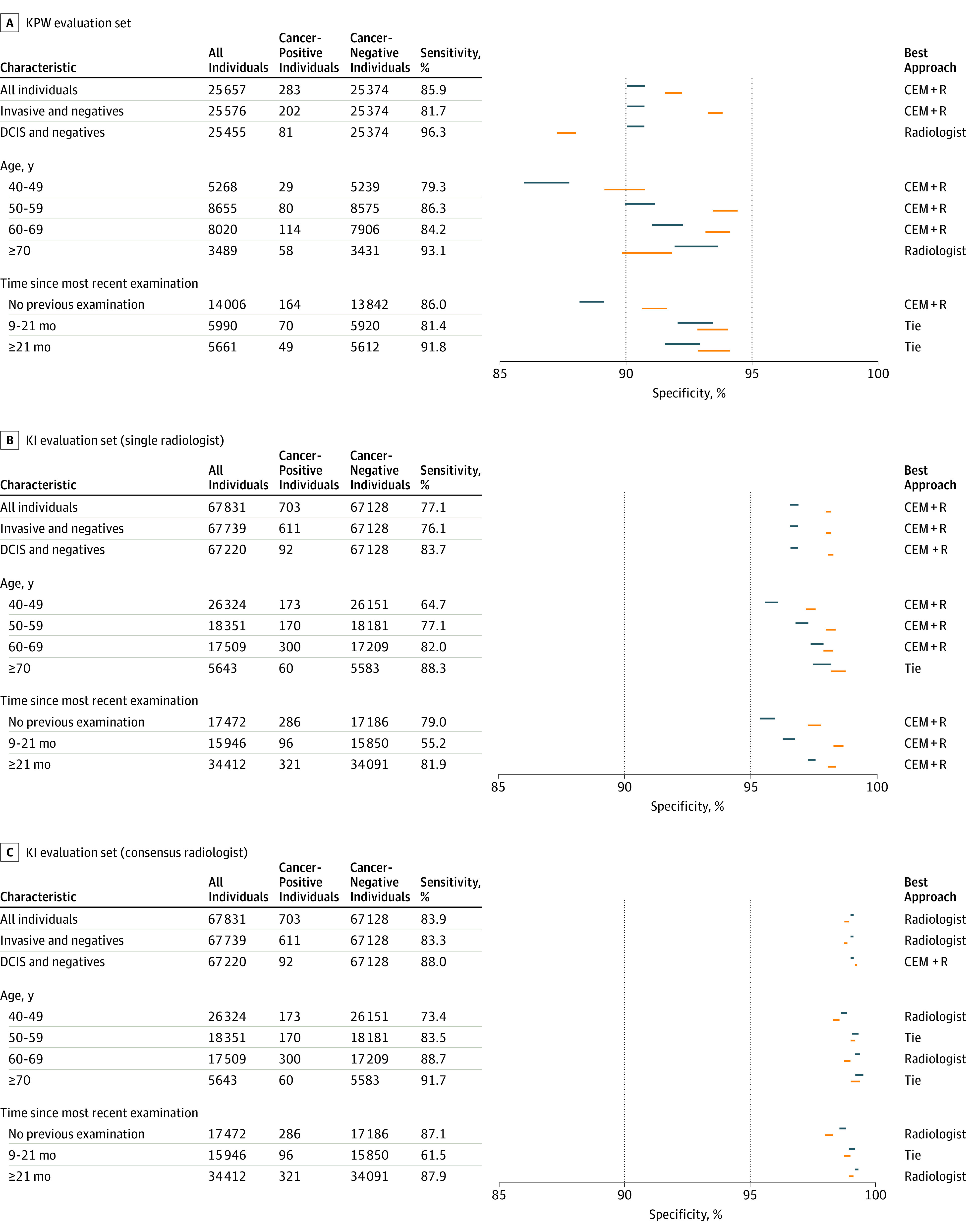
Importance: Mammography screening currently relies on subjective human interpretation. Artificial intelligence (AI) advances could be used to increase mammography screening accuracy by reducing missed cancers and false positives. Objective: To evaluate whether AI can overcome human mammography interpretation limitations with a rigorous, unbiased evaluation of machine learning algorithms. Design, Setting, and Participants: In this diagnostic accuracy study conducted between September 2016 and November 2017, an international, crowdsourced challenge was hosted to foster AI algorithm development focused on interpreting screening mammography. More than 1100 participants comprising 126 teams from 44 countries participated. Analysis began November 18, 2016. Main Outcomes and Measurements: Algorithms used images alone (challenge 1) or combined images, previous examinations (if available), and clinical and demographic risk factor data (challenge 2) and output a score that translated to cancer yes/no within 12 months. Algorithm accuracy for breast cancer detection was evaluated using area under the curve and algorithm specificity compared with radiologists' specificity with radiologists' sensitivity set at 85.9% (United States) and 83.9% (Sweden). An ensemble method aggregating top-performing AI algorithms and radiologists' recall assessment was developed and evaluated. Results: Overall, 144 231 screening mammograms from 85 580 US women (952 cancer positive ≤12 months from screening) were used for algorithm training and validation. A second independent validation cohort included 166 578 examinations from 68 008 Swedish women (780 cancer positive). The top-performing algorithm achieved an area under the curve of 0.858 (United States) and 0.903 (Sweden) and 66.2% (United States) and 81.2% (Sweden) specificity at the radiologists' sensitivity, lower than community-practice radiologists' specificity of 90.5% (United States) and 98.5% (Sweden). Combining top-performing algorithms and US radiologist assessments resulted in a higher area under the curve of 0.942 and achieved a significantly improved specificity (92.0%) at the same sensitivity. Conclusions and Relevance: While no single AI algorithm outperformed radiologists, an ensemble of AI algorithms combined with radiologist assessment in a single-reader screening environment improved overall accuracy. This study underscores the potential of using machine learning methods for enhancing mammography screening interpretation.
Overview
The claims and statistics of this paper haven’t been checked yet.
Run the check — free, no account needed. It reads the open-access text and checks the sources it cites.
Integrity signals
Registry
Cites
Statistics
Figures
Verification
Claims the paper makes
The claims and statistics of this paper haven’t been checked yet.
Run the check — free, no account needed. It reads the open-access text and checks the sources it cites.
Claims other papers make about this one
Each is checked against this paper’s own text: does it say what the citing paper says it does?
In the citing paper
“An early catalyst in this space was the Digital Mammography DREAM Challenge, an open AI competition where groups were able to benchmark and compare different AI approaches (8).”
From Artificial Intelligence in Oncology: Current Landscape, Challenges, and Future Directions (2024) · cites this paper as [8]
Self-check
Percentages, sums and reported differences re-computed from the paper’s own numbers.
The self-check needs the paper’s full text.
Citations
Cites
ImageNet classification with deep convolutional neural networks
Krizhevsky, A., Sutskever, I., & Hinton, G. E. (2017) · Communications of the ACM · doi:10.1145/3065386 · Cited by 112,155
Faster R-CNN: Towards Real-Time Object Detection with Region Proposal Networks
Ren, S., He, K., Girshick, R., & Sun, J. (2017) · IEEE Transactions on Pattern Analysis and Machine Intelligence · doi:10.1109/tpami.2016.2577031 · Cited by 73,472
Faster R-CNN: Towards Real-Time Object Detection with Region Proposal Networks
Ren, S., He, K., Girshick, R., & Sun, J. (2015) · arXiv · doi:10.48550/arxiv.1506.01497 · Cited by 70,812
Regularization Paths for Generalized Linear Models via Coordinate Descent
Friedman, J., Hastie, T., & Tibshirani, R. (2010) · Journal of Statistical Software · doi:10.18637/jss.v033.i01 · Cited by 19,927
Dermatologist-level classification of skin cancer with deep neural networks
Esteva, A., Kuprel, B., Novoa, R. A., Ko, J., Swetter, S. M., Blau, H. M., & Thrun, S. (2017) · Nature · doi:10.1038/nature21056 · Cited by 14,362
Wolpert, D. H. (1992) · Neural Networks · doi:10.1016/s0893-6080(05)80023-1 · Cited by 7,951
Predicting the future - big data, machine learning, and clinical medicine
Obermeyer, Z., & Emanuel, E. J. (2016) · New England Journal of Medicine · doi:10.1056/nejmp1606181 · Cited by 3,652
Prediction of cardiovascular risk factors from retinal fundus photographs via deep learning
Poplin, R., Varadarajan, A. V., Blumer, K., Liu, Y., McConnell, M. V., Corrado, G. S., Peng, L., & Webster, D. R. (2018) · Nature Biomedical Engineering · doi:10.1038/s41551-018-0195-0 · Cited by 1,865
STARD 2015: An Updated List of Essential Items for Reporting Diagnostic Accuracy Studies
Bossuyt, P. M., Reitsma, J. B., Bruns, D. E., Gatsonis, C., Glasziou, P., Irwig, L., Lijmer, J. G., Moher, D., Rennie, D., de Vet, H. C. W., et al. (2015) · Radiology · doi:10.1148/radiol.2015151516 · Cited by 1,360
Screening for Breast Cancer: An Update for the U.S. Preventive Services Task Force
Nelson, H. D., Tyne, K., Naik, A. M., Bougatsos, C., Chan, B. K. S., & Humphrey, L. L. (2009) · Annals of Internal Medicine · doi:10.7326/0003-4819-151-10-200911170-00009 · Cited by 1,328
Research works printed but not linked: 3
- Kuhn, A Short Introduction to the Caret Package
- SAGE Bionetworks. Digital mammography DREAM challenge. https://sagebionetworks.org/research-projects/digital-mammography-dream-challenge/. Accessed January 22, 2020.
- SAGE Bionetworks. Synapse. https://www.synapse.org/. Accessed January 22, 2020.
Cited by
Sorted by citations
Items 301–400 of 420
Artificial Intelligence Based Solutions in Disaster Literacy Training
Karacaoğlu, Ö. C., Karacaoğlu, D., & Özbaltan, N. (2025) · Atlantis highlights in social sciences, education and humanities/Atlantis Highlights in Social Sciences, Education and Humanities · doi:10.2991/978-94-6463-686-4_5 · Cited by 2
Mammografiprogrammet – tidligere, i dag og i fremtiden
Hofvind, S. S. H., Holen, Å. S., & Mangerud, G. (2022) · Norsk Epidemiologi · doi:10.5324/nje.v30i1-2.4979 · Cited by 2
Advances in breast imaging: a review on where we are and where we are going
Coelho, F. M. D. O., Gontijo, M. F. S. V., Loaiza, K. P. C., Franco, R. C. S., & de Avelar, J. T. C. (2024) · Mastology · doi:10.29289/2594539420230001 · Cited by 2
Munawir, A., Srigati, S. A., & Wulandari, P. (2023) · Ganesha Medicine · doi:10.23887/gm.v3i1.55575 · Cited by 2
AIM and Business Models of Healthcare
Dee, E. C., Yu, R. C., Celi, L. A., & Nehal, U. S. (2022) · Springer eBooks · doi:10.1007/978-3-030-64573-1_247 · Cited by 2
Artificial intelligence clinical applications in breast diagnostic imaging
Zarcaro, C., & Clauser, P. (2023) · Journal of Radiological Review · doi:10.23736/s2723-9284.23.00246-9 · Cited by 2
Wu, Q., Tan, H., Wu, Y., Dong, P., Che, J., Li, Z., Lei, C., Shen, D. G., Xue, Z., & Wang, M. (2022) · Lecture notes in computer science · doi:10.1007/978-3-031-21014-3_14 · Cited by 2
Ng, A., Glocker, B., Oberije, C., Fox, G., Nash, J., Karpati, E., Kerruish, S., & Kecskemethy, P. D. (2022) · medRxiv · doi:10.1101/2022.06.22.22276751 · Cited by 2
Digital public health technologies: rationale for a multidisciplinary research laboratoryPreprint
Андрейченко, А. Е., Gorbacheva, A. M., Denisova, D., Okhotin, A., Kriuchkova, A., Pushkina, N., & Барчук, А. А. (2022) · OSF Preprints (OSF Preprints) · doi:10.31219/osf.io/2kxbm · Cited by 2
Darmiati, S., Afifi, R., Billy, C. A., Panigoro, S. S., Kartini, D., & Prihartono, J. (2023) · Indonesian Journal of Cancer · doi:10.33371/ijoc.v17i4.1100 · Cited by 1
Yadav, D. K., Rathee, S., & Patil, U. K. (2024) · Current Cancer Therapy Reviews · doi:10.2174/0115733947301672240528061642 · Cited by 1
Kwon, M.-R., Kim, S. H., Park, G. E., Mun, H. S., Kang, B. J., Kim, Y. T., & Youn, I. (2025) · La radiologia medica · doi:10.1007/s11547-025-02033-8 · Cited by 1
Advancements and Challenges in AI-Driven Breast Cancer Detection: A Comprehensive Review
Kalsi, P., & Sharma, I. (2025) · International Conference on Computing for Sustainable Global Development · doi:10.23919/indiacom66777.2025.11115411 · Cited by 1
Pelka, O., Sigle, S., Werner, P., Schweizer, S. T., Iancu, A., Scherer, L., Kamzol, N. A., Eil, J. H., Apfelbacher, T., Seletkov, D., et al. (2025) · RöFo - Fortschritte auf dem Gebiet der Röntgenstrahlen und der bildgebenden Verfahren · doi:10.1055/a-2651-6653 · Cited by 1
Self-citationKim, E. Y., Park, E. K., Kwon, M.-R., Kim, M., Park, J., Kang, J., Cho, Y., Lee, S. H., Song, M., Kim, K. H., et al. (2025) · Radiology Artificial Intelligence · doi:10.1148/ryai.240804 · Cited by 1
Artificial Intelligence in Breast Cancer Diagnosis and Management
Rao, S. S., & Vidya, R. (2025) · British Journal of Hospital Medicine · doi:10.12968/hmed.2024.0786 · Cited by 1
AI for enhancing efficiency and effectiveness of population breast cancer screening
Marinovich, M. L., & Houssami, N. (2026) · The Lancet · doi:10.1016/s0140-6736(26)00092-9 · Cited by 1
A review of application of Artificial Intelligence in breast cancer detection and treatment
Siddiqui, A., Khobragade, K., Kautish, P., Siddiqui, M., & Marak, Z. R. (2026) · Discover Oncology · doi:10.1007/s12672-026-05010-3 · Cited by 1
Govindarajan, K., & Narayanasamy, D. (2024) · Traitement du signal · doi:10.18280/ts.410302 · Cited by 1
Zinchenko, V. V., Арсеньевич, Е. Р., & Арзамасов, К. М. (2025) · Modelirovanie, optimizaciâ i informacionnye tehnologii. · doi:10.26102/2310-6018/2025.48.1.027 · Cited by 1
Navarro, S. M., Atkinson, A. G., Donagay, E., Jabaay, M. J., Lund, S. B., Park, M. S., Loomis, E. A., Zietlow, J. M., Vu, T. N. D., Rivera, M., et al. (2025) · Healthcare · doi:10.3390/healthcare13243184 · Cited by 1
Применение искусственного интеллекта в биостатистике медицинского образования
Abdikadyr, Z., Мусина, А. А., Серик, Б., Сулейменова, Р. К., & Стогниев, И. М. (2025) · Journal of Health Development · doi:10.32921/2663-1776-2025-60-5-jhd025 · Cited by 1
Kim, H. J., Chae, E. Y., Eom, H. J., Choi, W. J., Shin, H. J., Cha, J. H., & Kim, H. H. (2026) · European Radiology · doi:10.1007/s00330-026-12493-9 · Cited by 1
Self-citationVuillaume, L. A., Carval, G., Grajoszex, M., Ouamara, N., & Salvestrini, J. (2022) · Annales françaises de médecine d’urgence · doi:10.3166/afmu-2022-0382 · Cited by 1
Dahlblom, V., Tingberg, A., Zackrisson, S., & Dustler, M. (2022) · International Workshop on Breast Imaging (IWBI) · doi:10.1117/12.2625731 · Cited by 1
Africano, G., Arponen, O., Rinta-Kiikka, I. C., & Pertuz, S. D. (2023) · Acta Radiologica · doi:10.1177/02841851231218960 · Cited by 1
Optimal linear ensemble of binary classifiers
Ahsen, M. E., VOGEL, R. M., & Stolovitzky, G. A. (2024) · Bioinformatics Advances · doi:10.1093/bioadv/vbae093 · Cited by 1
Self-citationPen, R. Z., Shapiro, I. L., & Marchenko, R. A. (2024) · Scientific Research of the SCO Countries: Synergy and Integration · doi:10.34660/inf.2023.61.79.295 · Cited by 1
Accuracy of ultrasonic artificial intelligence in diagnosing benign and malignant breast diseases
Liu, Q., Qu, M., Sun, L., & Wang, H. (2021) · Medicine · doi:10.1097/md.0000000000028289 · Cited by 1
Transfer Learning Pre-training Dataset Effect Analysis for Breast Cancer Imaging
Bulathsinghalage, C., & Liu, L. (2022) · EPiC series in computing · doi:10.29007/nns7 · Cited by 1
WRDet: a breast cancer detector for full-field digital mammograms
Vu, Y. N. T., Mombourquette, B., Matthews, T. P., Su, J., & Singh, S. (2022) · SPIE Medical Imaging: Computer-Aided Diagnosis · doi:10.1117/12.2611932 · Cited by 2
Breast Cancer Stage Identification Using Machine Learning
Arega, B. S., & Mengistu, T. M. (2024) · EAI/Springer Innovations in Communication and Computing · doi:10.1007/978-3-031-64151-0_3 · Cited by 1
Multi-Disease Classification of 13,667 Body CT Scans Using Weakly Supervised Deep LearningPreprint
Tushar, F. I., D’Anniballe, V. M., Hou, R., Mazurowski, M. A., Fu, W., Samei, E., Rubin, G. D., & Lo, J. Y.-C. (2020) · arXiv (Cornell University) · Cited by 1
Self-citationLa gynécologie-obstétrique à l’âge de l’Intelligence Artificielle
M’Barek, I. B., Jauvion, G., & Ceccaldi, P. (2022) · Gynécologie Obstétrique Fertilité & Sénologie · doi:10.1016/j.gofs.2022.02.075 · Cited by 1
Tan, H., Wu, Q., Wu, Y., Zheng, B., Wang, B., Chen, Y., Du, L., Zhou, J., Fu, F., Guo, H., et al. (2023) · Research Square · doi:10.21203/rs.3.rs-2489648/v1 · Cited by 1
Vo, H. Q., Vo, H. Q., Wang, L., Wong, K. K., Ezeana, C. F., Yu, X., Yang, W., Chang, J. C., Nguyen, H. V., & Wong, S. T. C. (2024) · doi:10.36227/techrxiv.170594643.34409575/v1 · Cited by 1
ARTIFICIAL INTELLIGENCE TO INTERPRET MAMMOGRAMS- ARE WE THERE YET?
Sethi, D., Sachdev, N., & Singh, Y. (2020) · INTERNATIONAL JOURNAL OF SCIENTIFIC RESEARCH · doi:10.36106/ijsr/6834881
Wang, L., Timsina, P., & Pandey, G. S. (2020) · IEEE International Conference on Big Data (BigData) · doi:10.1109/bigdata50022.2020.9378392
Self-citationAIM and Business Models of Healthcare
Dee, E. C., Yu, R. C., Celi, L. A., & Nehal, U. S. (2021) · Springer eBooks · doi:10.1007/978-3-030-58080-3_247-1
Bilder sind Daten: Eine Perspektive der Brustbildgebung auf ein zeitgenössisches Paradigma
Dietzel, M., Clauser, P., Kapetas, P., Schulz‐Wendtland, R., & Baltzer, P. (2021) · Senologie - Zeitschrift für Mammadiagnostik und -therapie · doi:10.1055/a-1557-1062
Возможности комплексного анализа каменного инвентаря (на примере раскопа VI стоянки Хуторская)
Леонидовна, Л. Е., & Николаевна, П. Г. (2011) · Vestnik arheologii, antropologii i ètnografii
Dai, X., Gao, X., & Liu, T. (2022) · International Conference on Smart Systems and Inventive Technology (ICSSIT) · doi:10.1109/icssit53264.2022.9716401
The Future is Bright for Collaboration Between AI and Breast Imagers
Massat, M. B. (2020) · Applied Radiology · doi:10.37549/ar2679
Lång, K., & Levy, M. S. (2022) · Springer eBooks · doi:10.1007/978-3-030-91170-6_9
Artificial intelligence (AI) in Mammography
Sidebottom, R., Lyburn, I. D., & Vinnicombe, S. (2022) · Springer eBooks · doi:10.1007/978-3-031-10898-3_19
AI applications in routine clinical imaging
Vaidyanathan, A. (2023) · Research Publications (Maastricht University) · doi:10.26481/dis.20230228av
Microcalcification Detection Using Ensemble Classifier
Vidivelli, S., & Devi, S. S. (2023) · Lecture notes in electrical engineering · doi:10.1007/978-981-19-7346-8_24
Artificial Intelligence Techniques for Fetal Medicine
Werner, H., Tonni, G., & Lopes, J. (2023) · Springer eBooks · doi:10.1007/978-3-031-14855-2_6
Artificial Intelligence in Breast Imaging
Bitencourt, A. G. V., & Pinker, K. (2023) · Springer eBooks · doi:10.1007/978-981-99-0035-0_36
Brion, A., Dang, L.-A., Poncelet, É., Ferret, L., Vermersch, M., & Nicolas, L. (2024) · Preprints.org · doi:10.20944/preprints202401.1706.v1
Mammographie de dépistage : quelle place pour l’intelligence artificielle ?
Boyer, B. (2024) · Imagerie de la Femme · doi:10.1016/j.femme.2023.12.002
Journal of Population and Therapeutics and Clinical Pharmacology
Alshahrani, A. A. A., alzarai, A. A. M., Alshehri, B. M. D., Ammar, M. H., Almalawi, A. Q., Alqoozy, N. A. H., Alotaibi, H. M. S., Alzahrani, R. A. H., Alharthi, K. E. H., Alshehri, T. A. A., et al. (2022) · doi:10.53555//jptcp.v29i04.5683
Artificial intelligence in radial diagnostics of breast cancer
Teodozova, E. L., & Khomutova, E. Y. (2023) · Scientific Bulletin of the Omsk State Medical University · doi:10.61634/2782-3024-2023-12-26-35
Evolution of Generative AI in Healthcare
Thakur, N. (2024) · Advances in medical technologies and clinical practice book series · doi:10.4018/979-8-3693-3731-8.ch002
Imaging phenotype evaluation from digital breast tomosynthesis data: A preliminary study
Isosalo, A., Inkinen, S. I., Prostredná, L., Heino, H., Ipatti, P. S., Reponen, J., & Nieminen, M. T. (2024) · Computers in Biology and Medicine · doi:10.1016/j.compbiomed.2024.109285
Arrighi, N., & Hofman, P. M. (2024) · Wiley eBooks · doi:10.1002/9781394332731.refs
MedHAI: Improved Framework for medical diagnosis using human experts
Gunika, & Sangal, A. L. (2024) · IEEE India Council International Subsections Conference (INDISCON) · doi:10.1109/indiscon62179.2024.10744362
ASSOCIATION OF CHRONIC KIDNEY DISEASE AND OBSTRUCTIVE SLEEP APNEA SYNDROME IN ELDERLY PATIENTS
Юрьевна, И. К., Khachaturov, A. N., & Гостева, Е. В. (2024) · Scientific Research of the SCO Countries: Synergy and Integration · doi:10.34660/inf.2023.39.64.293
Li, J. (2024) · Scientific Research of the SCO Countries: Synergy and Integration · doi:10.34660/inf.2023.90.68.287
Bykovsky, N. A., Kantor, E. A., & Fanakov, V. C. (2024) · Scientific Research of the SCO Countries: Synergy and Integration · doi:10.34660/inf.2023.24.17.297
Волоховский, В. Ю., & Воронцов, А. Н. (2024) · Scientific Research of the SCO Countries: Synergy and Integration · doi:10.34660/inf.2023.80.55.298
POTENTIAL FOR THE TOURISM CLUSTERS FUNCTIONING
Maliaronak, V. N. (2024) · Scientific Research of the SCO Countries: Synergy and Integration · doi:10.34660/inf.2023.71.17.279
PROXY WAR AND PLAUSIBLE DENIAL IN INTERNATIONAL LAW: THE CASE OF THE RUSSIAN INTERVENTION IN UKRAINE
Tshibola, A. M. L., Aboubacar, S. F., Yumba, M. T., & Namegabe, M. E. (2024) · Scientific Research of the SCO Countries: Synergy and Integration · doi:10.34660/inf.2023.94.21.280
BIOPHYSICS OF AMINO ACID SYNTHESIS
Makarov, L. M., Pozdnyakov, A. V., & Иванов, Д. О. (2024) · Scientific Research of the SCO Countries: Synergy and Integration · doi:10.34660/inf.2023.87.98.292
Rogozhin, A. N. (2024) · Scientific Research of the SCO Countries: Synergy and Integration · doi:10.34660/inf.2023.53.84.278
USE OF ARTIFICIAL INTELLIGENCE IN THE DIAGNOSIS OF BREAST CANCER
Zakharova, P. V., Kosova, V. A., & Alieva, S. (2024) · Scientific Research of the SCO Countries: Synergy and Integration · doi:10.34660/inf.2023.14.18.294
GAMIFICATION IN EDUCATION: A SOCIOLOGICAL ANALYSIS OF ITS EFFECTS ON LEARNING OUTCOMES
Bondareva, A. A., Mudarisova, A. I., & Mudarisov, R. R. (2024) · Scientific Research of the SCO Countries: Synergy and Integration · doi:10.34660/inf.2023.30.88.288
CASH MANAGEMENT AS AN ELEMENT OF THE INTERNAL PUBLIC FINANCIAL CONTROL SYSTEM
Koloskova, L. G. (2024) · Scientific Research of the SCO Countries: Synergy and Integration · doi:10.34660/inf.2023.53.82.277
Amenzade, R. (2024) · Scientific Research of the SCO Countries: Synergy and Integration · doi:10.34660/inf.2023.12.89.290
IS THERE ANY PROGRESS IN THE DEVELOPMENT OF CULTURE?
Chernyakova, N. S. (2024) · Scientific Research of the SCO Countries: Synergy and Integration · doi:10.34660/inf.2023.37.39.289
QUANTUM CONDUCTIVITY, BLOCH OSCILLATIONS, ELECTRONIC ENTANGLEMENT IN QUANTUM-SIZED NANOCRYSTALS
Zhukov, N. D. (2024) · Scientific Research of the SCO Countries: Synergy and Integration · doi:10.34660/inf.2023.27.78.300
INNOVATIONS IN THE TRAINING OF PR SPECIALISTS: REGIONAL ASPECT
Skripnikova, N. N., & Lutaya, K. A. (2024) · Scientific Research of the SCO Countries: Synergy and Integration · doi:10.34660/inf.2023.38.45.281
李, 禄. (2025) · Advances in Clinical Medicine · doi:10.12677/acm.2025.152503
Tichiwangana, T. R., Ji, Q., Fan, X., Ying, T., & Chen, X. (2025) · Expert Review of Medical Devices · doi:10.1080/17434440.2025.2514007
Applications of Artificial intelligence in the assessment of breast imaging
Grzebyk, M., Tokarska, A., Gawrońska, K., Rybak, J., Arnista, A., Kołodziejczyk, A., Waszczuk, A., Świątko, M., Sosnowski, P., & Łapiński, P. (2025) · Medical Science · doi:10.54905/disssi.v29i158.e69ms3574
De waarde van artificiële intelligentie bij mammografische borstkankerscreening
LASEURE, H., Ranschaert, E. R., & Verstraete, K. (2025) · Tijdschrift voor Geneeskunde · doi:10.47671/tvg.81.24.131
Ismail, F. A., Karim, M. K. A., Kechik, M. M. A., Kamal, I., Ismail, Z., & Suchah, N. S. F. M. (2025) · International Journal of Innovation and Industrial Revolution · doi:10.35631/ijirev.721001
Skarakhodava, H., Krzewska, K., Floriańczyk, A., Romanowicz, E., Kołdyj, A., Ozdarska, A., Biernat, A. K., Lampart, M., Rupińska, A., & Kozon, K. (2025) · International Journal of Innovative Technologies in Social Science · doi:10.31435/ijitss.3(47).2025.3500
Lin, L., Wang, G., Hao, L., & Yan, T.-B. (2025) · JMIR Medical Informatics · doi:10.2196/60207
Karami, A., Rezaeijo, S. M., Motamedfar, A., Shahin, B., Nazari, P., & Cheki, M. (2025) · The Egyptian Journal of Radiology and Nuclear Medicine · doi:10.1186/s43055-025-01588-w
Rungsinaporn, K., Issaragrisil, S., Kimhamanon, P., Chaiyungyuen, S., Prasawang, N., Chamveha, I., & Worakriangkrai, S. (2025) · THE BANGKOK MEDICAL JOURNAL · doi:10.31524/bkkmedj.2025.21.007
Artificial intelligence in paediatric respiratory medicine
Lawton, A., & Hughes, D. A. (2025) · Paediatric Respiratory Reviews · doi:10.1016/j.prrv.2025.10.001
Kharchenko, A., & Balabai, A. A. (2025) · Ukrainian Scientific Medical Youth Journal · doi:10.32345/usmyj.3(157).2025.72-81
Perry, H. (2025) · American Journal of Roentgenology · doi:10.2214/ajr.25.34036
Will Machines Take Over? Algorithms for Human-Machine Collaborative Decision Making in HealthcarePreprint
Ahsen, M. E., Ayvaci, M., & Mookerjee, R. V. (2025) · SSRN Electronic Journal · doi:10.2139/ssrn.5744362
Self-citationElhakim, M. T., Stougaard, S. W., Graumann, O., Nielsen, M., Lång, K., Gerke, O., Larsen, L. B., & Rasmussen, B. S. B. (2023) · Figshare · doi:10.6084/m9.figshare.24883584
Kwon, M.-R., Chang, Y., Ham, S.-Y., Cho, Y., Kim, E. Y., Kang, J., Park, E. K., Kim, K. H., Kim, M., Kim, T. S., et al. (2024) · Figshare · doi:10.6084/m9.figshare.25670105
Kayastha, V. R., Parmar, D., Jain, Q., Patel, H., & Mishra, S. (2025) · Communications in computer and information science · doi:10.1007/978-3-031-31723-1_4
Fairness in Breast Cancer Diagnosis with Deep Learning
Bot, L. M. L., Kwok, S. C., Amankwaa-Frempong, E., & Appiah, K. (2025) · IEEE International Conference on Computer Vision Workshops · doi:10.1109/iccvw69036.2025.00763
Beaumont, H., Iannessi, A., Louis, T., Pacilè, S., & Fillard, P. (2026) · Frontiers in Oncology · doi:10.3389/fonc.2026.1737940
Galappaththi, D., de Silva, M., Jawed, M., & Shahid, S. M. (2026) · Diagnostics · doi:10.3390/diagnostics16060873
Mamografi ve Dijital Meme Tomosentezinde Yapay Zekânın Rolü
Başar, Y. (2026) · Özgür Yayınları eBooks · doi:10.58830/ozgur.pub1254.c5098
Will machines take over? Algorithms for human–machine collaborative decision making in healthcare
Ahsen, M. E., Mookerjee, R. V., & Ayvaci, M. (2026) · Production and Operations Management · doi:10.1177/10591478261443194
Self-citationAPLICAÇÕES DE INTELIGÊNCIA ARTIFICIAL NO DIAGNÓSTICO PRECOCE DO CÂNCER: UMA REVISÃO SISTEMÁTICA
Borin, T. F., Rocha, J. S. D. S., Lima, D. D. S., de Oliveira, V. P., & Soares, L. R. (2026) · Revista Científica Ipedss · doi:10.55703/27644006060115
Global Trends and Thematic Evolution of Machine Learning in Breast Cancer Research
Yuliati, D., Irawan, M. I., & Mufid, M. S. (2025) · International Conference on Artificial Intelligence, Blockchain, Cloud Computing, and Data Analytics (ICoABCD) · doi:10.1109/icoabcd67551.2025.11470725
Empowering Women Healthcare by Leveraging Mendix and Generative AI Adoption
Agarwal, A., & Das, A. (2026) · SN Computer Science · doi:10.1007/s42979-026-05027-z
Koivogui, A., Abihsera, G., SAIFI, S., Deghaye, M., Lamarque, D., Chérel, P., Herpe, G., & SELLIER, N. (2026) · Cancer Epidemiology · doi:10.1016/j.canep.2026.103136
Gupta, A., Bhardwaj, M., Narang, M., Yadav, N., Boparai, G., & Rangarajan, K. (2026) · Indian journal of radiology and imaging - new series/Indian journal of radiology and imaging/Indian Journal of Radiology & Imaging · doi:10.1055/s-0046-1824317
Chaib, M. F. R., & Moller, S. (2026) · Congresso Brasileiro em Saúde da Mulher · doi:10.56161/sci.ed.20260527r45
Dias, L. C., Filho, C. D. P. A., Amador, A. E., Marques, M. V., de Souza, G. B., Karam, F. S., Silva, A. C. A. D. A., da Silva, A. A., & Ribeiro, M. (2026) · Congresso Brasileiro em Saúde da Mulher · doi:10.56161/sci.ed.20260527r48
Metadata
- Authors
- Schaffter, T., Buist, D. S. M., Lee, C. I., Nikulin, Y., Ribli, D., Guan, Y., Lotter, W., Jie, Z., Du, H., Wang, S., Feng, J., Feng, M., Kim, H.-E., Albiol, F., Albiol, A., Morrell, S., Wojna, Z., Ahsen, M. E., Asif, U., Jimeno Yepes, A., Yohanandan, S., Rabinovici-Cohen, S., Yi, D., Hoff, B., Yu, T., Chaibub Neto, E., Rubin, D. L., Lindholm, P., Margolies, L. R., McBride, R. B., Rothstein, J. H., Sieh, W., Ben-Ari, R., Harrer, S., Trister, A., Friend, S., Norman, T., Sahiner, B., Strand, F., Guinney, J., Stolovitzky, G., and the DM DREAM Consortium, Mackey, L., Cahoon, J., Shen, L., Sohn, J. H., Trivedi, H., Shen, Y., Buturovic, L., Pereira, J. C., Cardoso, J. S., Castro, E., Kalleberg, K. T., Pelka, O., Nedjar, I., Geras, K. J., Nensa, F., Goan, E., Koitka, S., Caballero, L., Cox, D. D., Krishnaswamy, P., Pandey, G., Friedrich, C. M., Perrin, D., Fookes, C., Shi, B., Cardoso Negrie, G., Kawczynski, M., Cho, K., Khoo, C. S., Lo, J. Y., Sorensen, A. G., & Jung, H.
- Affiliations
- Sage Bionetworks (US)
- Kaiser Permanente Washington Health Research Institute (US)
- University of Washington (US)
- Therapixel, Paris, France
- Department of Physics of Complex Systems, ELTE Eötvös Loránd University, Budapest, Hungary
- University of Michigan (US)
- IBM (United States) (US)
- Tencent (China) (CN)
- National University of Singapore (SG)
- Synapxe (SG)
- National University Health System (SG)
- Lunit Inc, Seoul, Korea
- Consejo Superior de Investigaciones Científicas (ES)
- Universitat Politècnica de València (ES)
- University College London (GB)
- NortonLifeLock (United States) (US)
- University of Illinois Urbana-Champaign (US)
- IBM Research - Australia (AU)
- IBM Research Australia, Melbourne, Australia
- IBM Research Haifa, Haifa University Campus, Mount Carmel, Haifa, Israel
- Stanford University (US)
- Computational Oncology, Sage Bionetworks, Seattle, Washington
- Karolinska Institutet (SE)
- Icahn School of Medicine at Mount Sinai (US)
- Fred Hutch Cancer Center (US)
- Gates Foundation (US)
- United States Food and Drug Administration (US)
- Karolinska University Hospital (SE)
- IBM Research - Thomas J. Watson Research Center (US)
- Microsoft (United States) (US)
- North Carolina State University (US)
- University of California, San Francisco (US)
- Emory University (US)
- New York University (US)
- Clinical Persona, East Palo Alto, California
- Institute for Systems and Computer Engineering, Technology and Science, Porto, Portugal
- INESC TEC (PT)
- KolibriFX, Oslo, Norway
- Dortmund University of Applied Sciences and Arts (DE)
- University of Abou Bekr Belkaïd (DZ)
- NYU Langone Health (US)
- Essen University Hospital (DE)
- Queensland University of Technology (AU)
- MIT-IBM Computing Research Lab (US)
- Agency for Science, Technology and Research (SG)
- Duke University (US)
- Satalia, London, United Kingdom
- Korea University (KR)
- Corresponding author
- Stolovitzky, Gustavo
- ORCID
- Schaffter, Thomas
- Buist, Diana S. M.
- Lee, Christoph I.
- Nikulin, Yaroslav
- Guan, Yuanfang
- Lotter, William
- Jie, Zequn
- Du, Hao
- Wang, Sijia
- Feng, Jiashi
- Feng, Mengling
- Albiol, Francisco
- Albiol, Alberto
- Morrell, Stephen
- Wojna, Zbigniew
- Ahsen, Mehmet Eren
- Asif, Umar
- Yi, Darvin
- Hoff, Bruce
- Yu, Thomas
- Rubin, Daniel L.
- Lindholm, Peter
- Margolies, Laurie R.
- McBride, Russell Bailey
- Rothstein, Joseph H.
- Sieh, Weiva
- Harrer, Stefan
- Trister, Andrew
- Friend, Stephen
- Norman, Thea
- Sahiner, Berkman
- Strand, Fredrik
- Guinney, Justin
- Stolovitzky, Gustavo
- Mackey, Lester
- Cahoon, Joyce
- Shen, Li
- Sohn, Jae Ho
- Trivedi, Hari
- Shen, Yiqiu
- Cardoso, Jaime S.
- Castro, Eduardo
- Kalleberg, Karl Trygve
- Pelka, Obioma
- Nedjar, Imane
- Geras, Krzysztof J.
- Nensa, Felix
- Goan, Ethan
- Koitka, Sven
- Caballero, Luis
- Cox, David D.
- Krishnaswamy, Pavitra
- Pandey, Gaurav
- Friedrich, Christoph M.
- Perrin, Dimitri
- Fookes, Clinton
- Shi, Bibo
- Cho, Kyunghyun
- Lo, Joseph Y.
- Sorensen, A. Gregory
- Jung, Hwejin
- Venue
- JAMA Network Open
- Publisher
- American Medical Association (AMA)
- Published
- 2020-03-02
- Volume
- 3
- Issue
- 3
- Pages
- e200265
- ISSN
- 2574-3805 (ISSN-L)
- PMC
- PMC7052735
- OpenAlex
- W3009692632
- Cited by
- 409
- References
- 32
- Citations by year
- 2026: 29 · 2025: 55 · 2024: 92 · 2023: 74 · 2022: 71 · 2021: 66 · 2020: 21
- FWCI
- 22.80
- Type
- journal article
- Language
- en
- Open access
- gold
- doi.org
- Licence
- cc-by
- Locations
- JAMA Network Open (publishedVersion, cc-by)
- PubMed Central (publishedVersion, cc-by)
- PubMed (publishedVersion)
- DIGITAL.CSIC (Spanish National Research Council (CSIC)) (acceptedVersion, other-oa)
- QUT ePrints (Queensland University of Technology) (submittedVersion)
- National University of Singapore (submittedVersion, cc-by)
- UCL Discovery (University College London) (submittedVersion)
- Minerva Access (University of Melbourne) (submittedVersion, cc-by)
- Funders
- Engineering and Physical Sciences Research Council (1482827)
- National Cancer Institute (P01CA154292, R37CA240403)
- National Institute of General Medical Sciences (R35GM133346)
- National Institute of Biomedical Imaging and Bioengineering (P41EB017183)
- Topics
- Physical Sciences › Computer Science › Artificial Intelligence › AI in cancer detection
- Health Sciences › Medicine › Radiology, Nuclear Medicine and Imaging › Digital Radiography and Breast Imaging
- Health Sciences › Medicine › Health Informatics › Artificial Intelligence in Healthcare and Education
- Keywords
- mammography screening, breast cancer detection, artificial intelligence, radiologist interpretation, ensemble learning, false positives, breast cancer risk factors
- MeSH terms
- Radiologists, Deep Learning, Adult, Aged, Algorithms, Artificial Intelligence, Breast Neoplasms, Female, Humans, Image Interpretation, Computer-Assisted, Mammography, Middle Aged, Radiology, Sensitivity and Specificity, Sweden, United States, Early Detection of Cancer
About these checks
Registry, retraction, Cites and statistics results are computed, not judged, with one exception: a printed reference that no index lists is read once by a model before it counts as a citation error. Claims are extracted by a model and kept only when their quote appears verbatim in the text; each cited source is then checked, with a model’s help, for whether it says what the claim relies on. Those verdicts can be wrong — read the claim’s quote and the cited source before relying on one.
Statistics and arithmetic are recomputed, not judged.
Confidence bands are shown only for fields calibrated on our annotated test set. None is calibrated yet, so no band is shown.
What counts as a claim ›How we measure confidence ›How we check this ›
Are you an author, or do you see a problem with this page? Request a review
