

The DOI matches its Crossref record; 24 of the 24 references it cites were found in a registry or an index.
Photoreversible Patterning of Biomolecules within Click‐Based Hydrogels
Open accessAbstract
Lighting the way: Biochemical cues have been reversibly patterned into hydrogels with full spatiotemporal control by using two photochemical reactions. The hydrogel is conjugated with a peptide by a thiol-ene photoaddition reaction that is initiated by visible light. Subsequent, selective photocleavage of an o-nitrobenzyl ether with UV light enables dynamic presentation of the peptide to cells with control in three dimensions.
Overview
The claims and statistics of this paper haven’t been checked yet.
Run the check — free, no account needed. It reads the open-access text and checks the sources it cites.
Integrity signals
Registry
Cites
Statistics
Figures
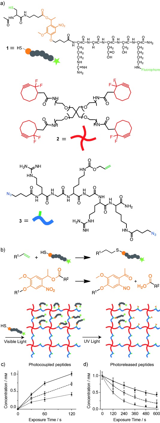
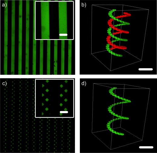
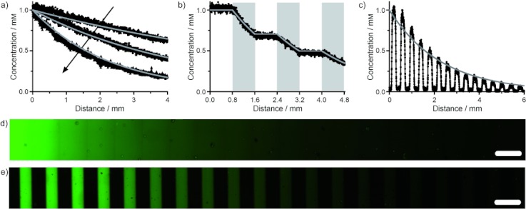
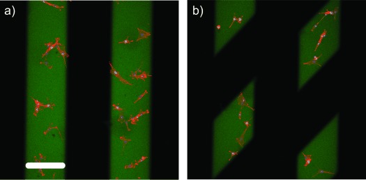
Verification
Claims the paper makes
The claims and statistics of this paper haven’t been checked yet.
Run the check — free, no account needed. It reads the open-access text and checks the sources it cites.
Claims other papers make about this one
Each is checked against this paper’s own text: does it say what the citing paper says it does?
In the citing paper
“Synthetic hydrogels can be functionalized with biologically active moieties that permit the growth and spread of cells146–151; concentration and spacing of these cues can be changed independently152–155.”
From Towards organoid culture without Matrigel (2021) · cites this paper as [153]
Self-check
Percentages, sums and reported differences re-computed from the paper’s own numbers.
The self-check needs the paper’s full text.
Citations
Cites
Alberts, B., Johnson, A., Lewis, J. H., Morgan, D. O., RAFF, M. C., Roberts, K. J., & Walter, P. (2017) · W.W. Norton eBooks · doi:10.1201/9781315735368 · Cited by 7,937
Hoyle, C. E., & Bowman, C. N. (2010) · Angewandte Chemie International Edition · doi:10.1002/anie.200903924 · Cited by 4,201
Hydrogels in Biology and Medicine: From Molecular Principles to Bionanotechnology
Peppas, N. A., Hilt, J. Z., Khademhosseini, A., & Langer, R. (2006) · Advanced Materials · doi:10.1002/adma.200501612 · Cited by 4,109
Hydrogels as extracellular matrix mimics for 3D cell culture
Tibbitt, M. W., & Anseth, K. S. (2009) · Biotechnology and Bioengineering · doi:10.1002/bit.22361 · Cited by 2,727
Self-citationPhotodegradable Hydrogels for Dynamic Tuning of Physical and Chemical Properties
Kloxin, A. M., Kasko, A. M., Salinas, C. N., & Anseth, K. S. (2009) · Science · doi:10.1126/science.1169494 · Cited by 1,748
Self-citationDesigning materials to direct stem-cell fate
Lütolf, M. P., Gilbert, P. M., & Blau, H. M. (2009) · Nature · doi:10.1038/nature08602 · Cited by 1,409
Sequential click reactions for synthesizing and patterning three-dimensional cell microenvironments
DeForest, C. A., Polizzotti, B. D., & Anseth, K. S. (2009) · Nature Materials · doi:10.1038/nmat2473 · Cited by 853
Self-citationBryant, S. J., Nuttelman, C. R., & Anseth, K. S. (2000) · Journal of Biomaterials Science Polymer Edition · doi:10.1163/156856200743805 · Cited by 775
Self-citationDeForest, C. A., & Anseth, K. S. (2011) · Nature Chemistry · doi:10.1038/nchem.1174 · Cited by 716
Self-citationA Versatile Synthetic Extracellular Matrix Mimic via Thiol‐Norbornene Photopolymerization
Fairbanks, B. D., Schwartz, M. P., Halevi, A. E., Nuttelman, C. R., Bowman, C. N., & Anseth, K. S. (2009) · Advanced Materials · doi:10.1002/adma.200901808 · Cited by 662
Self-citation
Cited by
Sorted by citations
Lanthanide Nanoparticles: From Design toward Bioimaging and Therapy
Dong, H., Du, S., Zheng, X.-Y., Lyu, G.-M., Sun, L., Li, L.-D., Zhang, P., Zhang, C., & Yan, C. (2015) · Chemical Reviews · doi:10.1021/acs.chemrev.5b00091 · Cited by 1,133
Functional and Biomimetic Materials for Engineering of the Three-Dimensional Cell Microenvironment
Huang, G., Li, F., Zhao, X., Ma, Y., Li, Y., Lin, M., Jin, G., Lu, T. J., Genin, G. M., & Xu, F. (2017) · Chemical Reviews · doi:10.1021/acs.chemrev.7b00094 · Cited by 825
The design of reversible hydrogels to capture extracellular matrix dynamics
Rosales, A. M., & Anseth, K. S. (2016) · Nature Reviews Materials · doi:10.1038/natrevmats.2015.12 · Cited by 770
Self-citationClick hydrogels, microgels and nanogels: Emerging platforms for drug delivery and tissue engineering
Jiang, Y., Chen, J., Deng, C., Suuronen, E. J., & Zhong, Z. (2014) · Biomaterials · doi:10.1016/j.biomaterials.2014.03.001 · Cited by 774
3D biofabrication strategies for tissue engineering and regenerative medicine
Bajaj, P., Schweller, R. M., Khademhosseini, A., West, J. L., & Bashir, R. (2014) · Annual Review of Biomedical Engineering · doi:10.1146/annurev-bioeng-071813-105155 · Cited by 632
Metadata
- Authors
- DeForest, C. A., & Anseth, K. S.
- Affiliations
- Howard Hughes Medical Institute (US)
- Corresponding author
- Anseth, Kristi S.
- Venue
- Angewandte Chemie International Edition
- Publisher
- Wiley
- Published
- 2011-12-08
- Volume
- 51
- Issue
- 8
- Pages
- 1816-1819
- ISSN
- 1433-7851 (ISSN-L)
- 1521-3773
- PMC
- PMC3430005
- OpenAlex
- W2109351872
- Cited by
- 315
- References
- 24
- Citations by year
- 2026: 9 · 2025: 7 · 2024: 12 · 2023: 11 · 2022: 21 · 2021: 23 · 2020: 20 · 2019: 18 · 2018: 38 · 2017: 27 · 2016: 18 · 2015: 22 · 2014: 33 · 2013: 35 · 2012: 21
- FWCI
- 12.99
- Type
- journal article
- Language
- en
- Open access
- bronze
- doi.org
- Locations
- Angewandte Chemie International Edition (publishedVersion)
- Europe PMC (PubMed Central) (publishedVersion, cc-by)
- PubMed Central (publishedVersion, cc-by)
- PubMed (publishedVersion)
- Funders
- National Science Foundation (1006711)
- Howard Hughes Medical Institute
- Topics
- Physical Sciences › Materials Science › Biomaterials › Hydrogels: synthesis, properties, applications
- Physical Sciences › Chemistry › Organic Chemistry › Photopolymerization techniques and applications
- Physical Sciences › Chemistry › Organic Chemistry › Click Chemistry and Applications
- Keywords
- hydrogels, click chemistry, peptide conjugation, photocleavage, spatiotemporal control, 3D cell culture
- MeSH terms
- Oligopeptides, Peptides, Molecular Structure, Hydrogels, Photochemical Processes, Click Chemistry
About these checks
Registry, retraction, Cites and statistics results are computed, not judged, with one exception: a printed reference that no index lists is read once by a model before it counts as a citation error. Claims are extracted by a model and kept only when their quote appears verbatim in the text; each cited source is then checked, with a model’s help, for whether it says what the claim relies on. Those verdicts can be wrong — read the claim’s quote and the cited source before relying on one.
Statistics and arithmetic are recomputed, not judged.
Confidence bands are shown only for fields calibrated on our annotated test set. None is calibrated yet, so no band is shown.
What counts as a claim ›How we measure confidence ›How we check this ›
Are you an author, or do you see a problem with this page? Request a review
