

The DOI matches its Crossref record; 45 of the 46 references it cites were found in a registry or an index.
Molecular Evolution, Structure, and Function of Peroxidasins
Open accessAbstract
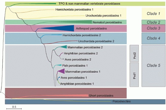
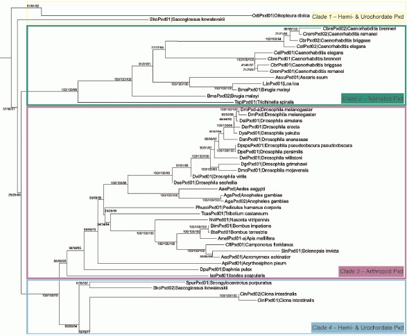
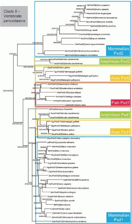
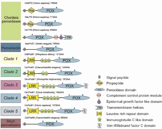
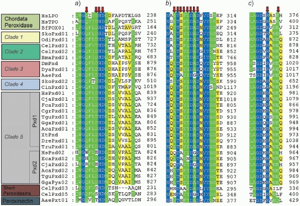
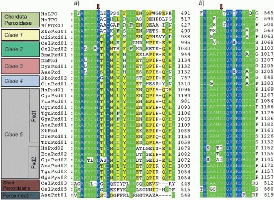
Peroxidasins represent the subfamily 2 of the peroxidase‐cyclooxygenase superfamily and are closely related to chordata peroxidases (subfamily 1) and peroxinectins (subfamily 3). They are multidomain proteins containing a heme peroxidase domain with high homology to human lactoperoxidase that mediates one‐ and two‐electron oxidation reactions. Additional domains of the secreted and glycosylated metalloproteins are type C‐like immunoglobulin domains, typical leucine‐rich repeats, as well as a von Willebrand factor C module. These are typical motifs of extracellular proteins that mediate protein–protein interactions. We have reconstructed the phylogeny of this new family of oxidoreductases and show the presence of four invertebrate clades as well as one vertebrate clade that includes also two different human representatives. The variability of domain assembly in the various clades was analyzed, as was the occurrence of relevant catalytic residues in the peroxidase domain based on the knowledge of catalysis of the mammalian homologues. Finally, the few reports on expression, localization, enzymatic activity, and physiological roles in the model organisms Drosophila melanogaster, Caenorhabditis elegans, and Homo sapiens are critically reviewed. Roles attributed to peroxidasins include antimicrobial defense, extracellular matrix formation, and consolidation at various developmental stages. Many research questions need to be solved in future, including detailed biochemical/physical studies and elucidation of the three dimensional structure of a model peroxidasin as well as the relation and interplay of the domains and the in vivo functions in various organisms including man.
Overview
The claims and statistics of this paper haven’t been checked yet.
Free: it reads the open-access text and checks the first 5 claims that cite a source we can check. Create a free account to check every claim.
Integrity signals
Registry
Cites
Statistics
Figures

Verification
Claims the paper makes
The claims and statistics of this paper haven’t been checked yet.
Free: it reads the open-access text and checks the first 5 claims that cite a source we can check. Create a free account to check every claim.
Claims other papers make about this one
Each is checked against this paper’s own text: does it say what the citing paper says it does?
In the citing paper
“However, the mechanisms behind peroxidasin-induced fibrosis regression does not require its crosslinking activity towards collagen IV (282); rather, peroxidasin deficiency produces a hypoxic environment that reduces the activation of HSCs and recruits pro-healing macrophages (281).”
From Found in translation—Fibrosis in metabolic dysfunction–associated steatohepatitis (MASH) (2023) · cites this paper as [282]
Self-check
Percentages, sums and reported differences re-computed from the paper’s own numbers.
The self-check needs the paper’s full text.
Citations
Cites
MEGA4: Molecular Evolutionary Genetics Analysis (MEGA) Software Version 4.0
Tamura, K., Dudley, J., Nei, M., & Kumar, S. (2007) · Molecular Biology and Evolution · doi:10.1093/molbev/msm092 · Cited by 28,730
Clustal W and Clustal X version 2.0
Larkin, M. A., Blackshields, G., Brown, N. P., Chenna, R., McGettigan, P. A., McWilliam, H., Valentin, F., Wallace, I. M., Wilm, A., Lopez, R., et al. (2007) · Bioinformatics · doi:10.1093/bioinformatics/btm404 · Cited by 28,358
The PSIPRED protein structure prediction server
McGuffin, L. J., Bryson, K., & Jones, D. T. (2000) · Bioinformatics · doi:10.1093/bioinformatics/16.4.404 · Cited by 3,838
Generation and initial analysis of more than 15,000 full-length human and mouse cDNA sequences
Mammalian Gene Collection (MGC) Program Team* (2002) · Proceedings of the National Academy of Sciences · doi:10.1073/pnas.242603899 · Cited by 1,749
The leucine-rich repeat as a protein recognition motif
Kobe, B. (2001) · Current Opinion in Structural Biology · doi:10.1016/s0959-440x(01)00266-4 · Cited by 1,679
ESyPred3D: Prediction of proteins 3D structures
Lambert, C., Léonard, N., De Bolle, X., & Depiereux, E. (2002) · Bioinformatics · doi:10.1093/bioinformatics/18.9.1250 · Cited by 611
Heinecke, J. W., Li, W., Daehnke, H. L., & Goldstein, J. A. (1993) · Journal of Biological Chemistry · doi:10.1016/s0021-9258(18)53581-x · Cited by 485
The leucine-rich repeat structure
Bella, J., Hindle, K. L., McEwan, P. A., & Lovell, S. C. (2008) · Cellular and Molecular Life Sciences · doi:10.1007/s00018-008-8019-0 · Cited by 466
Active site structure and catalytic mechanisms of human peroxidases
Furtmüller, P. G., Zederbauer, M., Jantschko, W., Helm, J., Bogner, M., Jakopitsch, C., & Obinger, C. (2006) · Archives of Biochemistry and Biophysics · doi:10.1016/j.abb.2005.09.017 · Cited by 358
Self-citationMembrane proteins with immunoglobulin-like domains—a master superfamily of interaction molecules
Barclay, A. N. (2003) · Seminars in Immunology · doi:10.1016/s1044-5323(03)00047-2 · Cited by 309
Research works printed but not linked: 1
- Mitchell M. S., 2000, Cancer Res., 60, 6448
Cited by
Independent evolution of four heme peroxidase superfamilies
Zámocký, M., Hofbauer, S., Schaffner, I., Gasselhuber, B., Nicolussi, A., Soudi, M., Pirker, K. F., Furtmüller, P. G., & Obinger, C. (2015) · Archives of Biochemistry and Biophysics · doi:10.1016/j.abb.2014.12.025 · Cited by 247
Self-citationA unique covalent bond in basement membrane is a primordial innovation for tissue evolution
Fidler, A. L., Vanacore, R. M., Chetyrkin, S. V., Pedchenko, V. K., Bhave, G., Yin, V. P., Stothers, C. L., Rose, K. L., McDonald, W. H., Clark, T. A., et al. (2014) · Proceedings of the National Academy of Sciences · doi:10.1073/pnas.1318499111 · Cited by 171
“Our study delineates peroxidasin and its domain structure from Protostoma (27) basally to Radiata and Placozoa.”
BackgroundInfluentialFound in translation—Fibrosis in metabolic dysfunction–associated steatohepatitis (MASH)
Wang, S., & Friedman, S. L. (2023) · Science Translational Medicine · doi:10.1126/scitranslmed.adi0759 · Cited by 158
“However, the mechanism behind peroxidasin-induced fibrosis regression does not require its cross-linking activity toward collagen IV ( 277 ); rather, peroxidasin deficiency produces a hypoxic environment that reduces the activation of HSCs and recruits proheal-ing macrophages ( 276 ).”
BackgroundDavies, M. J. (2021) · Pharmacology & Therapeutics · doi:10.1016/j.pharmthera.2020.107685 · Cited by 119
Genome-enabled insights into the biology of thrips as crop pests
Rotenberg, D., Baumann, A. A., Ben-Mahmoud, S. K., Christiaens, O., Dermauw, W., Ioannidis, P., Jacobs, C. G. C., Jentzsch, I. M. V., Oliver, J. E., Poelchau, M. F., et al. (2020) · BMC Biology · doi:10.1186/s12915-020-00862-9 · Cited by 106
Metadata
- Authors
- Soudi, M., Zamocky, M., Jakopitsch, C., Furtmüller, P. G., & Obinger, C.
- Affiliations
- BOKU University (AT)
- Corresponding author
- Obinger, Christian
- Venue
- Chemistry & Biodiversity
- Publisher
- Wiley
- Published
- 2012-09
- Volume
- 9
- Issue
- 9
- Pages
- 1776-1793
- ISSN
- 1612-1872 (ISSN-L)
- 1612-1880
- PMC
- PMC3533774
- OpenAlex
- W2036174322
- Cited by
- 58
- References
- 46
- Citations by year
- 2026: 1 · 2025: 5 · 2024: 2 · 2023: 4 · 2022: 3 · 2021: 2 · 2020: 7 · 2019: 3 · 2018: 9 · 2017: 5 · 2016: 1 · 2015: 4 · 2014: 7 · 2013: 5
- FWCI
- 2.23
- Type
- journal article
- Language
- en
- Open access
- green
- www.ncbi.nlm.nih.gov
- Locations
- Chemistry & Biodiversity (publishedVersion)
- PubMed Central (publishedVersion, cc-by)
- PubMed (publishedVersion)
- Topics
- Life Sciences › Immunology and Microbiology › Immunology › Neutrophil, Myeloperoxidase and Oxidative Mechanisms
- Life Sciences › Biochemistry, Genetics and Molecular Biology › Molecular Biology › Redox biology and oxidative stress
- Life Sciences › Immunology and Microbiology › Immunology › Invertebrate Immune Response Mechanisms
- Keywords
- peroxidasin, lactoperoxidase, leucine-rich repeats, protein domain architecture, phylogeny, catalytic residues, antimicrobial defense, extracellular matrix synthesis, Drosophila melanogaster, Caenorhabditis elegans, oxidative enzymes
- MeSH terms
- Peroxidasin, Amino Acid Sequence, Animals, Humans, Models, Molecular, Molecular Sequence Data, Peroxidase, Peroxidases, Phylogeny, Extracellular Matrix Proteins, Sequence Alignment, Evolution, Molecular, Chordata
About these checks
Registry, retraction, Cites and statistics results are computed, not judged, with one exception: a printed reference that no index lists is read once by a model before it counts as a citation error. Claims are extracted by a model and kept only when their quote appears verbatim in the text; each cited source is then checked, with a model’s help, for whether it says what the claim relies on. Those verdicts can be wrong — read the claim’s quote and the cited source before relying on one.
Statistics and arithmetic are recomputed, not judged.
Confidence bands are shown only for fields calibrated on our annotated test set. None is calibrated yet, so no band is shown.
What counts as a claim ›How we measure confidence ›How we check this ›
Are you an author, or do you see a problem with this page? Request a review
How to cite
Cite CoChat for the checks on this page, and the paper for its own findings.
- This page
- CoChat. (2026). Citation check: Molecular Evolution, Structure, and Function of Peroxidasins. https://app.cochat.ai/papers/10.1002/cbdv.201100438
- The paper
- Soudi, M., Zamocky, M., Jakopitsch, C., Furtmüller, P. G., & Obinger, C. (2012). Molecular Evolution, Structure, and Function of Peroxidasins. Chemistry & Biodiversity. https://doi.org/10.1002/cbdv.201100438
Data sources
Bibliographic data: Semantic Scholar, OpenAlex, Crossref.
Contains information from the Semantic Scholar Academic Graph Datasets, made available under the ODC Attribution License (ODC-BY 1.0).
Semantic Scholar’s data is described in: Kinney, R., et al. (2023). The Semantic Scholar Open Data Platform. arXiv:2301.10140.
