

The DOI matches its Crossref record; 96 of the 96 references it cites were found in a registry or an index.
Virtual Screening of Bacteriocins From Lactic Acid Bacteria Against Monkeypox DNA Polymerase: Sakacin P and Mundticin KS Emerge as Promising Candidates
Open accessPublished version of a preprint
Abstract
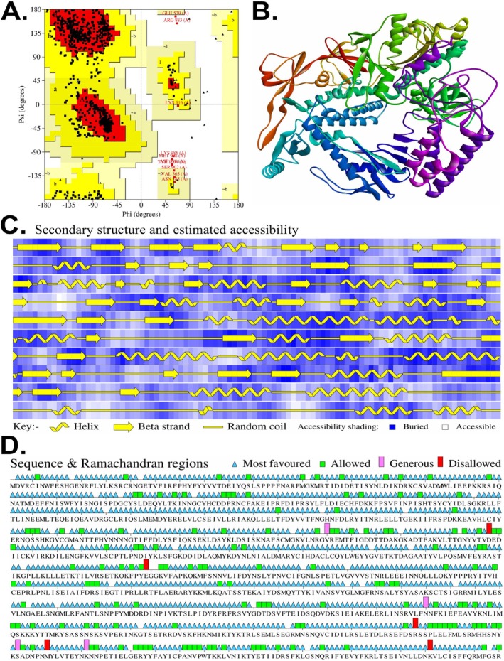
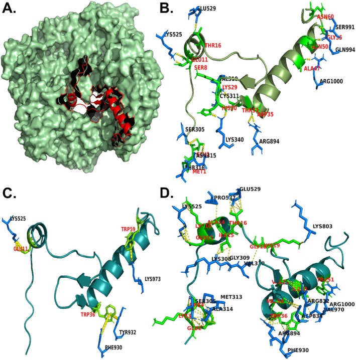
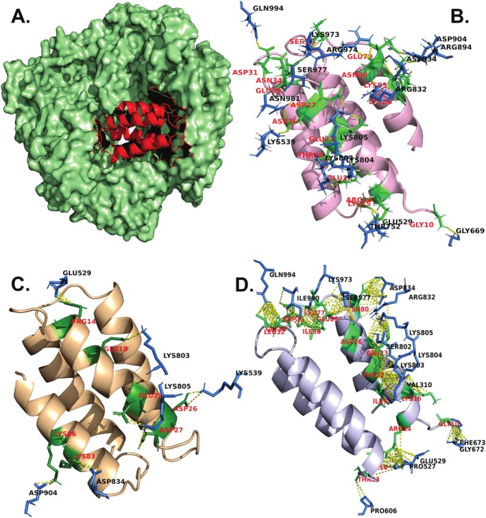
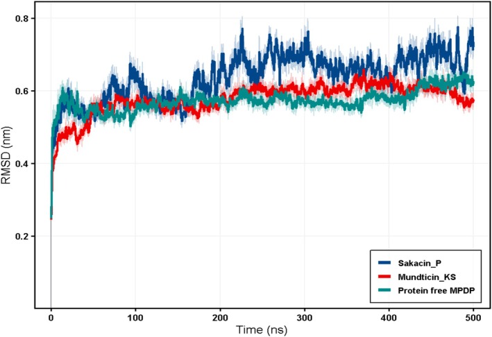
The monkeypox virus (MPXV) has recently risen to be a significant global health threat and currently there are no approved antiviral agents, which makes it necessary to develop new antiviral strategies. In this study, we employed in silico techniques to investigate whether bacteriocins could be potent inhibitors of the MPXV DNA polymerase (MPDP). At first, the MPXV DNA polymerase enzyme chain was extracted from the Cryo‐EM structure of MPXV DNA replication complex and was then assessed using SWISS‐MODEL/QMEAN for structural quality. The structures of selected bacteriocins were either retrieved from the Protein Data Bank or predicted using AlphaFold. The quality of these predicted models was measured by LGscore. Moreover, the physicochemical properties of the selected bacteriocins, such as Sakacin P and Mundticin KS, were analyzed to assess their molecular stability and compatibility with the molecular docking process. The results of protein‐peptide docking simulations on the HADDOCK platform showed that among all the bacteriocins tested, Sakacin P and Mundticin KS had the highest binding affinities toward MPXV DNA polymerase. The analysis of the docking experiments also revealed vital stabilizing interactions, such as the formation of hydrogen bonds, ionic linkages, and π–π stacking, which were crucial in the strength of the protein–ligand complexes. Subsequently, the molecular dynamics (MD) simulations further confirmed the stability of the protein–bacteriocin complexes. Our results indicate that bacteriocins, especially Sakacin P and Mundticin KS, can be considered potential antiviral agents against MPXV by interfering with its DNA replication mechanism. The present work shall serve as a reference for future laboratory testing and for the promising innovation of bacteriocin‐based drugs targeting MPXV.
Overview
The claims and statistics of this paper haven’t been checked yet.
Run the check — free, no account needed. It reads the open-access text and checks the sources it cites.
Integrity signals
Registry
Cites
Statistics
Figures
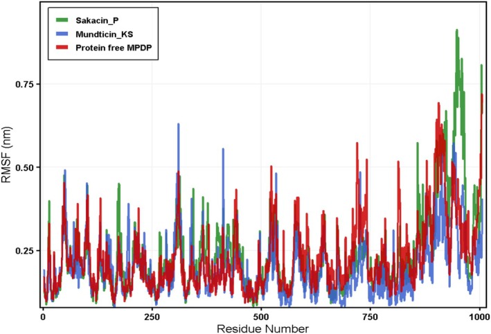
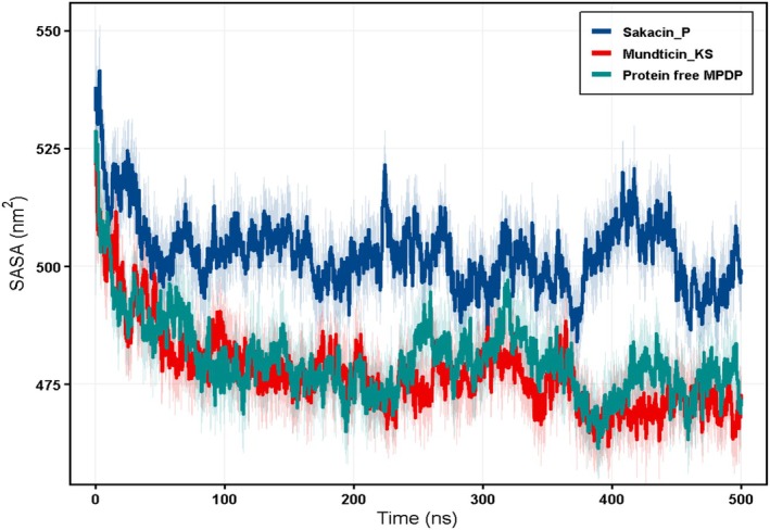
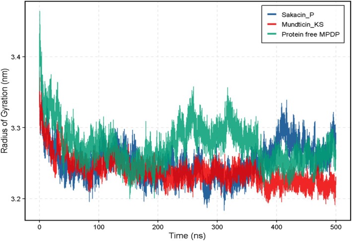
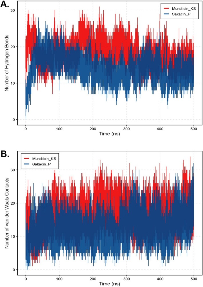
Verification
Claims the paper makes
The claims and statistics of this paper haven’t been checked yet.
Run the check — free, no account needed. It reads the open-access text and checks the sources it cites.
Self-check
Percentages, sums and reported differences re-computed from the paper’s own numbers.
The self-check needs the paper’s full text.
Citations
Cites
Highly accurate protein structure prediction with AlphaFold
Jumper, J., Evans, R., Pritzel, A., Green, T., Figurnov, M., Ronneberger, O., Tunyasuvunakool, K., Bates, R., Žídek, A., Potapenko, A., et al. (2021) · Nature · doi:10.1038/s41586-021-03819-2 · Cited by 43,764 · cited 1×
“…local geometry; therefore, final model selection for docking relied on orthogonal validation criteria, including Ramachandran stereochemical assessment and ProQ LGscore evaluation (Laskowski et al. 1993; Wallner and Elofsson 2003; Jumper et al. 2021; McDonald et al. 2023; Terwilliger et al. 2023).”
Comparison of simple potential functions for simulating liquid water
Jorgensen, W. L., Chandrasekhar, J., Madura, J. D., Impey, R. W., & Klein, M. L. (1983) · The Journal of Chemical Physics · doi:10.1063/1.445869 · Cited by 41,298
Abraham, M. J., Murtola, T., Schulz, R., Páll, S., Smith, J. C., Hess, B., & Lindahl, E. (2015) · SoftwareX · doi:10.1016/j.softx.2015.06.001 · Cited by 27,190
PROCHECK: a program to check the stereochemical quality of protein structures
Laskowski, R. A., MacArthur, M. W., Moss, D. S., & Thornton, J. M. (1993) · Journal of Applied Crystallography · doi:10.1107/s0021889892009944 · Cited by 24,581
Ryckaert, J.-P., Ciccotti, G., & Berendsen, H. J. C. (1977) · Journal of Computational Physics · doi:10.1016/0021-9991(77)90098-5 · Cited by 21,239
Development and testing of a general amber force field
Wang, J., Wolf, R. M., Caldwell, J. W., Kollman, P. A., & Case, D. A. (2004) · Journal of Computational Chemistry · doi:10.1002/jcc.20035 · Cited by 19,506
Accurate structure prediction of biomolecular interactions with AlphaFold 3
Abramson, J., Adler, J., Dunger, J., Evans, R., Green, T., Pritzel, A., Ronneberger, O., Willmore, L., Ballard, A. J., Bambrick, J., et al. (2024) · Nature · doi:10.1038/s41586-024-07487-w · Cited by 15,861
SWISS-MODEL: homology modelling of protein structures and complexes
Waterhouse, A., Bertoni, M., Bienert, S., Studer, G., Tauriello, G., Gumienny, R., Heer, F. T., de Beer, T. A. P., Rempfer, C., Bordoli, L., et al. (2018) · Nucleic Acids Research · doi:10.1093/nar/gky427 · Cited by 13,067
The MM/PBSA and MM/GBSA methods to estimate ligand-binding affinities
Genheden, S., & Ryde, U. (2015) · Expert Opinion on Drug Discovery · doi:10.1517/17460441.2015.1032936 · Cited by 5,769
gmx_MMPBSA: A New Tool to Perform End-State Free Energy Calculations with GROMACS
Valdés-Tresanco, M. S., Valdés-Tresanco, M. E., Valiente, P. A., & Moreno, E. (2021) · Journal of Chemical Theory and Computation · doi:10.1021/acs.jctc.1c00645 · Cited by 3,393
Toward the estimation of the absolute quality of individual protein structure models
Benkert, P., Biasini, M., & Schwede, T. F. (2010) · Bioinformatics · doi:10.1093/bioinformatics/btq662 · Cited by 2,321
Mariani, V., Biasini, M., Barbato, A., & Schwede, T. (2013) · Bioinformatics · doi:10.1093/bioinformatics/btt473 · Cited by 1,138
Van Oss, C. J., Good, R. J., & Chaudhury, M. K. (1986) · Journal of Colloid and Interface Science · doi:10.1016/0021-9797(86)90041-x · Cited by 949
Patil, R., Das, S., Stanley, A., Yadav, L., Sudhakar, A., & Varma, A. K. (2010) · PLoS ONE · doi:10.1371/journal.pone.0012029 · Cited by 802
The Continuing Story of Class IIa Bacteriocins
Drider, D., Fimland, G., Héchard, Y., McMullen, L. M., & Prévost, H. (2006) · Microbiology and Molecular Biology Reviews · doi:10.1128/mmbr.00016-05 · Cited by 750
Can correct protein models be identified?
Wallner, B., & Elofsson, A. (2003) · Protein Science · doi:10.1110/ps.0236803 · Cited by 710
The HADDOCK2.4 web server for integrative modeling of biomolecular complexes
Honorato, R. V., Trellet, M. E., Jiménez‐García, B., Schaarschmidt, J., Giulini, M., Reys, V., Koukos, P. I., Rodrigues, J., Karaca, E., van Zundert, G. C. P., et al. (2024) · Nature Protocols · doi:10.1038/s41596-024-01011-0 · Cited by 705
Vieco-Saiz, N., Belguesmia, Y., Raspoet, R., Auclair, E., Gancel, F., Kempf, I., & Drider, D. (2019) · Frontiers in Microbiology · doi:10.3389/fmicb.2019.00057 · Cited by 658
Monkeypox: disease epidemiology, host immunity and clinical interventions
Lum, F., Torres‐Ruesta, A., Tay, M. Z., Lin, R. T. P., Lye, D. C., Rénia, L., & Ng, L. F. P. (2022) · Nature reviews. Immunology · doi:10.1038/s41577-022-00775-4 · Cited by 548
Bacteriocins as a new generation of antimicrobials: toxicity aspects and regulations
Soltani, S., Hammami, R., Cotter, P. D., Rebuffat, S., Said, L. B., Gaudreau, H., Bédard, F., Biron, E., Drider, D., & Fliss, I. (2021) · FEMS Microbiology Reviews · doi:10.1093/femsre/fuaa039 · Cited by 544
Structure, Dynamics, Assembly, and Evolution of Protein Complexes
Marsh, J. A., & Teichmann, S. A. (2014) · Annual Review of Biochemistry · doi:10.1146/annurev-biochem-060614-034142 · Cited by 473
Assembly reflects evolution of protein complexes
Levy, E. D., Erba, E. B., Robinson, C. V., & Teichmann, S. A. (2008) · Nature · doi:10.1038/nature06942 · Cited by 419
Terwilliger, T. C., Liebschner, D., Croll, T. I., Williams, C. J., McCoy, A. J., Poon, B. K., Afonine, P. V., Oeffner, R. D., Richardson, J. S., Read, R. J., et al. (2024) · Nature Methods · doi:10.1038/s41592-023-02087-4 · Cited by 411
A unified statistical framework for sequence comparison and structure comparison
Levitt, M., & Gerstein, M. (1998) · Proceedings of the National Academy of Sciences · doi:10.1073/pnas.95.11.5913 · Cited by 300
Bioprotective mechanisms of lactic acid bacteria against fungal spoilage of food
Siedler, S., Balti, R., & Neves, A. R. (2018) · Current Opinion in Biotechnology · doi:10.1016/j.copbio.2018.11.015 · Cited by 259
Antiviral Potential of Lactic Acid Bacteria and Their Bacteriocins
Al Kassaa, I., Hober, D., Hamze, M., Chihib, N. E., & Drider, D. (2014) · Probiotics and Antimicrobial Proteins · doi:10.1007/s12602-014-9162-6 · Cited by 217
Antiviral activity of antimicrobial cationic peptides against Junin virus and herpes simplex virus
Matanic, V. C. A., & Castilla, V. (2004) · International Journal of Antimicrobial Agents · doi:10.1016/j.ijantimicag.2003.07.022 · Cited by 207
Holo, H., Jeknić, Z., Daeschel, M. A., Stevanović, S., & Nes, I. F. (2001) · Microbiology · doi:10.1099/00221287-147-3-643 · Cited by 203
Characterisation of an antiviral pediocin-like bacteriocin produced by Enterococcus faecium
Todorov, S. D., Wachsman, M. B., Tomè, E., Dousset, X., Destro, M. T., Dicks, L. M. T., Franco, B. D. G. D. M., Vaz‐Velho, M., & Drider, D. (2010) · Food Microbiology · doi:10.1016/j.fm.2010.05.001 · Cited by 189
Geometric criteria of hydrogen bonds in proteins and identification of `bifurcated' hydrogen bonds
Torshin, I. Y., Weber, I. T., & Harrison, R. W. (2002) · Protein Engineering Design and Selection · doi:10.1093/protein/15.5.359 · Cited by 177
Férir, G., Petrova, M., Andreï, G., Huskens, D., Hoorelbeke, B., Snoeck, R., Vanderleyden, J., Balzarini, J. M., Bartoschek, S., Brönstrup, M., et al. (2013) · PLoS ONE · doi:10.1371/journal.pone.0064010 · Cited by 172
Enterocin CRL35 inhibits late stages of HSV-1 and HSV-2 replication in vitro
Wachsman, M. B., Castilla, V., Holgado, A. P. D. R., Torres, R. A. D., Sesma, F., & Coto, C. E. (2003) · Antiviral Research · doi:10.1016/s0166-3542(02)00099-2 · Cited by 161
Antiviral effects of probiotic metabolites on COVID-19
Anwar, F., Altayb, H. N., Al-Abbasi, F. A., Al‐Malki, A. L., Kamal, M. A., & Kumar, V. (2020) · Journal of Biomolecular Structure and Dynamics · doi:10.1080/07391102.2020.1775123 · Cited by 155
Cation-π Interactions and their Functional Roles in Membrane Proteins
Infield, D. T., Rasouli, A., Galles, G. D., Chipot, C. J., Tajkhorshid, E., & Ahern, C. A. (2021) · Journal of Molecular Biology · doi:10.1016/j.jmb.2021.167035 · Cited by 153
QMEANDisCo—distance constraints applied on model quality estimation
Studer, G., Rempfer, C., Waterhouse, A. M., Gumienny, R., Haas, J., & Schwede, T. F. (2020) · Bioinformatics · doi:10.1093/bioinformatics/btaa058 · Cited by 153
Benchmarking AlphaFold2 on peptide structure prediction
McDonald, E. F., Jones, T., Plate, L., Meiler, J., & Gulsevin, A. (2023) · Structure · doi:10.1016/j.str.2022.11.012 · Cited by 152
Melittin: a venom-derived peptide with promising anti-viral properties
Memariani, H., Memariani, M., Moravvej, H., & Shahidi‐Dadras, M. (2019) · European Journal of Clinical Microbiology & Infectious Diseases · doi:10.1007/s10096-019-03674-0 · Cited by 143
Visual dynamics: a WEB application for molecular dynamics simulation using GROMACS
Vieira, I. H. P., Botelho, E. B., Gomes, T. J. D. S., Kist, R., Caceres, R. A., & Zanchi, F. B. (2023) · BMC Bioinformatics · doi:10.1186/s12859-023-05234-y · Cited by 141
Self-citationZhao, Y., Li, J., Gu, H., Wei, D., XU, Y. ‐., Fu, W., & Yu, Z. (2015) · Interdisciplinary Sciences Computational Life Sciences · doi:10.1007/s12539-015-0263-z · Cited by 135
Bacteriocin: A natural approach for food safety and food security
Lahiri, D., Nag, M., Dutta, B., Sarkar, T., Pati, S., Basu, D., Kari, Z. A., Wei, L. S., Smaoui, S., Goh, K. W., et al. (2022) · Frontiers in Bioengineering and Biotechnology · doi:10.3389/fbioe.2022.1005918 · Cited by 128
Kawamoto, S., Shima, J., Sato, R., Eguchi, T., Ohmomo, S., Shibato, J., Horikoshi, N., Takeshita, K., & Sameshima, T. (2002) · Applied and Environmental Microbiology · doi:10.1128/aem.68.8.3830-3840.2002 · Cited by 127
Conte, A. D., Camagni, G. F., Clementel, D., Minervini, G., Monzón, A. M., Ferrari, C., Piovesan, D., & Tosatto, S. C. E. (2024) · Nucleic Acids Research · doi:10.1093/nar/gkae337 · Cited by 125
Daba, G. M., & Elkhateeb, W. A. (2020) · Biocatalysis and Agricultural Biotechnology · doi:10.1016/j.bcab.2020.101750 · Cited by 117
GMXPBSA 2.1: A GROMACS tool to perform MM/PBSA and computational alanine scanning
Paissoni, C., Spiliotopoulos, D., Musco, G., & Spitaleri, A. (2014) · Computer Physics Communications · doi:10.1016/j.cpc.2014.09.010 · Cited by 90
Katla, T., Møretrø, T., Sveen, I., et al. (2002) · Journal of Applied Microbiology · doi:10.1046/j.1365-2672.2002.01675.x · Cited by 89
Bacteriocins: potentials and prospects in health and agrifood systems
Reuben, R. C., & Torres, C. (2024) · Archives of Microbiology · doi:10.1007/s00203-024-03948-y · Cited by 89
Antiviral activity of enterocin CRL35 against herpesviruses
Wachsman, M. B., Farías, M. E., Takeda, E., Sesma, F. J. M., Holgado, A. P. D. R., de Torres, R. A., & Coto, C. E. (1999) · International Journal of Antimicrobial Agents · doi:10.1016/s0924-8579(99)00078-3 · Cited by 83
Multilevel Summation Method for Electrostatic Force Evaluation
Hardy, D. J., Wu, Z., Phillips, J. C., Stone, J. E., Skeel, R. D., & Schulten, K. J. (2014) · Journal of Chemical Theory and Computation · doi:10.1021/ct5009075 · Cited by 80
Theory of Polymer–Nanopore Interactions Refined Using Molecular Dynamics Simulations
Balijepalli, A. K., Robertson, J. W. F., Reiner, J. E., et al. (2013) · Journal of the American Chemical Society · doi:10.1021/ja4026193 · Cited by 72
Letafati, A., & Sakhavarz, T. (2023) · Microbial Pathogenesis · doi:10.1016/j.micpath.2023.106027 · Cited by 70
Barbosa, M. D. S., Todorov, S. D., Иванова, И., Belguesmia, Y., Choiset, Y., Rabesona, H., Chobert, J., Haertlé, T., & Franco, B. D. G. D. M. (2015) · Food Control · doi:10.1016/j.foodcont.2015.07.029 · Cited by 69
Mathiesen, G., Huehne, K., Kröckel, L., Axelsson, L. T., & Eijsink, V. G. H. (2005) · Applied and Environmental Microbiology · doi:10.1128/aem.71.7.3565-3574.2005 · Cited by 68
Antiinfluenza virus activity of a bacteriocin produced by Lactobacillus delbrueckii
Serkedjieva, J., Danova, S., & Ivanova, I. (2000) · Applied Biochemistry and Biotechnology · doi:10.1385/abab:88:1-3:285 · Cited by 64
Human monkeypox virus: Epidemiologic review and research progress in diagnosis and treatment
Sun, Y., Nie, W., Tian, D., & Ye, Q. (2024) · Journal of Clinical Virology · doi:10.1016/j.jcv.2024.105662 · Cited by 62
The “hydrophobic effect”: Essentially a van der Waals interaction
van Oss, C. J., Absolom, D. R., & Neumann, A. W. (1980) · Colloid & Polymer Science · doi:10.1007/bf01480835 · Cited by 61
Islam, M. A., Mumin, J., Haque, M. M., Haque, M. A., Khan, A., Bhattacharya, P., & Haque, M. A. (2023) · Infectious Medicine · doi:10.1016/j.imj.2023.11.001 · Cited by 57
Schelegueda, L. I., Vallejo, M., Gliemmo, M. F., Marguet, E. R., & Campos, C. A. (2015) · LWT · doi:10.1016/j.lwt.2015.06.017 · Cited by 56
Inhibition of Human Cytomegalovirus DNA Polymerase by C-Terminal Peptides from the UL54 Subunit
Loregian, A., Rigatti, R., Murphy, M., Schievano, E., Palù, G., & Marsden, H. S. (2003) · Journal of Virology · doi:10.1128/jvi.77.15.8336-8344.2003 · Cited by 53
Bacteriocin producing microbes with bactericidal activity against multidrug resistant pathogens
Leslie, V. A., Alarjani, K. M., Malaisamy, A., & Balasubramanian, B. (2021) · Journal of Infection and Public Health · doi:10.1016/j.jiph.2021.09.029 · Cited by 52
PRODIGY-crystal: a web-tool for classification of biological interfaces in protein complexes
Jiménez‐García, B., Elez, K., Koukos, P. I., Bonvin, A. M. J. J., & Vangone, A. (2019) · Bioinformatics · doi:10.1093/bioinformatics/btz437 · Cited by 50
Daba, G. M., & Elkhateeb, W. A. (2023) · International Journal of Biological Macromolecules · doi:10.1016/j.ijbiomac.2023.128325 · Cited by 50
DRAVP: A Comprehensive Database of Antiviral Peptides and Proteins
Liu, Y., Zhu, Y., Sun, X., Ma, T., Lao, X., & Zheng, H. (2023) · Viruses · doi:10.3390/v15040820 · Cited by 48
Tenea, G. N., & Pozo, T. D. (2019) · Journal of Microbiology and Biotechnology · doi:10.4014/jmb.1904.04063 · Cited by 47
Manna, S., Chowdhury, T., Chakraborty, R., & Mandal, S. M. (2020) · Probiotics and Antimicrobial Proteins · doi:10.1007/s12602-020-09727-7 · Cited by 45
Bacteriocins future perspectives: Substitutes to antibiotics
Cheruvari, A., & Kammara, R. (2024) · Food Control · doi:10.1016/j.foodcont.2024.110834 · Cited by 45
Exploring ligand binding pathways on proteins using hypersound-accelerated molecular dynamics
Araki, M., Matsumoto, S., Bekker, G., Isaka, Y., Sagae, Y., Kamiya, N., & Okuno, Y. (2021) · Nature Communications · doi:10.1038/s41467-021-23157-1 · Cited by 44
Emerging lactic acid bacteria bacteriocins as anti-cancer and anti-tumor agents for human health
Niamah, A. K., Al‐Sahlany, S. T. G., Verma, D. K., Shukla, R. M., Patel, A. R., Tripathy, S., Sıngh, S., Baranwal, D., Singh, A. K., Utama, G. L., et al. (2024) · Heliyon · doi:10.1016/j.heliyon.2024.e37054 · Cited by 37
In Silico Analysis of Bacteriocins from Lactic Acid Bacteria Against SARS-CoV-2
Erol, İ., Kotil, S., Fidan, Ö., Yetiman, A. E., Durdağı, S., & Ortakcı, F. (2021) · Probiotics and Antimicrobial Proteins · doi:10.1007/s12602-021-09879-0 · Cited by 36
Self-citationDevelopment of novel broad-spectrum antimicrobial lipopeptides derived from plantaricin NC8 β
Wiman, E., Zattarin, E., Aili, D., Bengtsson, T., Selegård, R., & Khalaf, H. (2023) · Scientific Reports · doi:10.1038/s41598-023-31185-8 · Cited by 36
Unwrapping NPT Simulations to Calculate Diffusion Coefficients
Bullerjahn, J. T., von Bülow, S., Heidari, M., H'enin, J., & Hummer, G. (2023) · Journal of Chemical Theory and Computation · doi:10.1021/acs.jctc.3c00308 · Cited by 36
Jhun, J., Min, H. K., Ryu, J., Lee, S.-Y., Ryu, J., Choi, J. W., Na, H. S., Lee, S. Y., Jung, Y., Park, S.-J., et al. (2020) · Journal of Translational Medicine · doi:10.1186/s12967-020-02477-8 · Cited by 33
Monkeypox virus: Future role in Human population
Farasani, A. M. (2022) · Journal of Infection and Public Health · doi:10.1016/j.jiph.2022.10.002 · Cited by 31
Krishna, S., Kurrey, C., Yadav, M. C., Mahilkar, S., Sonkar, S. C., Vishvakarma, N. K., Sonkar, A., Chandra, L., & Koner, B. C. (2024) · Infectious Medicine · doi:10.1016/j.imj.2024.100105 · Cited by 31
The 2024 Public Health Emergency of International Concern: A Global Failure to Control Mpox
Zumla, A. I., Rosenthal, P. J., Sam-Agudu, N. A., Ogoina, D., Kingebeni, P. M., Ntoumi, F., Nakouné, E. R., Njouom, R., Ndembi, N., Mills, E. J., et al. (2024) · American Journal of Tropical Medicine and Hygiene · doi:10.4269/ajtmh.24-0606 · Cited by 29
Cryo-EM structures of human monkeypox viral replication complexes with and without DNA duplex
Xu, Y., Wu, Y., Zhang, Y., Fan, R., Yang, Y., Li, D., Zhu, S., Yang, B., Zhang, Z., & Dong, C. (2023) · Cell Research · doi:10.1038/s41422-023-00796-1 · Cited by 25
Akkus, E., Tayfuroğlu, Ö., Yıldız, M., & Kocak, A. (2023) · The Journal of Physical Chemistry B · doi:10.1021/acs.jpcb.3c00834 · Cited by 24
The untapped potential of actinobacterial lanthipeptides as therapeutic agents
Ramírez-Rendón, D., Chavez, F. F. G., García-Ausencio, C., Rodrı́guez-Sanoja, R., & Sánchez, S. (2023) · Molecular Biology Reports · doi:10.1007/s11033-023-08880-w · Cited by 24
Daba, G. M., Elnahas, M. O., & Elkhateeb, W. A. (2021) · Biocatalysis and Agricultural Biotechnology · doi:10.1016/j.bcab.2021.102248 · Cited by 23
Omer, A. A. M., Hinkula, J., Tran, P.-T.-H., Melik, W., Zattarin, E., Aili, D., Selegård, R., Bengtsson, T., & Khalaf, H. (2022) · PLoS ONE · doi:10.1371/journal.pone.0278419 · Cited by 22
Monkeypox: An outbreak of a rare viral disease
Luo, Y., Zhang, T., Cao, J., Hou, W.-S., Wang, A., & Jin, C. (2023) · Journal of Microbiology Immunology and Infection · doi:10.1016/j.jmii.2023.12.006 · Cited by 19
Iorio, R., Petricca, S., Luzi, C., Bellio, P., Cristiano, L., Festuccia, C., Amicosante, G., & Celenza, G. (2021) · Translational Vision Science & Technology · doi:10.1167/tvst.10.6.8 · Cited by 16
Dicks, L. M. T., & Grobbelaar, M. J. (2021) · Microorganisms · doi:10.3390/microorganisms9081565 · Cited by 15
Constant pH Simulation with FMM Electrostatics in GROMACS. (A) Design and Applications
Briand, E., Kohnke, B., Kutzner, C., & Grubmüller, H. (2025) · Journal of Chemical Theory and Computation · doi:10.1021/acs.jctc.4c01318 · Cited by 15
Human monkeypox virus: A systematic critical review during the pandemic peak
Derhab, N. (2024) · Indian Journal of Medical Microbiology · doi:10.1016/j.ijmmb.2024.100704 · Cited by 14
Saeed, S., Rasool, S. A., Ahmed, S. A., ., T. K., Khan, M. B., Abbasi, A., & Ali, S. A. (2006) · World Journal of Microbiology and Biotechnology · doi:10.1007/s11274-005-9095-z · Cited by 13
Preservative Effectiveness of Lactic Acid Bacteria on Fruits and Vegetables
Amenu, D., Nugusa, A., & Tafesse, T. (2025) · International Journal of Cell Biology · doi:10.1155/ijcb/5833236 · Cited by 13
Kumari, R., Singhvi, N., Sharma, P., Choudhury, C., & Shakya, R. (2023) · Journal of Biomolecular Structure and Dynamics · doi:10.1080/07391102.2023.2167121 · Cited by 11
Tajdozian, H., Seo, H., Jeong, Y., Ghorbanian, F., Park, C.-E., Sarafraz, F., Rahim, M. A., Lee, Y., Kim, S., Lee, S., et al. (2024) · Annals of Microbiology · doi:10.1186/s13213-024-01773-8 · Cited by 9
Rather, I. A., Lew, L.-C., Kamli, M. R., Hakeem, K. R., Sabir, J. S. M., Park, Y.-H., & Hor, Y.-Y. (2022) · Vaccines · doi:10.3390/vaccines10122106 · Cited by 7
Kim, H., Oh, H., Kim, B., Ji, Y., Holzapfel, W. H., Kang, H., & Arellano, K. (2023) · Scientific Reports · doi:10.1038/s41598-023-45043-0 · Cited by 7
Yashavarddhan, M. H., Bohra, D., Rana, R., Tuli, H. S., Ranjan, V., Rana, D. S., & Ganguly, N. K. (2023) · Microbiological Research · doi:10.1016/j.micres.2023.127504 · Cited by 6
DRAVP 2.0: A Curated and Genomically Annotated Database of Antiviral Peptides and Proteins
Nawaz, M., Yao, H., Liu, H., Akhtar, F., Ma, T., Chen, Y. J., Hua, Z., & Zheng, H. (2026) · Journal of Molecular Biology · doi:10.1016/j.jmb.2026.169628 · Cited by 6
Fidan, Ö., & Mujwar, S. (2024) · Bitlis Eren Üniversitesi Fen Bilimleri Dergisi · doi:10.17798/bitlisfen.1388403 · Cited by 5
Self-citationKameli, N., Algaissi, A. A., Taha, M. M. E., et al. (2024) · Journal of Infection and Public Health · doi:10.1016/j.jiph.2024.102593 · Cited by 5
The Diversity of Bacteriocin and Its Antiviral Potential: An Overview
Elalem, N. (2021) · The Egyptian Journal of Medical Microbiology · doi:10.21608/ejmm.2021.203649 · Cited by 4
Design and Antibacterial Mechanism of Peptides Derived from Sakacin P
Chang, B., Ma, W., Lu, Z., Lv, F., Meng, F., Zheng, L., & Bie, X. (2022) · Russian Journal of Bioorganic Chemistry · doi:10.1134/s1068162022020054 · Cited by 1
Research works printed but not linked: 2
- Qureshi H., 2006, Coliphage Hsa as a Model for Antiviral Studies/Spectrum by Some Indigenous Bacteriocin Like Inhibitory Substances (BLIS), Pakistan Journal of Pharmaceutical Sciences, 19, 182
- Yang S. C., 2014, Antibacterial Activities of Bacteriocins: Application in Foods and Pharmaceuticals, Frontiers in Microbiology, 5
Metadata
- Authors
- Karaman, M. Z., Goel, K., Zanchi, F. B., Ozdarendeli, A., Mujwar, S., Yetiman, A. E., & Fidan, O.
- Affiliations
- Abdullah Gül University (TR)
- Chitkara University (IN)
- Fiocruz Rondônia (BR)
- Department of Microbiology, Faculty of Medicine Erciyes University Kayseri Kayseri Türkiye
- Erciyes University (TR)
- Department of Bioengineering, Faculty of Life and Natural Sciences Abdullah Gul University Kayseri Türkiye
- Corresponding author
- Yetiman, Ahmet E.
- ORCID
- Venue
- Food Science & Nutrition
- Publisher
- Wiley
- Published
- 2026-08
- Volume
- 14
- Issue
- 8
- Pages
- e72219-e72219
- ISSN
- 2048-7177 (ISSN-L)
- PMC
- PMC13447968
- OpenAlex
- W7197002866
- Cited by
- 1
- References
- 96
- Citations by year
- 2026: 1
- FWCI
- 3.63
- Type
- journal article
- Language
- en
- Open access
- hybrid
- doi.org
- Licence
- cc-by
- creativecommons.org/licenses/by/4.0/ (vor, 5 d)
- doi.wiley.com/10.1002/tdm_license_1.1 (tdm)
- Locations
- Food Science & Nutrition (publishedVersion, cc-by)
- Research Square (acceptedVersion, cc-by)
- PubMed (publishedVersion)
- PubMed Central (publishedVersion, cc-by)
- Erciyes University - AVESIS (submittedVersion, cc-by-nc-sa)
- Funders
- Türkiye Bilimsel ve Teknolojik Araştırma Kurumu (2210A)
- Topics
- Life Sciences › Pharmacology, Toxicology and Pharmaceutics › Drug Discovery › Computational Drug Discovery Methods
- Life Sciences › Immunology and Microbiology › Microbiology › Antimicrobial Peptides and Activities
- Life Sciences › Biochemistry, Genetics and Molecular Biology › Molecular Biology › vaccines and immunoinformatics approaches
- Keywords
- monkeypox virus, bacteriocins, sakacin P, lactic acid bacteria, antiviral agents, DNA replication inhibition, protein-peptide docking, protein-ligand binding affinity, molecular dynamics simulation, virtual screening
About these checks
Registry, retraction, Cites and statistics results are computed, not judged, with one exception: a printed reference that no index lists is read once by a model before it counts as a citation error. Claims are extracted by a model and kept only when their quote appears verbatim in the text; each cited source is then checked, with a model’s help, for whether it says what the claim relies on. Those verdicts can be wrong — read the claim’s quote and the cited source before relying on one.
Statistics and arithmetic are recomputed, not judged.
Confidence bands are shown only for fields calibrated on our annotated test set. None is calibrated yet, so no band is shown.
What counts as a claim ›How we measure confidence ›How we check this ›
Are you an author, or do you see a problem with this page? Request a review
Data sources
Bibliographic data: Semantic Scholar, OpenAlex, Crossref.
Contains information from the Semantic Scholar Academic Graph Datasets, made available under the ODC Attribution License (ODC-BY 1.0).
Semantic Scholar’s data is described in: Kinney, R., et al. (2023). The Semantic Scholar Open Data Platform. arXiv:2301.10140.
