

The DOI matches its Crossref record; 16 of the 16 references it cites were found in a registry or an index.
Layer‐Dependent Effect of Aβ‐Pathology on Cortical Microstructure With Ex Vivo Human Brain Diffusion MRI at 7 Tesla
Abstract
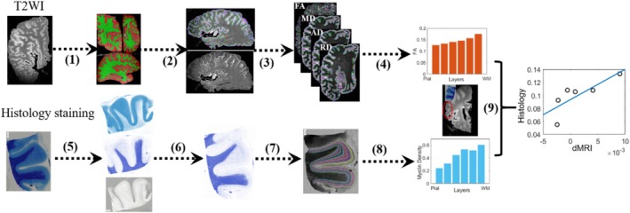
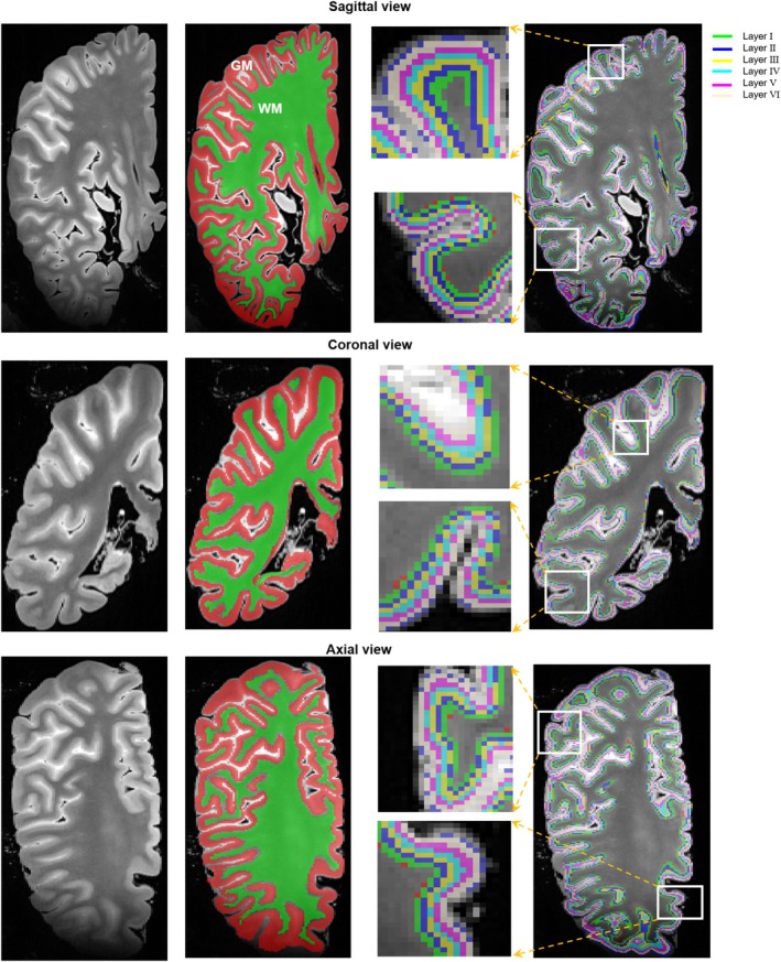
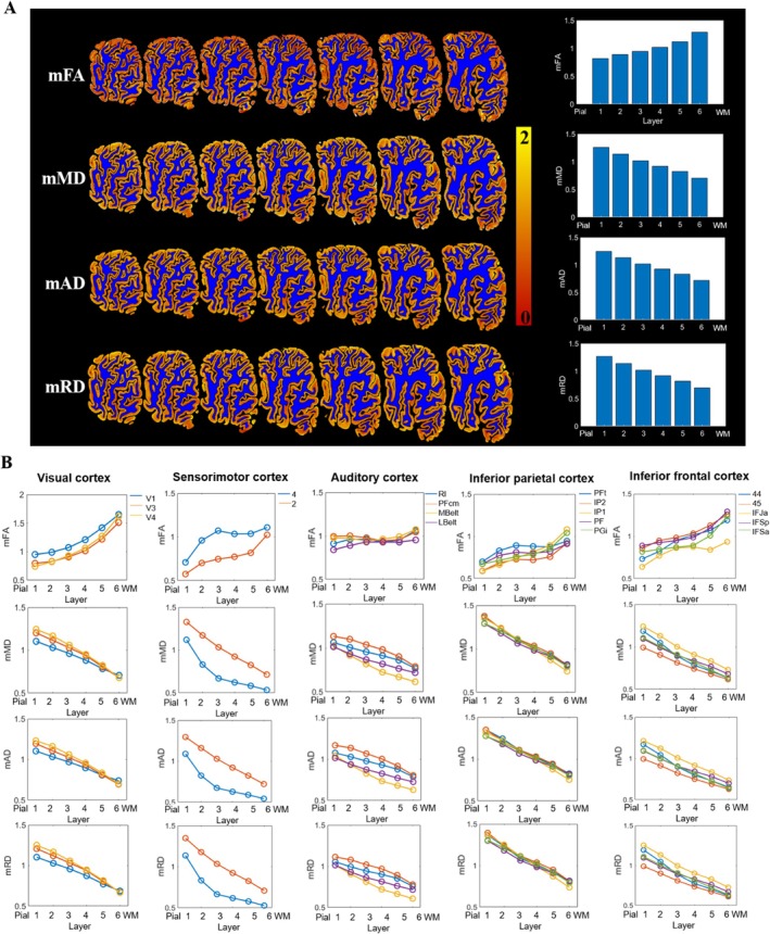
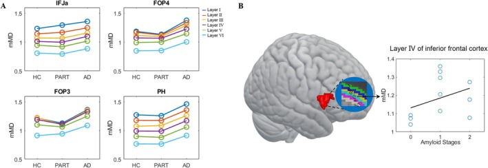
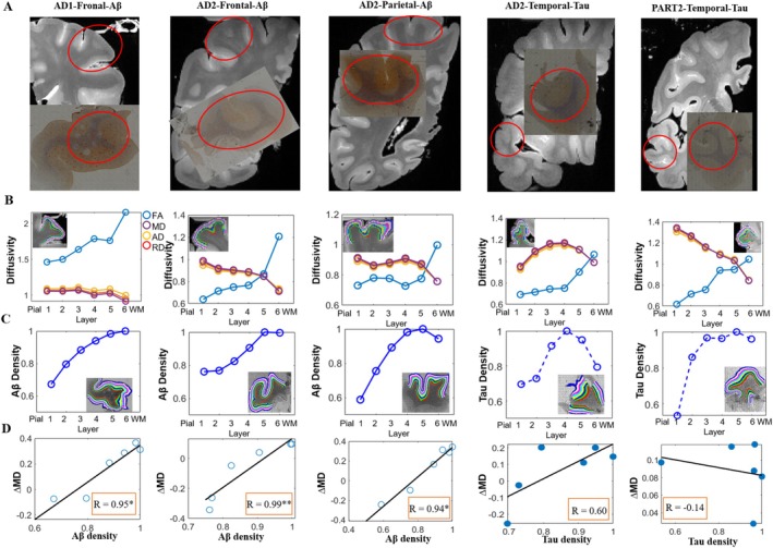
The laminar‐specific distributions of Aβ and Tau deposition in the neocortex of Alzheimer's disease (AD) have been established. However, direct evidence about the effect of AD pathology on cortical microstructure is lacking in human studies. We performed high‐resolution T2‐weighted and diffusion‐weighted MRI (dMRI) on 15 ex vivo whole‐hemisphere specimens, including eight cases with low AD neuropathologic change, three cases with primary age‐related tauopathy (PART), and four healthy controls (HCs). Using the diffusion tensor model, we evaluated microstructure patterns in six layers of gray matter cortex and performed MRI‐histology correlation analysis across cortical layers. Aβ‐positive cases exhibited higher diffusivity than Aβ‐negative cases (PART and HC) in selected cortical regions, particularly in the inferior frontal cortex. Both Aβ/Tau depositions and dMRI‐based microstructural markers demonstrated distinct cortical layer‐dependent and region‐specific patterns. A significant positive correlation was observed between increased diffusivity and Aβ burden across six cortical layers but not with Tau burden. Furthermore, the mean diffusivity in layer V of the inferior frontal cortex significantly increased with the Amyloid stage. Our findings demonstrate a layer‐dependent effect of Aβ pathology on cortical microstructure of the human brain, which may be used to serve as a marker of low AD neuropathologic change.
Overview
The claims and statistics of this paper haven’t been checked yet.
Run the check — free, no account needed. It reads the open-access text and checks the sources it cites.
Integrity signals
Registry
Cites
Statistics
Figures
Verification
Claims the paper makes
The claims and statistics of this paper haven’t been checked yet.
Run the check — free, no account needed. It reads the open-access text and checks the sources it cites.
Claims other papers make about this one
Each is checked against this paper’s own text: does it say what the citing paper says it does?
In the citing paper
“which has also been reported in the human brain.61”
From A spatial imaging-transcriptomics paradigm for deciphering the molecular basis of microscopic MRI in the normal brain and Alzheimer’s disease (2025) · cites this paper as [61]
Self-check
Percentages, sums and reported differences re-computed from the paper’s own numbers.
The self-check needs the paper’s full text.
Citations
Cites
NODDI: Practical in vivo neurite orientation dispersion and density imaging of the human brain
Zhang, H., Schneider, T., Wheeler-Kingshott, C. A., & Alexander, D. C. (2012) · NeuroImage · doi:10.1016/j.neuroimage.2012.03.072 · Cited by 3,288
Profound Loss of Layer II Entorhinal Cortex Neurons Occurs in Very Mild Alzheimer’s Disease
Gómez-Isla, T., Price, J. L., McKeel Jr., D. W., Morris, J. C., Growdon, J. H., & Hyman, B. T. (1996) · The Journal of Neuroscience · doi:10.1523/jneurosci.16-14-04491.1996 · Cited by 1,796 · cited 1×
“…dependence, with layers II–III and V of the frontotemporal cortex (Bussière et al. 2003; Hof et al. 1990) and layer II of the entorhinal cortex (Gómez-Isla et al. 1996) being vulnerable to the laminar distribution of NFTs. Similarly, our study observed layer-dependent nonlinear changes from…”
A meta-analysis of diffusion tensor imaging in mild cognitive impairment and Alzheimer's disease
Sexton, C. E., Kalu, U. G., Filippini, N., Mackay, C. E., & Ebmeier, K. P. (2011) · Neurobiology of Aging · doi:10.1016/j.neurobiolaging.2010.05.019 · Cited by 410
Diffusion tensor imaging of post mortem multiple sclerosis brain
Schmierer, K., Wheeler-Kingshott, C. A. M., Boulby, P. A., Scaravilli, F., Altmann, D. R., Barker, G. J., Tofts, P. S., & Miller, D. H. (2007) · NeuroImage · doi:10.1016/j.neuroimage.2006.12.010 · Cited by 395
Hof, P. R., Morrison, J. H., & Cox, K. (1990) · Journal of Comparative Neurology · doi:10.1002/cne.903010105 · Cited by 365 · cited 1×
“Tau accumulation in AD also showed layer dependence, with layers II–III and V of the frontotemporal cortex (Bussière et al. 2003; Hof et al. 1990) and layer II of the entorhinal cortex (Gómez-Isla et al. 1996) being vulnerable to the laminar distribution of NFTs. Similarly, our study observed…”
Molecular signatures underlying neurofibrillary tangle susceptibility in Alzheimer’s disease
Otero-Garcia, M., Mahajani, S. U., Wakhloo, D., Tang, W., Xue, Y.-Q., Morabito, S., Pan, J., Oberhauser, J., Madira, A. E., Shakouri, T., et al. (2022) · Neuron · doi:10.1016/j.neuron.2022.06.021 · Cited by 262
Wagstyl, K., Larocque, S., Cucurull, G., Lepage, C., Cohen, J. P., Bludau, S., Palomero-Gallagher, N., Lewis, L. B., Funck, T., Spitzer, H., et al. (2020) · PLOS Biology · doi:10.1371/journal.pbio.3000678 · Cited by 218
Weston, P. S. J., Simpson, I. J. A., Ryan, N. S., Ourselin, S., & Fox, N. C. (2015) · Alzheimer's Research & Therapy · doi:10.1186/s13195-015-0132-3 · Cited by 214
Cortical microstructural changes along the Alzheimer’s disease continuum
Montal, V., Vilaplana, E., Alcolea, D., Pegueroles, J., Pasternak, O., González‐Ortiz, S., Clarimón, J., Carmona‐Iragui, M., Illán‐Gala, I., Morenas‐Rodríguez, E., et al. (2018) · Alzheimer's & Dementia · doi:10.1016/j.jalz.2017.09.013 · Cited by 204
Bussière, T., Giannakopoulos, P., Bouras, C., Perl, D. P., Morrison, J. H., & Hof, P. R. (2003) · Journal of Comparative Neurology · doi:10.1002/cne.10760 · Cited by 185 · cited 1×
“Tau accumulation in AD also showed layer dependence, with layers II–III and V of the frontotemporal cortex (Bussière et al. 2003; Hof et al. 1990) and layer II of the entorhinal cortex (Gómez-Isla et al. 1996) being vulnerable to the laminar distribution of NFTs. Similarly, our study observed…”
Research works printed but not linked: 70
- 10.1016/j.neuroimage.2014.10.053 · DOI
- 10.1002/mrm.29413 · DOI
- 10.1016/j.neuroimage.2022.119653 · DOI
- 10.1016/j.pscychresns.2020.111184 · DOI
- 10.1371/journal.pone.0193839 · DOI
- 10.1002/mrm.28419 · DOI
- 10.1002/mrm.23061 · DOI
- 10.1002/hbm.25237 · DOI
- 10.1007/978-3-7091-6467-9_11 · DOI
- 10.1016/j.nicl.2023.103365 · DOI
- Casamitjana A. M.Mancini E.Robinson et al.2024.“A Next‐Generation Histological Atlas of the Human Brain and Its Application to Automated Brain MRI Segmentation.”bioRxiv 2024.02.05.579016.https://doi.org/10.1101/2024.02.05.579016. · DOI
- 10.1016/j.neuroimage.2015.10.043 · DOI · Paper page
- 10.1016/j.neuroimage.2006.12.028 · DOI
- 10.1007/BF00305876 · DOI
- 10.1073/pnas.1721653115 · DOI · Paper page
- 10.1007/s00401‐015‐1390‐7 · DOI
- 10.1002/alz.12150 · DOI
- 10.1073/pnas.200033797 · DOI
- 10.1007/s11065-014-9263-8 · DOI
- 10.1038/nature18933 · DOI · Paper page
- 10.1371/journal.pbio.3000362 · DOI
- 10.1016/j.jneumeth.2024.110059 · DOI
- 10.7554/eLife.78756 · DOI
- 10.1371/journal.pone.0173982 · DOI
- 10.1016/j.neuroimage.2021.118091 · DOI
- 10.1016/j.neuroimage.2022.119792 · DOI
- 10.1016/j.jalz.2011.10.007 · DOI · Paper page
- 10.1038/s41592-020-01008-z · DOI · Paper page
- 10.1073/pnas.2113896119 · DOI
- 10.1007/s00401‐015‐1407‐2 · DOI · Paper page
- 10.1162/imag_a_00171 · DOI
- 10.1016/j.cortex.2012.11.015 · DOI · Paper page
- 10.1097/00005072-195312040-00008 · DOI · Paper page
- 10.1007/s00401‐012‐1041‐1 · DOI
- 10.1186/s12883-020-01939-2 · DOI
- 10.1111/ene.14609 · DOI
- 10.1093/cercor/bhs311 · DOI
- 10.1016/j.cell.2019.09.001 · DOI · Paper page
- 10.1016/j.neuroimage.2023.119993 · DOI
- 10.1073/pnas.85.16.6182 · DOI
- 10.3233/JAD-2012-129012 · DOI
- 10.1002/hbm.25595 · DOI
- 10.1523/JNEUROSCI.2362-20.2021 · DOI
- 10.1016/j.jneumeth.2021.109226 · DOI
- Otero‐Garcia M. Y.‐Q.Xue T.Shakouri et al.2020.“Single‐Soma Transcriptomics of Tangle‐Bearing Neurons in Alzheimer's Disease Reveals the Signatures of Tau‐Associated Synaptic Dysfunction.”bioRxiv. 2020.2005. 2011.088591. · DOI
- 10.1148/radiol.2021210383 · DOI
- 10.1007/s12264‐018‐0306‐7 · DOI
- 10.1002/mrm.25012 · DOI · Paper page
- 10.1016/j.biopsych.2016.07.012 · DOI
- Ruifrok A. C., 2001, Quantification of Histochemical Staining by Color Deconvolution, Analytical and Quantitative Cytology and Histology, 23, 291
- 10.1002/alz.12884 · DOI
- 10.3174/ajnr.A6168 · DOI
- 10.1148/radiology.217.2.r00nv24331 · DOI
- 10.3389/fmed.2018.00031 · DOI
- 10.1093/brain/awac343 · DOI
- 10.3389/fphys.2020.00116 · DOI
- 10.1093/cercor/bhad052 · DOI
- 10.1038/s41374‐019‐0186‐0 · DOI · Paper page
- 10.1016/j.nicl.2021.102913 · DOI
- 10.1002/hbm.25821 · DOI
- 10.5214/ans.0972.7531.190410 · DOI
- 10.1016/0734-189X(85)90133-1 · DOI
- Von Economo C., 1925, The Cytoarchitectonics of the Adult Human Cortex
- 10.1093/cercor/bhab337 · DOI
- Zeng X. O.Puonti A.Sayeed et al.2023.“Segmentation of Supragranular and Infragranular Layers in Ultra‐High Resolution 7T Ex Vivo MRI of the Human Cerebral Cortex.”bioRxiv 2023.12.06.570416.https://doi.org/10.1101/2023.12.06.570416. · DOI
- 10.1002/hbm.25998 · DOI
- 10.1016/j.neurobiolaging.2021.06.013 · DOI
- 10.1002/hbm.26062 · DOI
- 10.1016/j.neuroimage.2021.118584 · DOI
- 10.1016/j.neuroimage.2020.117313 · DOI
Cited by
Sorted by citations
Shen, Y., Shen, Y., Wang, M., Jin, K., Yang, P., Cao, Z., Zhu, Q., Zhao, Z., Li, H., Han, L., et al. (2025) · Cell Reports · doi:10.1016/j.celrep.2025.116073 · Cited by 16
Self-citation
Metadata
- Authors
- Zhao, Z., Cao, Z., Zhu, Q., Xu, H., Li, S., Zhu, L., Xu, G., Zhu, K., Zhang, J., & Wu, D.
- Affiliations
- Children's Hospital Zhejiang University School of Medicine, National Clinical Research Center for Child Health Hangzhou China
- Key Laboratory for Biomedical Engineering of Ministry of Education, Department of Biomedical Engineering College of Biomedical Engineering & Instrument Science, Zhejiang University Hangzhou China
- National Human Brain Bank for Health and Disease Zhejiang University Hangzhou China
- ORCID
- Venue
- Human Brain Mapping
- Publisher
- Wiley
- Published
- 2025-05
- Volume
- 46
- Issue
- 7
- ISSN
- 1065-9471
- 1097-0193
- PMC
- PMC12046383
- Cited by
- 8
- References
- 16
- Language
- en
- Funders
- National Natural Science Foundation of China (81971184, 81971606, 82122032)
- Science and Technology Department of Zhejiang Province (202006140, 2022C03057)
About these checks
Registry, retraction, Cites and statistics results are computed, not judged, with one exception: a printed reference that no index lists is read once by a model before it counts as a citation error. Claims are extracted by a model and kept only when their quote appears verbatim in the text; each cited source is then checked, with a model’s help, for whether it says what the claim relies on. Those verdicts can be wrong — read the claim’s quote and the cited source before relying on one.
Statistics and arithmetic are recomputed, not judged.
Confidence bands are shown only for fields calibrated on our annotated test set. None is calibrated yet, so no band is shown.
What counts as a claim ›How we measure confidence ›How we check this ›
Are you an author, or do you see a problem with this page? Request a review
How to cite
Cite CoChat for the checks on this page, and the paper for its own findings.
- This page
- CoChat. (2026). Citation check: Layer‐Dependent Effect of Aβ‐Pathology on Cortical Microstructure With Ex Vivo Human Brain Diffusion MRI at 7 Tesla. https://app.cochat.ai/papers/10.1002/hbm.70222
- The paper
- Zhao, Z., Cao, Z., Zhu, Q., Xu, H., Li, S., Zhu, L., Xu, G., Zhu, K., Zhang, J., & Wu, D. (2025). Layer‐Dependent Effect of Aβ‐Pathology on Cortical Microstructure With Ex Vivo Human Brain Diffusion MRI at 7 Tesla. Human Brain Mapping. https://doi.org/10.1002/hbm.70222
