

The DOI matches its Crossref record; 1 of the 1 references it cites were found in a registry or an index.
HPV16 E5 expression induces switching from FGFR2b to FGFR2c and epithelial-mesenchymal transition
Abstract
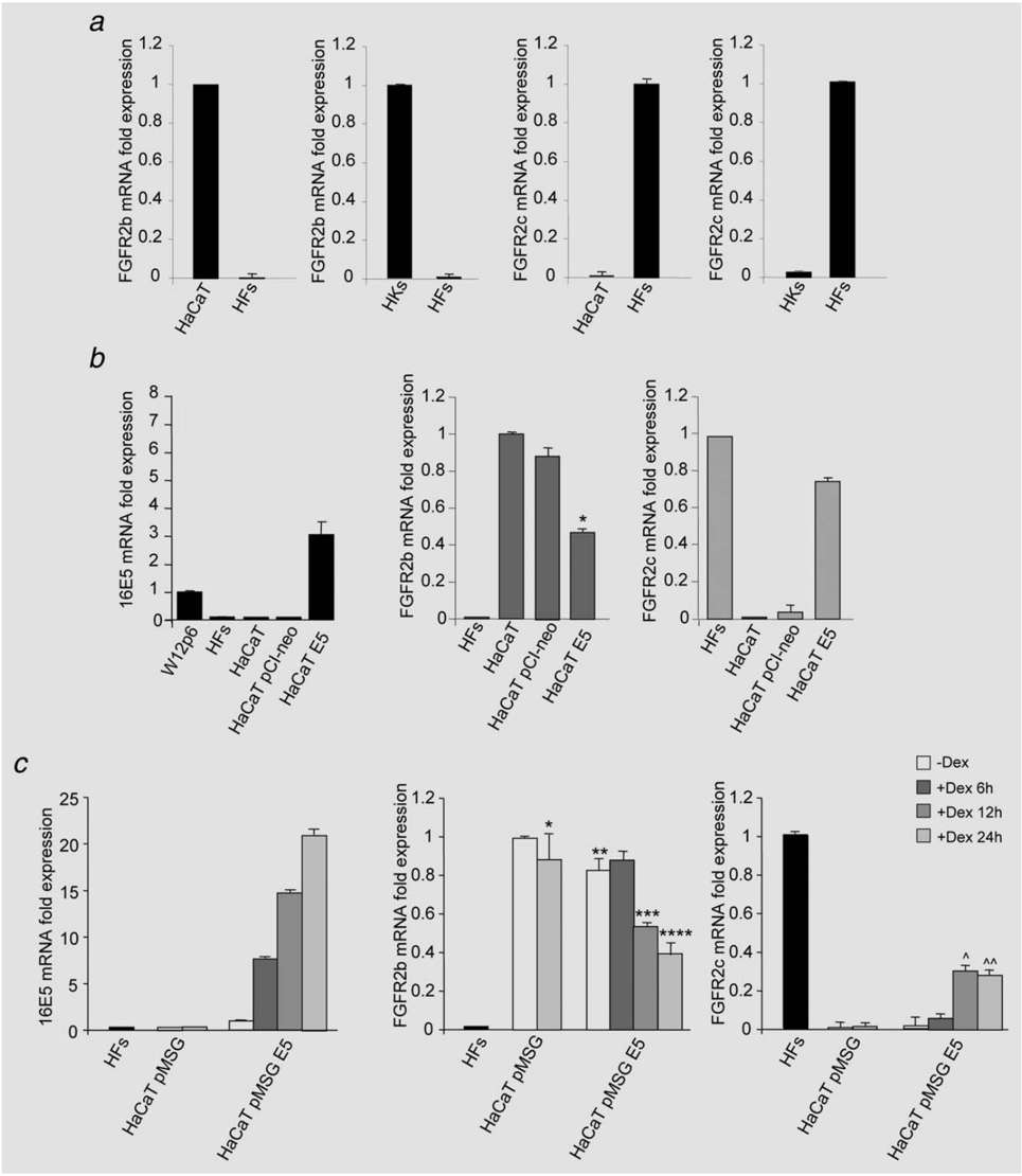
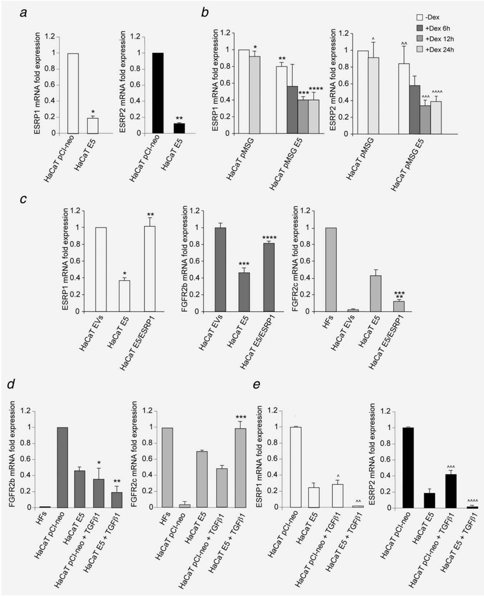
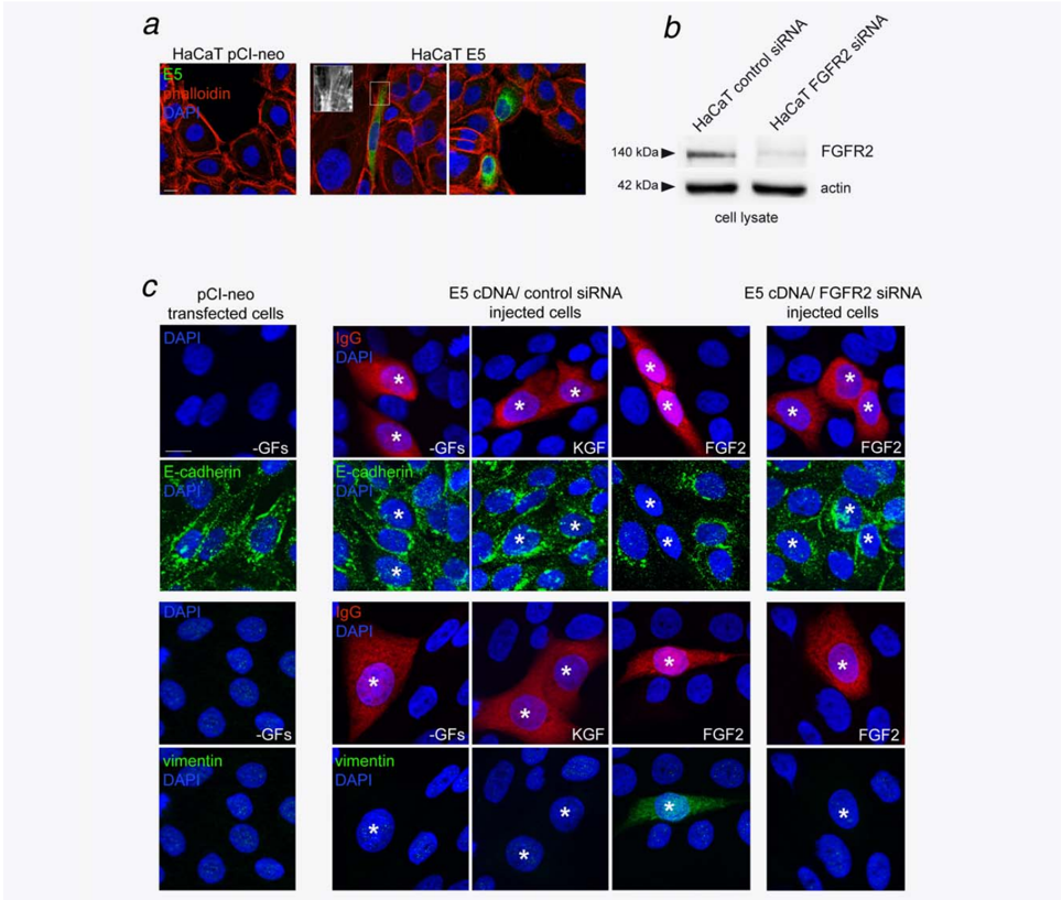
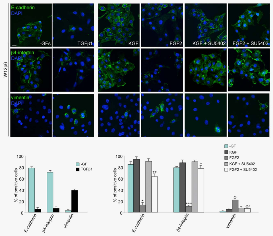
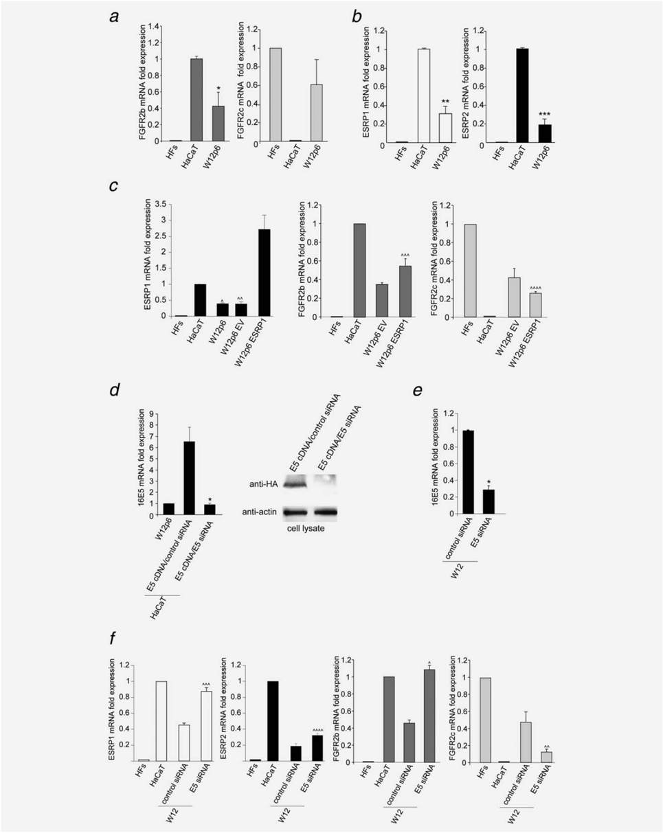
The E5 oncoprotein of the human papillomavirus type 16 (HPV16 E5) deregulates epithelial homeostasis through the modulation of receptor tyrosine kinases and their signaling. Accordingly, the fibroblast growth factor receptor 2b (FGFR2b/KGFR), epithelial splicing transcript variant of the FGFR2, is down‐modulated by the viral protein expression, leading to impairment of keratinocyte differentiation. Here, we report that, in cell models of transfected human keratinocytes as well as in cervical epithelial cells containing episomal HPV16, the down‐regulation of FGFR2b induced by 16E5 is associated with the aberrant expression of the mesenchymal FGFR2c isoform as a consequence of splicing switch: in fact, quantitative RT‐PCR analysis showed that this molecular event is transcriptionally regulated by the epithelial splicing regulatory proteins 1 and 2 (ESRP1 and ESRP2) and is able to produce effects synergistic with those caused by TGFβ treatment. Immunofluorescence analysis revealed that this altered FGFR2 splicing leads to changes in the specificity for the ligands FGFs and in the cellular response, triggering epithelial‐mesenchymal transition (EMT). Through 16E5 or FGFR2 silencing as well as inhibition of FGFR2 activity we demonstrated the direct role of the viral protein in the receptor isoform switching and EMT, suggesting that these early molecular events during HPV infection might represent additional mechanisms driving cervical transformation and tumor progression.
Overview
The claims and statistics of this paper haven’t been checked yet.
Run the check — free, no account needed. It reads the open-access text and checks the sources it cites.
Integrity signals
Registry
Cites
Statistics
Figures
Verification
Claims the paper makes
The claims and statistics of this paper haven’t been checked yet.
Run the check — free, no account needed. It reads the open-access text and checks the sources it cites.
Claims other papers make about this one
Each is checked against this paper’s own text: does it say what the citing paper says it does?
In the citing paper
“The expression of HPV-16 E5 downregulates the expression of KGFR2b, enhancing the aberrant expression of the mesenchymal FGFR2c isoform, which promotes the epithelial–mesenchymal transition (EMT)”
From The Not-So-Good, the Bad and the Ugly: HPV E5, E6 and E7 Oncoproteins in the Orchestration of Carcinogenesis (2021) · cites this paper as [219]
Self-check
Percentages, sums and reported differences re-computed from the paper’s own numbers.
The self-check needs the paper’s full text.
Citations
Cites
Normal keratinization in a spontaneously immortalized aneuploid human keratinocyte cell line.
Boukamp, P., Petrussevska, R. T., Breitkreutz, D., Hornung, J., Markham, A., & Fusenig, N. E. (1988) · The Journal of cell biology · doi:10.1083/jcb.106.3.761 · Cited by 4,170
Research works printed but not linked: 32
- 10.1038/nrc2780 · DOI
- 10.1038/nrm3528 · DOI
- 10.1158/1541-7786.MCR-10-0168 · DOI
- 10.1158/1078-0432.CCR-11-0699 · DOI
- 10.1016/j.molcel.2009.01.025 · DOI
- 10.1038/emboj.2010.351 · DOI
- Feng S, 1997, Fibroblast growth factor receptor 2 limits and receptor 1 accelerates tumorigenicity of prostate epithelial cells, Cancer Res, 58, 1509
- 10.1073/pnas.191377098 · DOI
- 10.1038/nrc798 · DOI
- 10.1038/nrc2886 · DOI
- 10.1186/1476-4598-10-140 · DOI
- 10.1038/onc.2011.203 · DOI · Paper page
- 10.18632/oncotarget.803 · DOI
- Oelze I, 1995, Human papillomavirus type 16 E5 protein affects cell‐cell communication in an epithelial cell line, J Virol, 69, 4489, 10.1128/jvi.69.7.4489-4494.1995 · DOI
- 10.1002/ijc.2910430422 · DOI
- 10.1158/0008-5472.CAN-03-3214 · DOI
- 10.1158/0008-5472.CAN-09-3335 · DOI
- 10.1111/j.0022-202X.2005.23929.x · DOI · Paper page
- 10.1007/s00418-012-0947-y · DOI
- 10.1002/ijc.20558 · DOI · Paper page
- 10.1093/carcin/bgn236 · DOI
- 10.1186/1476-4598-12-38 · DOI
- 10.4161/rna.6.5.9606 · DOI
- 10.1038/onc.2011.493 · DOI
- 10.1016/j.virol.2009.05.036 · DOI · Paper page
- 10.1016/j.bbrc.2013.04.060 · DOI
- 10.1186/1476-4598-7-3 · DOI
- 10.1016/j.humimm.2012.06.003 · DOI
- 10.1016/j.virol.2013.08.034 · DOI
- 10.1371/journal.pone.0021646 · DOI
- 10.1099/vir.0.016295-0 · DOI
- 10.1038/sj.onc.1206554 · DOI
Cited by
Basukala, O., & Banks, L. (2021) · Viruses · doi:10.3390/v13101892 · Cited by 137
Metadata
- Authors
- Ranieri, D., Belleudi, F., Magenta, A., & Torrisi, M. R.
- Affiliations
- Istituto Pasteur‐Fondazione Cenci Bolognetti Dipartimento di Medicina Clinica e Molecolare Sapienza Università di Roma Italy
- Venue
- International Journal of Cancer
- Publisher
- Wiley
- Published
- 2014-12-13
- Volume
- 137
- Issue
- 1
- Pages
- 61-72
- ISSN
- 0020-7136
- 1097-0215
- Cited by
- 54
- References
- 1
- Language
- en
- Funders
- MIUR
- Associazione Italiana per la Ricerca sul Cancro (AIRC), Italy (IG 15858)
About these checks
Registry, retraction, Cites and statistics results are computed, not judged, with one exception: a printed reference that no index lists is read once by a model before it counts as a citation error. Claims are extracted by a model and kept only when their quote appears verbatim in the text; each cited source is then checked, with a model’s help, for whether it says what the claim relies on. Those verdicts can be wrong — read the claim’s quote and the cited source before relying on one.
Statistics and arithmetic are recomputed, not judged.
Confidence bands are shown only for fields calibrated on our annotated test set. None is calibrated yet, so no band is shown.
What counts as a claim ›How we measure confidence ›How we check this ›
Are you an author, or do you see a problem with this page? Request a review
