

The DOI matches its Crossref record; 152 of the 152 references it cites were found in a registry or an index.
Cellular and molecular mechanisms of sarcopenia: the S100B perspective
Open accessAbstract
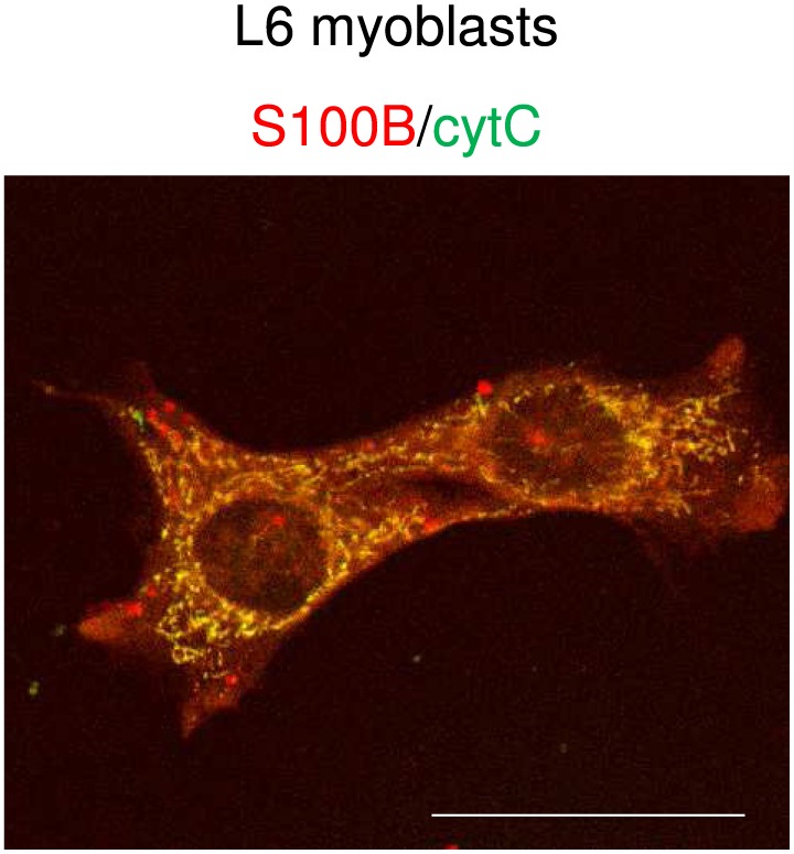
Abstract Primary sarcopenia is a condition of reduced skeletal muscle mass and strength, reduced agility, and increased fatigability and risk of bone fractures characteristic of aged, otherwise healthy people. The pathogenesis of primary sarcopenia is not completely understood. Herein, we review the essentials of the cellular and molecular mechanisms of skeletal mass maintenance; the alterations of myofiber metabolism and deranged properties of muscle satellite cells (the adult stem cells of skeletal muscles) that underpin the pathophysiology of primary sarcopenia; the role of the Ca 2+ ‐sensor protein, S100B, as an intracellular factor and an extracellular signal regulating cell functions; and the functional role of S100B in muscle tissue. Lastly, building on recent results pointing to S100B as to a molecular determinant of myoblast–brown adipocyte transition, we propose S100B as a transducer of the deleterious effects of accumulation of reactive oxygen species in myoblasts and, potentially, myofibers concurring to the pathophysiology of sarcopenia.
Overview
The claims and statistics of this paper haven’t been checked yet.
Run the check — free, no account needed. It reads the open-access text and checks the sources it cites.
Integrity signals
Registry
Cites
Statistics
Figures
Verification
Claims the paper makes
The claims and statistics of this paper haven’t been checked yet.
Run the check — free, no account needed. It reads the open-access text and checks the sources it cites.
Claims other papers make about this one
Each is checked against this paper’s own text: does it say what the citing paper says it does?
In the citing paper
“Second, gApN induces the expression of MyoD that allows myoblast proliferation and differentiation [56,69,70].”
From Adiponectin and Its Mimics on Skeletal Muscle: Insulin Sensitizers, Fat Burners, Exercise Mimickers, Muscling Pills … or Everything Together? (2020) · cites this paper as [70]
Self-check
Percentages, sums and reported differences re-computed from the paper’s own numbers.
The self-check needs the paper’s full text.
Citations
Cites
1 retracted
Immunohistochemical detection of interleukin‐6 in human skeletal muscle fibers following exercise
Penkowa, M., Keller, C., Keller, P., Jauffred, S., & Pedersen, B. K. (2003) · The FASEB Journal · doi:10.1096/fj.03-0311fje · Cited by 157
RetractedSandri, M., Sandri, C., Gilbert, A., Skurk, C., Calabria, E., Picard, A., Walsh, K., Schiaffino, S., Lecker, S. H., & Goldberg, A. L. (2004) · Cell · doi:10.1016/s0092-8674(04)00400-3 · Cited by 3,012 · cited 1×
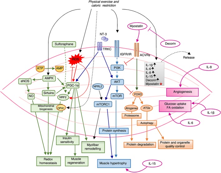
“They are activated in multiple models of muscle atrophy and are both suf fi cient and required for muscle atrophy 38 ( Figure 2).”
Cellular and Molecular Regulation of Muscle Regeneration
CHARGÉ, S. B. P., & Rudnicki, M. A. (2004) · Physiological Reviews · doi:10.1152/physrev.00019.2003 · Cited by 2,691 · cited 1×
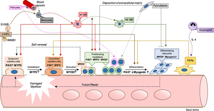
“…released from damaged muscle tissue — collectively known as damage-associated molecular patterns (DAMPs) — or secreted by in fi ltrating immune cells (i.e. hormones, growth factors, and cytokines) and components of the extracellular matrix 17 – 24 ( Figure 1) acting via cell surface receptors.”
Transcriptional co-activator PGC-1α drives the formation of slow-twitch muscle fibres
Lin, J., Wu, H., Tarr, P. T., Zhang, C.-Y., Wu, Z., Boss, O., Michael, L. F., Puigserver, P., Isotani, E., Olson, E. N., et al. (2002) · Nature · doi:10.1038/nature00904 · Cited by 2,546
Muscle as an Endocrine Organ: Focus on Muscle-Derived Interleukin-6
Pedersen, B. K., & Febbraio, M. A. (2008) · Physiological Reviews · doi:10.1152/physrev.90100.2007 · Cited by 2,388
Rejuvenation of aged progenitor cells by exposure to a young systemic environment
Conboy, I. M., Conboy, M. J., Wagers, A. J., Girma, E. R., Weissman, I. L., & Rando, T. A. (2005) · Nature · doi:10.1038/nature03260 · Cited by 2,287 · cited 1×
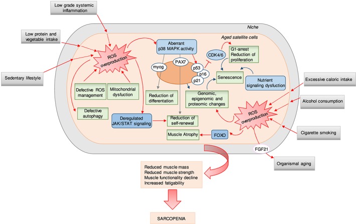
“From the side of SCs, extrinsic, niche-related factors and intrinsic, cell-autonomous factors concur to determine changes in SCs with aging ultimately leading to reduced SCs ’ ability to maintain muscle mass 5,18,66 – 73 ( Figure 3).”
Nrf2-Keap1 signaling in oxidative and reductive stress
Bellezza, I., Giambanco, I., Minelli, A., & Donato, R. (2018) · Biochimica et Biophysica Acta (BBA) - Molecular Cell Research · doi:10.1016/j.bbamcr.2018.02.010 · Cited by 2,134 · cited 1×
“sue (reviewed by Bellezza et al.(43)), an event counteracted by a”
Arnold, L., Henry, A., Poron, F., Baba-Amer, Y., van Rooijen, N., Plonquet, A., Gherardi, R. K., & Chazaud, B. (2007) · The Journal of Experimental Medicine · doi:10.1084/jem.20070075 · Cited by 2,002
RAGE Mediates a Novel Proinflammatory Axis
Hofmann, M. A., Drury, S., Fu, C., Qu, W., Taguchi, A., Lu, Y., Avila, C., Kambham, N., Bierhaus, A., Nawroth, P., et al. (1999) · Cell · doi:10.1016/s0092-8674(00)80801-6 · Cited by 1,864
Muscle injury activates resident fibro/adipogenic progenitors that facilitate myogenesis
Joe, A. W. B., Yi, L., Natarajan, A., Le Grand, F., So, L., Wang, J., Rudnicki, M. A., & Rossi, F. M. V. (2010) · Nature Cell Biology · doi:10.1038/ncb2015 · Cited by 1,690
Cited by
Sorted by citations
Cruz-Jentoft, A. J., & Sayer, A. A. (2019) · The Lancet · doi:10.1016/s0140-6736(19)31138-9 · Cited by 3,905
Sarcopenia: A Contemporary Health Problem among Older Adult Populations
Papadopoulou, S. Κ. (2020) · Nutrients · doi:10.3390/nu12051293 · Cited by 672
Rheumatoid sarcopenia: loss of skeletal muscle strength and mass in rheumatoid arthritis
Bennett, J. L., Pratt, A. G., Dodds, R. M., Sayer, A. A., & Isaacs, J. D. (2023) · Nature Reviews Rheumatology · doi:10.1038/s41584-023-00921-9 · Cited by 174
Myostatin as a Biomarker of Muscle Wasting and other Pathologies-State of the Art and Knowledge Gaps
Bączek, J., Silkiewicz, M., & Wojszel, Z. B. (2020) · Nutrients · doi:10.3390/nu12082401 · Cited by 166
“sarcopenia, the result of chronic inflammatory statuses, diabetes, hormonal alterations, vascular disturbances, renal, respiratory and/or cardiac failure and immobilization [29,30].”
BackgroundInflammaging: The ground for sarcopenia?
Liang, Z., Zhang, T., Liu, H., Li, Z., Peng, L., Wang, C., & Wang, T. (2022) · Experimental Gerontology · doi:10.1016/j.exger.2022.111931 · Cited by 156
Hyatt, H. W., & Powers, S. K. (2021) · Antioxidants · doi:10.3390/antiox10040588 · Cited by 134
Biological Aspects of Selected Myokines in Skeletal Muscle: Focus on Aging
Mancinelli, R., Checcaglini, F., Coscia, F., Gigliotti, P. V., Fulle, S., & Fanò, G. (2021) · International Journal of Molecular Sciences · doi:10.3390/ijms22168520 · Cited by 113
Lei, Y., Gan, M., Qiu, Y., Chen, Q., Wang, X., Liao, T., Zhao, M., Chen, L., Zhang, S., Zhao, Y., et al. (2024) · Cellular & Molecular Biology Letters · doi:10.1186/s11658-024-00572-y · Cited by 111
Mitochondrial Impairment in Sarcopenia
Bellanti, F., Buglio, A. L., & Vendemiale, G. (2021) · Biology · doi:10.3390/biology10010031 · Cited by 104
Sarcopenia: Diagnosis and Management, State of the Art and Contribution of Ultrasound
Giovannini, S., Brau, F., Forino, R., Berti, A., D’Ignazio, F., Loreti, C., Bellieni, A., D’Angelo, E., Caro, F. D., Biscotti, L., et al. (2021) · Journal of Clinical Medicine · doi:10.3390/jcm10235552 · Cited by 100
Anti‐diabetic drugs and sarcopenia: emerging links, mechanistic insights, and clinical implications
Zhang, X., Zhao, Y., Chen, S., & Shao, H. (2021) · Journal of Cachexia Sarcopenia and Muscle · doi:10.1002/jcsm.12838 · Cited by 96
Multi-omics research in sarcopenia: Current progress and future prospects
Liu, J., Dong, S., Shen, H., Yang, D.-Y., Chen, B., Ma, X., Peng, Y., Xiao, H., & Deng, H. (2022) · Ageing Research Reviews · doi:10.1016/j.arr.2022.101576 · Cited by 83
Muscular Atrophy and Sarcopenia in the Elderly: Is There a Role for Creatine Supplementation?
Dolan, E., Artioli, G. G., Pereira, R. M. R., & Gualano, B. (2019) · Biomolecules · doi:10.3390/biom9110642 · Cited by 72
“An in-depth description of these factors is beyond the scope of this piece, but a number of comprehensive reviews are available on the topic [10,14].”
BackgroundAbou-Samra, M., Selvais, C. M., Dubuisson, N., & Brichard, S. M. (2020) · International Journal of Molecular Sciences · doi:10.3390/ijms21072620 · Cited by 72
RAGE and its ligands: from pathogenesis to therapeutics
Jangde, N., Ray, R., & Rai, V. (2020) · Critical Reviews in Biochemistry and Molecular Biology · doi:10.1080/10409238.2020.1819194 · Cited by 81
Treatment of sarcopenia: the road to the future
Morley, J. E. (2018) · Journal of Cachexia Sarcopenia and Muscle · doi:10.1002/jcsm.12386 · Cited by 75
S100 proteins in obesity: liaisons dangereuses
Riuzzi, F., Chiappalupi, S., Arcuri, C., Giambanco, I., Sorci, G., & Donato, R. (2019) · Cellular and Molecular Life Sciences · doi:10.1007/s00018-019-03257-4 · Cited by 70
The association of lipid metabolism and sarcopenia among older patients: a cross-sectional study
Jiang, Y., Xu, B., Zhang, K., Zhu, W., Lian, X., Xu, Y., Chen, Z., Liu, L., & Guo, Z. (2023) · Scientific Reports · doi:10.1038/s41598-023-44704-4 · Cited by 74
Pathogenesis, Intervention, and Current Status of Drug Development for Sarcopenia: A Review
Jang, J. Y., Kim, D.-H., & Kim, N. D. (2023) · Biomedicines · doi:10.3390/biomedicines11061635 · Cited by 66
“Insulin resistance, elevated proinflammatory cytokines, genetic factors, and poor nutritional status (including insufficient energy and protein intake leading to weight loss and decreased levels of vitamin D) can further accelerate muscle breakdown [56,58].”
BackgroundIncreased Serum S100B Levels in Patients With Epilepsy: A Systematic Review and Meta-Analysis Study
Liang, K.-G., Mu, R.-Z., Liu, Y., Jiang, D., Jia, T., & Huang, Y. (2019) · Frontiers in Neuroscience · doi:10.3389/fnins.2019.00456 · Cited by 58
“S100 proteins are involved in the regulation of a number of cellular processes such as signal regulating, cell cycle progression, and act as a stimulator of cell proliferation and migration and as an inhibitor of apoptosis and differentiation (Donato et al., 2009; Riuzzi et al., 2018; Michetti et al., 2019).”
BackgroundKoeppel, M., Mathis, K. M., Schmitz, K. H., & Wiskemann, J. (2021) · Critical Reviews in Oncology/Hematology · doi:10.1016/j.critrevonc.2021.103371 · Cited by 62
Post‐translational regulation of muscle growth, muscle aging and sarcopenia
Zhong, Q., Zheng, K., Li, W., An, K., Liu, Y., Xiao, X., Hai, S., Dong, B., Li, S., An, Z., et al. (2023) · Journal of Cachexia Sarcopenia and Muscle · doi:10.1002/jcsm.13241 · Cited by 59
“This intricate process is orchestrated by a series of transcription factors, including PAX7, Myf5, MyoD, myogenin and MRF4, that are sequentially activated.(11)”
BackgroundMinniti, G., Pescinini-Salzedas, L. M., Minniti, G. A. D. S., Laurindo, L. F., Barbalho, S. M., Sinatora, R. V., Sloan, L. A., Haber, R. S. D. A., Araújo, A. C., Quesada, K. R., et al. (2022) · International Journal of Molecular Sciences · doi:10.3390/ijms232113452 · Cited by 53
Cheng, Y., Lin, S., Cao, Z., Yu, R., Fan, Y., & Chen, J. (2025) · International Immunopharmacology · doi:10.1016/j.intimp.2025.114056 · Cited by 55
Hämäläinen, O., Tirkkonen, A., Savikangas, T., Alén, M., Sipilä, S., & Hautala, A. J. (2024) · BMC Geriatrics · doi:10.1186/s12877-024-04764-1 · Cited by 47
Stuck, A. K., Tsai, L.-C., Freystaetter, G., Vellas, B. J., Kanis, J. А., Rizzoli, R., Kressig, R. W., Armbrecht, G., Silva, J. A. P. D., Dawson‐Hughes, B., et al. (2023) · The journal of nutrition health & aging · doi:10.1007/s12603-023-1888-y · Cited by 46
Čamernik, K., Mihelič, A., Mihalič, R., Haring, G., Herman, S., Marolt, D., Janež, A., Trebše, R., Marc, J., & Zupan, J. (2020) · Stem Cell Research & Therapy · doi:10.1186/s13287-020-01657-z · Cited by 46
Circulating Mediators of Apoptosis and Inflammation in Aging; Physical Exercise Intervention
Morawin, B., Tylutka, A., Chmielowiec, J., & Zembroń-Łacny, A. (2021) · International Journal of Environmental Research and Public Health · doi:10.3390/ijerph18063165 · Cited by 42
“Due to the increasing life expectancy in developed countries, sarcopenia is prognosed to become an even more serious and global health problem and a significant economic burden on healthcare systems [7,8].”
BackgroundCurcumin as a Therapeutic Agent for Sarcopenia
Gany, S. L. S., Chin, K., Tan, J. K., Aminuddin, A., & Makpol, S. (2023) · Nutrients · doi:10.3390/nu15112526 · Cited by 42
“The pathogenesis of sarcopenia includes impaired myofiber metabolism and adverse changes to muscle satellite cells, which lead to defective myogenesis and the loss of skeletal muscle homeostasis [13].”
BackgroundUnraveling the Links Underlying Arterial Stiffness, Bone Demineralization, and Muscle Loss
Tap, L., Kirkham, F. A., Mattace-Raso, F. U. S., Joly, L., Rajkumar, C., & Bénétos, A. (2020) · Hypertension · doi:10.1161/hypertensionaha.120.15184 · Cited by 40
“Increased CRP has been shown to predict loss of muscle,(18) while imbalanced production of reactive oxygen species can impair muscle cell maintenance.(90) Inflammatory mediators can act directly on muscle receptors to increase breakdown or indirectly by impairing production of anabolic proteins such as growth hormone.”
BackgroundOris, C., Bouillon‐Minois, J., Pinguet, J., Kahouadji, S., Durif, J., Meslé, V., Pereira, B., Schmidt, J., Sapin, V., & Bouvier, D. (2021) · The Journals of Gerontology Series A · doi:10.1093/gerona/glab055 · Cited by 37
Park, H., Jung, W., Kim, S.-W., & Lim, K. (2021) · Frontiers in Physiology · doi:10.3389/fphys.2021.706259 · Cited by 37
“Reduced exercise, lack of dietary intake, decreased type 2 muscle fibers in the skeletal muscle, and decreased secretion of insulin-like growth factor-1 (IGF-1) contribute to the development of sarcopenia (Riuzzi et al., 2018; Carter et al., 2019).”
BackgroundMitochondrial DNA variants in inclusion body myositis characterized by deep sequencing
Hedberg‐Oldfors, C., Lindgren, U., Basu, S., Visuttijai, K., Lindberg, C., Falkenberg, M., Larsson, E., & Oldfors, A. (2020) · Brain Pathology · doi:10.1111/bpa.12931 · Cited by 35
“The cause of sarcopenia is manifold but chronic inflammation as part of aging, so called “inflamm-aging” or “senoinflammation” (31–33), is considered an essential factor leading to sarcopenia (34,35).”
BackgroundRole of Adipokines in Sarcopenia
Lu, W., Feng, W., Lai, J., Yuan, D., Xiao, W., & Li, Y. (2023) · Chinese Medical Journal · doi:10.1097/cm9.0000000000002255 · Cited by 34
Factors inducing transdifferentiation of myoblasts into adipocytes
Wang, L., & Shan, T. (2020) · Journal of Cellular Physiology · doi:10.1002/jcp.30074 · Cited by 36
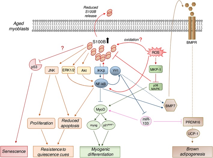
“At the molecular level, oxidative stress and ROS induces S100B accumulation, which as a molecular determinant of driving myoblasts differentiation into brown adipocytes through the NF‐κB/YY1 (Yin Yang 1)/miR‐133 pathway and NF‐κB/Ying Yang 1/BMP‐7 pathway (Morozzi et al., 2017; Riuzzi et al., 2018).”
BackgroundGinseng and ginsenosides: Therapeutic potential for sarcopenia
Zha, W., Sun, Y., Gong, W., Li, L., Kim, W., & Li, H. (2022) · Biomedicine & Pharmacotherapy · doi:10.1016/j.biopha.2022.113876 · Cited by 31
Methylglyoxal Induces Inflammation, Metabolic Modulation and Oxidative Stress in Myoblast Cells
Todoriki, S., Hosoda, Y., Yamamoto, T., Watanabe, M., Sekimoto, A., Satō, H., Mori, T., Miyazaki, M., Takahashi, N., & Sato, E. (2022) · Toxins · doi:10.3390/toxins14040263 · Cited by 28
“In addition, when skeletal muscle is damaged, myoblast cells differentiate into myotube cells and fuse to the damaged area for repair to maintain skeletal muscle [41].”
BackgroundGuo, R., Wu, Z., Liu, A., Li, Q., Han, T., & Shen, C. (2024) · International Immunopharmacology · doi:10.1016/j.intimp.2024.113143 · Cited by 24
Potential therapeutic targets for sarcopenia identified by Mendelian randomisation
Jiang, W., Zhan, W., Zhou, L., Dong, M., Liu, L., Xu, X. S., & Cao, Z. (2023) · Age and Ageing · doi:10.1093/ageing/afad024 · Cited by 23
“complex mechanisms, and the pathogenesis was not wholly understood [55].”
BackgroundWalking down Skeletal Muscle Lane: From Inflammasome to Disease
Dubuisson, N., Versele, R., de Carrizosa, M. A. D., Selvais, C. M., Brichard, S. M., & Abou‐Samra, M. (2021) · Cells · doi:10.3390/cells10113023 · Cited by 23
Impaired expression of BCAT1 relates to muscle atrophy of mouse model of sarcopenia
Ouyang, H., Gao, X., & Zhang, J. (2022) · BMC Musculoskeletal Disorders · doi:10.1186/s12891-022-05332-7 · Cited by 22
“However, the underlying mechanisms of muscle atrophy in sarcopenia are not fully understood [1].”
BackgroundRole of the S100 protein family in liver disease (Review)
Yao, S., Yang, X., An, J., Jin, H. D., Wen, G., Wang, H., & Tuo, B. (2021) · International Journal of Molecular Medicine · doi:10.3892/ijmm.2021.4999 · Cited by 20
EBENBICHLER, G. R., Habenicht, R., Ziegelbecker, S., Kollmitzer, J., Mair, P., & Kienbacher, T. (2019) · GeroScience · doi:10.1007/s11357-019-00134-7 · Cited by 20
Comfort, N. T., Gade, M., Strait, M. D., Merwin, S. J., Αντωνίου, Δ., Parodi, C., Marcinczyk, L., Jean‐Francois, L., Bloomquist, T. R., Memou, A., et al. (2023) · Journal of Cachexia Sarcopenia and Muscle · doi:10.1002/jcsm.13204 · Cited by 16
Bijeh, N., Ahmadi, M. M., Moghadam, B. H., Eskandari, M., & Golestani, F. (2022) · Biological Research For Nursing · doi:10.1177/10998004211073123 · Cited by 15
“It was demonstrated that myostatin inhibits myoblast differentiation and blocks the activation of the PI3K/AKT hypertrophy signaling pathway (Riuzzi et al., 2018).”
BackgroundCombinatorial interventions in aging
Parkhitko, A. A., Filine, E., & Tatar, M. (2023) · Nature Aging · doi:10.1038/s43587-023-00489-9 · Cited by 17
“Manipulating protein damage and clearing senescent cells (features of molecular and hallmark manifestations) may alter sarcopenia (physiological manifestation) 10,11 .”
BackgroundYogesh, M., Patel, M., Makwana, H., & Kalariya, H. (2024) · Clinical Diabetes and Endocrinology · doi:10.1186/s40842-024-00179-4 · Cited by 16
“The mechanisms underlying the development of sarcopenia are multifactorial and involve a complex interplay of factors such as sedentary lifestyles, nutritional deficiencies, chronic inflammation, and insulin resistance [15–17].”
BackgroundGalluzzo, V., Zazzara, M. B., Ciciarello, F., Savera, G., Pais, C., Calvani, R., Picca, A., Marzetti, E., Landi, F., Tosato, M., et al. (2022) · Clinical Nutrition ESPEN · doi:10.1016/j.clnesp.2022.08.029 · Cited by 15
“Changes in diet and physical activity routines due to restrictions put in place to reduce the spread of the virus during the pandemic might have altered individuals’ metabolism homeostasis, favoring the unbalance between anabolism and catabolism [53].”
BackgroundHashemi, P., Mardani, P., Raz, Z. E., Saedi, A., Fatahi, E., Izapanah, E., & Ahmadi, S. (2024) · Molecular Neurobiology · doi:10.1007/s12035-024-04407-x · Cited by 14
“S100-B is a calcium-binding protein that plays a role in regulating a cascade of physiological processes, including the inhibition of apoptosis and differentiation, signal regulation, cell cycle development, and cell proliferation and migration [10].”
Sarcopenia—a geriatric pandemic
Köller, M. D. (2022) · Wiener Medizinische Wochenschrift · doi:10.1007/s10354-022-00927-0 · Cited by 16
“S-100 modulates contractile activity and force generation, and also acts on myogenesis [22, 25].”
BackgroundDaneshvar, N., Tatsumi, R., Peeler, J. D., & Anderson, J. E. (2020) · American Journal of Physiology-Cell Physiology · doi:10.1152/ajpcell.00121.2020 · Cited by 14
Sharma, A. R., Chatterjee, S., Lee, Y., & Lee, S. (2023) · Aging and Disease · doi:10.14336/ad.2023.00903 · Cited by 15
“Increased levels of myogenin, an essential factor for myoblast differentiation, are responsible for the loss of muscle mass in many circumstances like starvation, atrophy, denervation, etc. [34].”
BackgroundHernández-Arciga, U., Hernández-Álvarez, D., López-Cervantes, S. P., López‐Díazguerrero, N. E., Alarcón‐Aguilar, A., Luna‐López, A., & Königsberg, M. (2020) · Biogerontology · doi:10.1007/s10522-020-09894-8 · Cited by 12
Ostarine-Induced Myogenic Differentiation in C2C12, L6, and Rat Muscles
Leciejewska, N., Kolodziejski, P. A., Sassek, M., Nogowski, L., Małek, E., & Pruszyńska‐Oszmałek, E. (2022) · International Journal of Molecular Sciences · doi:10.3390/ijms23084404 · Cited by 11
“Thus, new muscle fibers are formed by fusing mononuclear cells that form multinucleated myotubes, which then mature into muscle fibers with hundreds of cell nuclei [19].”
BackgroundSarcopenia and sarcopenic obesity among older adults in the nordic countries: a scoping review
Baygi, F., Buhl, S. F., Thilsing, T., Søndergaard, J., & Nielsen, J. B. (2024) · BMC Geriatrics · doi:10.1186/s12877-024-04970-x · Cited by 10
“Acute and chronic disease may increase the inflammatory response and accelerate age-related loss of muscle mass and increase risk of sarcopenia [68, 69].”
BackgroundStuck, A. K., Molino, C. D. G. R. C., Freystaetter, G., Vellas, B. J., Kanis, J. А., Rizzoli, R., Kressig, R. W., Armbrecht, G., da Silva, J. A. P., Dawson-Hughes, B. F., et al. (2024) · Journal of the American Geriatrics Society · doi:10.1111/jgs.19266 · Cited by 11
“…that there may be a response of the muscle to the interventions on a microstructural level that cannot be pictured by currently standard methods to measure muscle mass. 31 In addition, for clinical practice it is key to elaborate on the interventions for prevention and treatment of sarcopenia.”
Sarcopenia in the setting of nonalcoholic fatty liver
Arrese, M. A., Cabello‐Verrugio, C., Arab, J. P., Francisco Barrera, M., Baudrand, R. F., Guarda, F. J., Gul, I., & Cabrera, D. (2022) · Metabolism and Target Organ Damage · doi:10.20517/mtod.2021.16 · Cited by 10
Reza, M. S., Qiu, C., Lin, X., Su, K., Liu, A., Zhang, X., Gong, Y., Luo, Z., Tian, Q., Nwadiugwu, M. C., et al. (2025) · Journal of Cachexia Sarcopenia and Muscle · doi:10.1002/jcsm.13661 · Cited by 11
“Additionally, the underlying causes (pathophysiology) of sarcopenia are complex and not fully understood [27].”
Xing, W.-Y., Tang, L. M., Zheng, Y., Bai, Y.-W., Jiang, X., Bi, X., & Wang, X. (2025) · BMC Public Health · doi:10.1186/s12889-025-22723-2 · Cited by 9
Taengthong, P., Phoungpetchara, I., Khongsombat, O., & Tunsophon, S. (2022) · Journal of Functional Foods · doi:10.1016/j.jff.2022.105318 · Cited by 9
Sarcopenia as a non-motor symptom of Parkinson’s disease
Жукова, Н. Г., Матвеева, М. В., Kazantseva, P., Самойлова, Ю. Г., Masenko, A. Y., Gaponova, O. V., & Zhukova, I. A. (2024) · S S Korsakov Journal of Neurology and Psychiatry · doi:10.17116/jnevro202412409115 · Cited by 8
Zhang, Y., Wu, Q., Wang, Y., Chen, Q., Han, S., Li, B., Zhao, Q., Wang, Q., Wang, Y., & Gao, Y. (2025) · Interactive Journal of Medical Research · doi:10.2196/64456 · Cited by 8
“Moreover, an imbalance of oxygen in the body may contribute to the development of sarcopenia as well [10].”
Antioxidant and anti-ageing effects of oleuropein aglycone in canine skeletal muscle cells
Polacchini, G., Venerando, A., & Colitti, M. (2024) · Tissue and Cell · doi:10.1016/j.tice.2024.102369 · Cited by 8
Lipid metabolites and sarcopenia-related traits: a Mendelian randomization study
Liu, J., Wang, S., Shen, Y., Shi, H., & Han, L. (2024) · Diabetology & Metabolic Syndrome · doi:10.1186/s13098-024-01465-y · Cited by 7
Farhana, F., Sultana, M. A., Hia, R. A., & Hegde, V. K. (2025) · Neuroscience & Biobehavioral Reviews · doi:10.1016/j.neubiorev.2025.106501 · Cited by 8
Fragilidad, sarcopenia y osteoporosis
Olmos, J. M., Martínez, P. H., & Macías, J. G. (2024) · Medicina Clínica · doi:10.1016/j.medcli.2024.03.004 · Cited by 7
Intracellular Protein S-Nitrosylation—A Cells Response to Extracellular S100B and RAGE Receptor
Zaręba-Kozioł, M., Burdukiewicz, M., & Wysłouch‐Cieszyńska, A. (2022) · Biomolecules · doi:10.3390/biom12050613 · Cited by 7
“S100B interactions with intracellular proteins are regulators of cell proliferation, differentiation, shape, and the dynamics of microtubules and type III intermediate filaments [1,12].”
BackgroundMatsuo, H., Yoshimura, Y., Maeno, Y., & Tanaka, S. (2024) · Clinical Nutrition ESPEN · doi:10.1016/j.clnesp.2024.06.060 · Cited by 6
Primary sarcopenia is associated with elevated spontaneous NET formation
Balázs, I., Stelzer, M., Traub, J., Horvath, A., Feldbacher, N., & Stadlbauer, V. (2024) · Frontiers in Cell and Developmental Biology · doi:10.3389/fcell.2024.1347495 · Cited by 6
Therapeutic targeting of GDF11 in muscle atrophy: Insights and strategies
Wang, C., Liu, X., Hu, X., Wu, T., & Duan, R. (2024) · International Journal of Biological Macromolecules · doi:10.1016/j.ijbiomac.2024.135321 · Cited by 6
Frailty, sarcopenia and osteoporosis
Olmos, J. M., Martínez, P. H., & Macías, J. G. (2024) · Medicina Clínica (English Edition) · doi:10.1016/j.medcle.2024.03.004 · Cited by 6
Mishra, D., & Mohapatra, L. (2026) · Biochemical Pharmacology · doi:10.1016/j.bcp.2026.117813 · Cited by 5
Sarcopenia as a Prognostic Factor for the Outcomes of Surgical Treatment of Colorectal Carcinoma
Leščák, Š., Košíková, M., & Jenčová, S. (2025) · Healthcare · doi:10.3390/healthcare13070726 · Cited by 5
Hamrah, M. P., Rezaei–Tavirani, M., Movahedi, M., & Karvigh, S. A. (2020) · Archives of Academic Emergency Medicine · doi:10.22037/aaem.v8i1.931 · Cited by 5
“S100-B protein is mainly produced by astrocytes that are involved in diverse biological events such as cell proliferation, differentiation, apoptosis, and migration (36, 37).”
BackgroundFogarty, M. J., Dasgupta, D., Christensen, T. A., Hernandez-Vizcarrondo, G. A., Bhat, S. M., Rooker, K. D., & Sieck, G. C. (2026) · GeroScience · doi:10.1007/s11357-026-02226-7 · Cited by 4
Sarcopenia influences the kinetic growth rate after ALPPS
Reese, T., Galavics, C., Schneider, M. A., Brüning, R., & Oldhafer, K. J. (2022) · Surgery · doi:10.1016/j.surg.2022.04.022 · Cited by 4
Using measures of sarcopenia to predict recurrent cerebrovascular events in stroke and TIA patients
Kirkham, F. A., Rankin, P., Bunting, E., Ali, K., & Rajkumar, C. (2023) · Journal of Stroke and Cerebrovascular Diseases · doi:10.1016/j.jstrokecerebrovasdis.2023.106979 · Cited by 4
Bontempo, C., Gallo, L. P., Mattiacci, M., Valente, S., Astolfi, A., Conte, M. A., Manna, G. L., Pasquinelli, G., Gargiulo, M., Olivieri, F., et al. (2025) · Ageing Research Reviews · doi:10.1016/j.arr.2025.103003 · Cited by 3
Kim, J. W., Yun, S., Park, M., Song, E., Jang, S., Jang, A., Choi, K. M., Baik, S. H., Hwang, H., & Yoo, H. J. (2025) · Journal of Cachexia Sarcopenia and Muscle · doi:10.1002/jcsm.13805 · Cited by 3
“Muscle regeneration, or myogenesis, is the process of repairing and growing multinucleated myofibers in damaged skeletal muscles [6].”
InfluentialLin, S., Cheng, Y., Chen, X., Yang, F., Fan, Y., & Yang, S. (2025) · Food Science & Nutrition · doi:10.1002/fsn3.70166 · Cited by 3
“…in sex and growth hormone levels, insulin resistance, imbalances in protein metabolism, mitochondrial dysfunction, chronic inflammation, and increased oxidative stress (Marty et al. 2017; Riuzzi et al. 2018; Alizadeh et al. 2022; Zhao et al. 2023; Barbiera et al. 2020; Hashemi et al. 2016).”
The Arianna thread: the matching of S-100 family with the RyR’s muscle receptor
Fulle, S., Belia, S., & Illic, G. F. (2020) · European Journal of Translational Myology · doi:10.4081/ejtm.2019.8839 · Cited by 3
“To date, a considerable amount of data has accumulated to hypothesize a functional role for S100A in cardiac muscle, but research on a possible role for S100 in skeletal muscle is still incomplete.(15) The first experiments involving protein intervention in the Fig 1.”
BackgroundStaxen, C. S., Andersen, S. E., Pedersen, L. M., Poulsen, C. B., & Andersen, J. R. (2024) · Nutrients · doi:10.3390/nu16020283 · Cited by 3
“Conventionally, sarcopenia is caused by many factors, such as decrease in PAL, loss of neuromuscular junctions, changes in hormones, and chronic diseases with inflammation [58,59].”
BackgroundJiang, Y., Xu, B., Zhang, K., Zhu, W., Lian, X., Xu, Y., Chen, Z., Liu, L., & Guo, Z. (2023) · Research Square · doi:10.21203/rs.3.rs-2738812/v1 · Cited by 3
From aging to space: A comparative biology of skeletal muscle degeneration
Qaisar, R. (2025) · Biochimica et Biophysica Acta (BBA) - Molecular Basis of Disease · doi:10.1016/j.bbadis.2025.168058 · Cited by 3
Isolated fatty infiltration of the gastrocnemius medial head, a cadaveric case study
Rabey, K. N., Satkunam, L. E., Webber, C. A., & Hocking, J. C. (2021) · Human Pathology Case Reports · doi:10.1016/j.ehpc.2021.200480 · Cited by 2
A clinician's guide to the management of geriatric musculoskeletal disease: Part 2 – Sarcopenia
Tripodi, N., Wright, B., Lawton, A., Zanker, J., & Feehan, J. (2022) · International journal of osteopathic medicine · doi:10.1016/j.ijosm.2022.05.003 · Cited by 2
Suplementação de creatina em idosos: efeitos e benefícios
Pessoa, J. P. A., Campos, A. L. B., Silva, A. E. P. M., de Matos, A. M., Brandão, L. F. P., de Paiva, A. C., & de Faria, M. L. (2023) · Research Society and Development · doi:10.33448/rsd-v12i2.39822 · Cited by 2
Bondi, D., Bevere, M., Piccirillo, R., Sorci, G., Felice, V. D., Cecconi, A. D. R., Damico, D. C. S., Pietrangelo, T., & Fulle, S. (2023) · Molecular Genetics and Metabolism · doi:10.1016/j.ymgme.2023.107705 · Cited by 2
Carmeli, E. (2020) · IntechOpen eBooks · doi:10.5772/intechopen.93418 · Cited by 5
Impaired expression of BCAT1 relates to muscle atrophy of mouse model of sarcopeniaPreprint
Ouyang, H., Gao, X., & Zhang, J. (2022) · Research Square · doi:10.21203/rs.3.rs-1180536/v1 · Cited by 3
Low Serum Beta-2 Microglobulin Level: A Possible Biomarker for Sarcopenia in the Elderly Population
Kanat, C., Mete, B., Demırhındı, H., Öztürk, G. Y., Bulut, E. A., Kozanoğlu, E., Dağlıoğlu, G., Kaya, B., & Bayram, E. (2024) · Medicina · doi:10.3390/medicina60111879 · Cited by 1
Li, S., Zhang, Y., Li, Q., Liu, W., & Wu, Y. (2025) · Healthcare · doi:10.3390/healthcare13080910 · Cited by 1
“The mechanisms of sarcopenia include inflammation, oxidative stress, imbalances be-tween protein synthesis and degradation or primary sarcopenia in healthy older adults [8–10].”
Impact of exercise-induced DNA damage repair on age-related muscle weakness and sarcopenia
朱, 玲., Su, Y., & Su, Z. (2026) · Frontiers in Genetics · doi:10.3389/fgene.2025.1639224 · Cited by 2
“This directly mitigates the muscle weakness and fatigue characteristic of sarcopenia (Riuzzi et al., 2018).”
Rahaman, K. A., Kim, M.-J., Jung, Y., Kang, S., & Han, H. (2026) · Bioactive Materials · doi:10.1016/j.bioactmat.2026.01.035 · Cited by 2
Inthachat, W., Wudtiwai, B., Umsumarng, S., Buacheen, P., Thangsiri, S., Suttisansanee, U., Pitchakarn, P., & Temviriyanukul, P. (2026) · Journal of Agriculture and Food Research · doi:10.1016/j.jafr.2026.103318 · Cited by 1
Peng, J., Zhang, H., Wang, H., Meng, Q., Li, D., Gao, M., Li, Y., Ma, X., Li, X., Xu, R., et al. (2025) · Medicine · doi:10.1097/md.0000000000041917 · Cited by 1
“[39,40] OLFM4 is an antiapoptotic protein that has been reported to play a role in cell growth and inflammation, potentially promoting fibroblast-like synov-iocytes proliferation or antiapoptosis, exacerbating synovial inflammation.”
Ahmad, A. B., Gbonjubola, Y. T., & Adamu, R. I. (2025) · Adesh University Journal of Medical Sciences & Research · doi:10.25259/aujmsr_34_2025 · Cited by 1
Background and Management of Muscular Atrophy
Cseri, J. (2020) · IntechOpen eBooks · doi:10.5772/intechopen.90959 · Cited by 1
Investigating the Roles of UVRAG and EGFR in Skeletal Muscle Homeostasis
Febbraro, D. (2020) · TSpace (University of Toronto)
Myopathies Associated with Systemic Disorders and Ageing
(2020) · Elsevier eBooks · doi:10.1016/b978-0-7020-7471-4.00019-7
Metadata
- Authors
- Riuzzi, F., Sorci, G., Arcuri, C., Giambanco, I., Bellezza, I., Minelli, A., & Donato, R.
- Affiliations
- University of Perugia (IT)
- Corresponding author
- Donato, Rosario
- ORCID
- Venue
- Journal of Cachexia Sarcopenia and Muscle
- Publisher
- Wiley
- Published
- 2018-11-30
- Volume
- 9
- Issue
- 7
- Pages
- 1255-1268
- ISSN
- 2190-5991 (ISSN-L)
- 2190-6009
- PMC
- PMC6351675
- OpenAlex
- W2903461337
- Cited by
- 114
- References
- 152
- Citations by year
- 2026: 7 · 2025: 17 · 2024: 22 · 2023: 16 · 2022: 17 · 2021: 13 · 2020: 16 · 2019: 5 · 2018: 1
- FWCI
- 0.86
- Type
- journal article
- Language
- en
- Open access
- gold
- doi.org
- Licence
- Locations
- Journal of Cachexia Sarcopenia and Muscle (publishedVersion, cc-by-nc-nd)
- Europe PMC (PubMed Central) (publishedVersion, other-oa)
- PubMed Central (publishedVersion, cc-by-nc-nd)
- PubMed (publishedVersion)
- DOAJ (DOAJ: Directory of Open Access Journals) (submittedVersion, cc-by-sa)
- Funders
- Fondazione Cassa di Risparmio di Perugia (2016‐0136.021, 2012.0241.021, 2015.0325.021)
- Associazione Italiana per la Ricerca sul Cancro (17581)
- French Muscular Dystrophy Association (16812, 12992)
- Topics
- Life Sciences › Biochemistry, Genetics and Molecular Biology › Molecular Biology › Muscle Physiology and Disorders
- Health Sciences › Medicine › Physiology (medical) › Nutrition and Health in Aging
- Life Sciences › Biochemistry, Genetics and Molecular Biology › Molecular Biology › S100 Proteins and Annexins
- Keywords
- sarcopenia, S100B, satellite cells, calcium-sensing proteins, reactive oxygen species, aging, muscle stem cells
- MeSH terms
- Humans, Biomarkers, Muscle, Skeletal, Muscle Fibers, Skeletal, Satellite Cells, Skeletal Muscle, Sarcopenia, S100 Calcium Binding Protein beta Subunit
About these checks
Registry, retraction, Cites and statistics results are computed, not judged, with one exception: a printed reference that no index lists is read once by a model before it counts as a citation error. Claims are extracted by a model and kept only when their quote appears verbatim in the text; each cited source is then checked, with a model’s help, for whether it says what the claim relies on. Those verdicts can be wrong — read the claim’s quote and the cited source before relying on one.
Statistics and arithmetic are recomputed, not judged.
Confidence bands are shown only for fields calibrated on our annotated test set. None is calibrated yet, so no band is shown.
What counts as a claim ›How we measure confidence ›How we check this ›
Are you an author, or do you see a problem with this page? Request a review
