

The DOI matches its Crossref record; 152 of the 152 references it cites were found in a registry or an index.
Cellular and molecular mechanisms of sarcopenia: the S100B perspective
Open accessAbstract
Abstract Primary sarcopenia is a condition of reduced skeletal muscle mass and strength, reduced agility, and increased fatigability and risk of bone fractures characteristic of aged, otherwise healthy people. The pathogenesis of primary sarcopenia is not completely understood. Herein, we review the essentials of the cellular and molecular mechanisms of skeletal mass maintenance; the alterations of myofiber metabolism and deranged properties of muscle satellite cells (the adult stem cells of skeletal muscles) that underpin the pathophysiology of primary sarcopenia; the role of the Ca 2+ ‐sensor protein, S100B, as an intracellular factor and an extracellular signal regulating cell functions; and the functional role of S100B in muscle tissue. Lastly, building on recent results pointing to S100B as to a molecular determinant of myoblast–brown adipocyte transition, we propose S100B as a transducer of the deleterious effects of accumulation of reactive oxygen species in myoblasts and, potentially, myofibers concurring to the pathophysiology of sarcopenia.
Overview
The claims and statistics of this paper haven’t been checked yet.
Run the check — free, no account needed. It reads the open-access text and checks the sources it cites.
Integrity signals
Registry
Cites
Statistics
Figures
Verification
Claims the paper makes
The claims and statistics of this paper haven’t been checked yet.
Run the check — free, no account needed. It reads the open-access text and checks the sources it cites.
Claims other papers make about this one
Each is checked against this paper’s own text: does it say what the citing paper says it does?
In the citing paper
“Second, gApN induces the expression of MyoD that allows myoblast proliferation and differentiation [56,69,70].”
From Adiponectin and Its Mimics on Skeletal Muscle: Insulin Sensitizers, Fat Burners, Exercise Mimickers, Muscling Pills … or Everything Together? (2020) · cites this paper as [70]
Self-check
Percentages, sums and reported differences re-computed from the paper’s own numbers.
The self-check needs the paper’s full text.
Citations
Cites
1 retracted
Immunohistochemical detection of interleukin‐6 in human skeletal muscle fibers following exercise
Penkowa, M., Keller, C., Keller, P., Jauffred, S., & Pedersen, B. K. (2003) · The FASEB Journal · doi:10.1096/fj.03-0311fje · Cited by 157
RetractedSandri, M., Sandri, C., Gilbert, A., Skurk, C., Calabria, E., Picard, A., Walsh, K., Schiaffino, S., Lecker, S. H., & Goldberg, A. L. (2004) · Cell · doi:10.1016/s0092-8674(04)00400-3 · Cited by 3,012 · cited 1×
“They are activated in multiple models of muscle atrophy and are both suf fi cient and required for muscle atrophy 38 ( Figure 2).”
Cellular and Molecular Regulation of Muscle Regeneration
CHARGÉ, S. B. P., & Rudnicki, M. A. (2004) · Physiological Reviews · doi:10.1152/physrev.00019.2003 · Cited by 2,691 · cited 1×
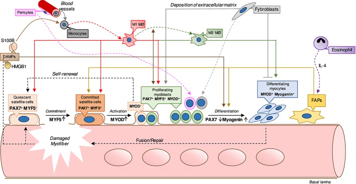
“…released from damaged muscle tissue — collectively known as damage-associated molecular patterns (DAMPs) — or secreted by in fi ltrating immune cells (i.e. hormones, growth factors, and cytokines) and components of the extracellular matrix 17 – 24 ( Figure 1) acting via cell surface receptors.”
Transcriptional co-activator PGC-1α drives the formation of slow-twitch muscle fibres
Lin, J., Wu, H., Tarr, P. T., Zhang, C.-Y., Wu, Z., Boss, O., Michael, L. F., Puigserver, P., Isotani, E., Olson, E. N., et al. (2002) · Nature · doi:10.1038/nature00904 · Cited by 2,546
Muscle as an Endocrine Organ: Focus on Muscle-Derived Interleukin-6
Pedersen, B. K., & Febbraio, M. A. (2008) · Physiological Reviews · doi:10.1152/physrev.90100.2007 · Cited by 2,388
Rejuvenation of aged progenitor cells by exposure to a young systemic environment
Conboy, I. M., Conboy, M. J., Wagers, A. J., Girma, E. R., Weissman, I. L., & Rando, T. A. (2005) · Nature · doi:10.1038/nature03260 · Cited by 2,287 · cited 1×
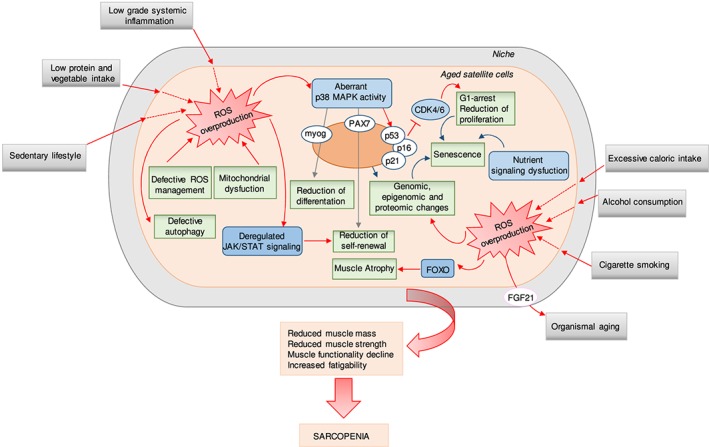
“From the side of SCs, extrinsic, niche-related factors and intrinsic, cell-autonomous factors concur to determine changes in SCs with aging ultimately leading to reduced SCs ’ ability to maintain muscle mass 5,18,66 – 73 ( Figure 3).”
Nrf2-Keap1 signaling in oxidative and reductive stress
Bellezza, I., Giambanco, I., Minelli, A., & Donato, R. (2018) · Biochimica et Biophysica Acta (BBA) - Molecular Cell Research · doi:10.1016/j.bbamcr.2018.02.010 · Cited by 2,134 · cited 1×
“sue (reviewed by Bellezza et al.(43)), an event counteracted by a”
Arnold, L., Henry, A., Poron, F., Baba-Amer, Y., van Rooijen, N., Plonquet, A., Gherardi, R. K., & Chazaud, B. (2007) · The Journal of Experimental Medicine · doi:10.1084/jem.20070075 · Cited by 2,002
RAGE Mediates a Novel Proinflammatory Axis
Hofmann, M. A., Drury, S., Fu, C., Qu, W., Taguchi, A., Lu, Y., Avila, C., Kambham, N., Bierhaus, A., Nawroth, P., et al. (1999) · Cell · doi:10.1016/s0092-8674(00)80801-6 · Cited by 1,864
Muscle injury activates resident fibro/adipogenic progenitors that facilitate myogenesis
Joe, A. W. B., Yi, L., Natarajan, A., Le Grand, F., So, L., Wang, J., Rudnicki, M. A., & Rossi, F. M. V. (2010) · Nature Cell Biology · doi:10.1038/ncb2015 · Cited by 1,690
Donato, R. (2001) · The International Journal of Biochemistry & Cell Biology · doi:10.1016/s1357-2725(01)00046-2 · Cited by 1,580
Increased Wnt signaling during aging alters muscle stem cell fate and increases fibrosis
Brack, A. S., Conboy, M. J., Roy, S., Lee, M., Kuo, C. J., Keller, C., & Rando, T. A. (2007) · Science · doi:10.1126/science.1144090 · Cited by 1,564
Mice lacking mitochondrial uncoupling protein are cold-sensitive but not obese
Enerbäck, S., Jacobsson, A., Simpson, E. M., Guerra, C., Yamashita, H., Harper, M.-E., & Kozak, L. P. (1997) · Nature · doi:10.1038/387090a0 · Cited by 1,379 · cited 1×
“cytes express uncoupling protein 1 (UCP-1), which targets mitochondria where it uncouples oxidative phosphorylation to produce heat.(145) ROS/S100B-stimulated NF-κB also up-”
Mitchell, W. K., Williams, J., Atherton, P., Larvin, M., Lund, J., & Narici, M. (2012) · Frontiers in Physiology · doi:10.3389/fphys.2012.00260 · Cited by 1,377 · cited 1×
“Sarcopenia may be either secondary to chronic inflammatory statuses, diabetes, hormonal alterations, vascular disturbances, renal, respiratory and/or cardiac failure, and immobilization(3) or primary, occurring in otherwise healthy, usually aged persons.(1,2) The pathogenesis of primary”
Autophagy maintains stemness by preventing senescence
García-Prat, L., Martínez-Vicente, M., Perdiguero, E., Ortet, L., Rodríguez-Ubreva, J., Rebollo, E., Ruiz-Bonilla, V., Gutarra, S., Ballestar, E., Serrano, A. L., et al. (2016) · Nature · doi:10.1038/nature16187 · Cited by 1,355 · cited 2×
“…of the cell proliferation inhibitor, p16 INK4a , 69 deregulated janus kinase/signal transducer and activator of transcription (JAK-STAT) signalling, 70 and defective autophagy 73 detected in aged SCs and suggested to be responsible for their altered proliferation and differentiation properties.”
Donato, R., Cannon, B. R., Sorci, G., Riuzzi, F., Hsu, K., Weber, D. J., & Geczy, C. L. (2012) · Current Molecular Medicine · doi:10.2174/156652413804486214 · Cited by 1,354 · cited 1×
“Alzheimer’s disease), S100B invariably sustains inflammation with resultant direct or indirect killing of neurons and activation of astrocytes and microglia.(88,89) In”
Senescence and aging: Causes, consequences, and therapeutic avenues
McHugh, D., & Gil, J. (2018) · Journal of Cell Biology · doi:10.1083/jcb.201708092 · Cited by 1,233 · cited 2×
“…as the cell ’ s inability to proliferate in response to appropriate stimuli owing to the hyper-activity of two main signalling pathways, p16 INK4a /Rb and p53/p21 CIP1 , which concur to repress cyclin-dependent kinase 4/6 (CDK4/6) activity ultimately leading to stem cell exhaustion 79 ( Figure 3).”
Transcriptional Control of Brown Fat Determination by PRDM16
Seale, P., Kajimura, S., Yang, W., Chin, S., Rohas, L. M., Uldry, M., Tavernier, G., Langin, D., & Spiegelman, B. M. (2007) · Cell Metabolism · doi:10.1016/j.cmet.2007.06.001 · Cited by 1,174
Regulatory interactions between muscle and the immune system during muscle regeneration
Tidball, J. G., & Villalta, S. A. (2010) · American Journal of Physiology-Regulatory, Integrative and Comparative Physiology · doi:10.1152/ajpregu.00735.2009 · Cited by 1,105 · cited 2×
“In fi ltrating immune cells, among which macrophages play an essential role, 9 – 12 vascular pericytes 13,14 and fi bro/adipogenic precursors (FAPs) 15,16 participate in muscle regeneration ( Figure 1).”
New role of bone morphogenetic protein 7 in brown adipogenesis and energy expenditure
Tseng, Y.-H., Kokkotou, E., Schulz, T. J., Huang, T. L., Winnay, J. N., Taniguchi, C. M., Tran, T. T., Suzuki, R., Espinoza, D. O., Yamamoto, Y., et al. (2008) · Nature · doi:10.1038/nature07221 · Cited by 1,099 · cited 1×
“…NF-κ B also up-regulates, in a YY1-dependent manner, bone morphogenetic protein 7 (BMP7), 137 which, once secreted, causes myoblast progenitors 146,147 and cultured myoblasts 137 to differentiate into brown adipocytes by repressing the expression of the adipogenic inhibitors, necdin and…”
Muscle wasting in disease: molecular mechanisms and promising therapies
Cohen, S., Nathan, J. A., & Goldberg, A. L. (2015) · Nature Reviews Drug Discovery · doi:10.1038/nrd4467 · Cited by 1,089 · cited 2×
“…and deranged satellite cell properties Sarcopenia may be either secondary to chronic in fl ammatory statuses, diabetes, hormonal alterations, vascular disturbances, renal, respiratory and/or cardiac failure, and immobilization 3 or primary, occurring in otherwise healthy, usually aged persons.”
Pericytes of human skeletal muscle are myogenic precursors distinct from satellite cells
Dellavalle, A., Sampaolesi, M., Tonlorenzi, R., Tagliafico, E., Sacchetti, B., Perani, L., Innocenzi, A., Gálvez, B. G., Messina, G., Roberta Morosetti, A., et al. (2007) · Nature Cell Biology · doi:10.1038/ncb1542 · Cited by 1,021 · cited 1×
“In fi ltrating immune cells, among which macrophages play an essential role, 9 – 12 vascular pericytes 13,14 and fi bro/adipogenic precursors (FAPs) 15,16 participate in muscle regeneration ( Figure 1).”
Geriatric muscle stem cells switch reversible quiescence into senescence
Sousa-Victor, P., Gutarra, S., García-Prat, L., Rodriguez-Ubreva, J., Ortet, L., Ruiz-Bonilla, V., Jardí, M., Ballestar, E., González, S., Serrano, A. L., et al. (2014) · Nature · doi:10.1038/nature13013 · Cited by 1,010 · cited 1×
“of senescent cells being proliferation and differentiation defective.(69,135,136) It is presently unknown whether the elevated levels of S100B found in senescent myoblasts(135) are a consequence or a cause of senescence.”
Role of Myokines in Exercise and Metabolism
Pedersen, B. K., Åkerström, T. C. A., Nielsen, A. R., & Fischer, C. P. (2007) · Journal of Applied Physiology · doi:10.1152/japplphysiol.00080.2007 · Cited by 841
Asakura, A., Rudnicki, M. A., & Komaki, M. (2001) · Differentiation · doi:10.1046/j.1432-0436.2001.680412.x · Cited by 839
Restoring systemic GDF11 levels reverses age-related dysfunction in mouse skeletal muscle
Sinha, M., Jang, Y. C., Oh, J., Khong, D., Wu, E. Y., Manohar, R., Miller, C., Regalado, S. G., Loffredo, F. S., Pancoast, J. R., et al. (2014) · Science · doi:10.1126/science.1251152 · Cited by 824 · cited 1×
“…nding that growth differentiation factor 11 (GDF11), a myostatin homologue, might support muscle trophism and regeneration and contrast muscle aging 80 was dampened by reports showing that, on the contrary, GDF11 levels increases with age and inhibits muscle regeneration, behaving like…”
Type 2 Innate Signals Stimulate Fibro/Adipogenic Progenitors to Facilitate Muscle Regeneration
Heredia, J. E., Mukundan, L., Chen, F. M., Mueller, A. A., Deo, R. C., Locksley, R. M., Rando, T. A., & Chawla, A. (2013) · Cell · doi:10.1016/j.cell.2013.02.053 · Cited by 818
Exercise is the real polypill.
Fiuza-Luces, C., Garatachea, N., Berger, N. A., & Lucia, A. (2013) · Physiology · doi:10.1152/physiol.00019.2013 · Cited by 791
S100B's double life: Intracellular regulator and extracellular signal
Donato, R., Sorci, G., Riuzzi, F., Arcuri, C., Bianchi, R., Brozzi, F., Tubaro, C., & Giambanco, I. (2008) · Biochimica et Biophysica Acta (BBA) - Molecular Cell Research · doi:10.1016/j.bbamcr.2008.11.009 · Cited by 747 · cited 2×
“hand type and is endowed with intracellular and extracellular regulatory functions.(88) S100B is a Ca-sensor protein”
Regulation of Muscle Growth and Regeneration by the Immune System
Tidball, J. G. (2017) · Nature Reviews Immunology · doi:10.1038/nri.2016.150 · Cited by 740 · cited 1×
“In fi ltrating immune cells, among which macrophages play an essential role, 9 – 12 vascular pericytes 13,14 and fi bro/adipogenic precursors (FAPs) 15,16 participate in muscle regeneration ( Figure 1).”
Milan, G., Romanello, V., Pescatore, F., Armani, A., Paik, J.-H., Frasson, L., Seydel, A., Zhao, J., Abraham, R., Goldberg, A. L., et al. (2015) · Nature Communications · doi:10.1038/ncomms7670 · Cited by 709 · cited 1×
“FoxOs co-ordinate a variety of stress-response genes, including autophagy and ROS detoxification, during catabolic conditions.(39) However, sarcopenia was suggested to be not”
A PGC-1α isoform induced by resistance training regulates skeletal muscle hypertrophy
Ruas, J. L., White, J. P., Rao, R. R., Kleiner, S., Brannan, K. T., Harrison, B. C., Greene, N. P., Wu, J., Estall, J. L., Irving, B. A., et al. (2012) · Cell · doi:10.1016/j.cell.2012.10.050 · Cited by 677
Rejuvenation of the muscle stem cell population restores strength to injured aged muscles
Cosgrove, B. D., Gilbert, P. M., Porpiglia, E., Mourkioti, F., Lee, S. P., Corbel, S. Y., Llewellyn, M. E., Delp, S. L., & Blau, H. M. (2014) · Nature Medicine · doi:10.1038/nm.3464 · Cited by 677
Mitochondrial dysfunction and sarcopenia of aging: from signaling pathways to clinical trials
Marzetti, E., Calvani, R., Cesari, M., Buford, T. W., Lorenzi, M., Behnke, B. J., & Leeuwenburgh, C. (2013) · The International Journal of Biochemistry & Cell Biology · doi:10.1016/j.biocel.2013.06.024 · Cited by 627 · cited 1×
“ROS overproduction, owing to altered mitochondrial function and/or defective ROS management, might be one cause of sarcopenia.(41,57,74,75) ROS imbalance might determine the ab-”
Austin, S., & St‐Pierre, J. (2012) · Journal of Cell Science · doi:10.1242/jcs.113662 · Cited by 617
Huttunen, H. J., Kuja‐Panula, J., Sorci, G., Agneletti, A. L., Donato, R., & Rauvala, H. (2000) · Journal of Biological Chemistry · doi:10.1074/jbc.m006993200 · Cited by 578 · cited 1×
“…effects of S100B are largely brought about by its interaction with RAGE (receptor for advanced glycation end products, encoded by AGER/ Ager ) 109,110 ; however, at least in the case of muscle injury, S100B can activate either RAGE or the basic fi broblast growth factor (bFGF)/FGF receptor 1…”
Bernet, J. D., Doles, J. D., Hall, J. K., Kelly Tanaka, K., Carter, T. A., & Olwin, B. B. (2014) · Nature Medicine · doi:10.1038/nm.3465 · Cited by 573 · cited 1×
“41,57,74,75 ROS imbalance might determine the aberrant p38 mitogen-activated protein kinase (MAPK) activity, 67,68 deregulated expression of the cell proliferation inhibitor, p16 INK4a , 69 deregulated janus kinase/signal transducer and activator of transcription (JAK-STAT) signalling, 70 and…”
Tezze, C., Romanello, V., Desbats, M. A., Fadini, G. P., Albiero, M., Favaro, G., Ciciliot, S., Soriano, M. E., Morbidoni, V., Cerqua, C., et al. (2017) · Cell Metabolism · doi:10.1016/j.cmet.2017.04.021 · Cited by 568
GDF11 increases with age and inhibits skeletal muscle regeneration
Egerman, M. A., Cadena, S. M., Gilbert, J. A., Meyer, A., Nelson, H. N., Swalley, S. E., Mallozzi, C., Jacobi, C., Jennings, L. L., Clay, I., et al. (2015) · Cell Metabolism · doi:10.1016/j.cmet.2015.05.010 · Cited by 527 · cited 1×
“…levels increases with age and inhibits muscle regeneration, behaving like transforming growth factor-β and sharing with it intracellular signalling pathways, 81 and is not a rejuvenator of aged skeletal muscle SCs. 82 Instead, GDF11 is risk factor for age-related frailty and disease in humans.”
Satellite cells, the engines of muscle repair
Wang, Y. X., & Rudnicki, M. A. (2011) · Nature Reviews Molecular Cell Biology · doi:10.1038/nrm3265 · Cited by 493 · cited 1×
“…myoblasts (Myf5, PAX7), (ii) myoblast proliferation and the return of a minority of proliferating myoblasts to the state of quiescent SCs (PAX7), (iii) myoblast proliferation and differentiation (MyoD), and (iv) myoblast differentiation into fusion-competent myocytes (myogenin) 7 ( Figure 1).”
Intrinsic and extrinsic mechanisms regulating satellite cell function
Dumont, N. A., Wang, Y. X., & Rudnicki, M. A. (2015) · Development · doi:10.1242/dev.114223 · Cited by 491 · cited 1×
“between the sarcolemma and the basal lamina and essential for the maintenance of muscle mass.(6) Normally quiescent, in”
Myogenin and Class II HDACs Control Neurogenic Muscle Atrophy by Inducing E3 Ubiquitin Ligases
Moresi, V., Williams, A. H., Meadows, E., Flynn, J. M., Potthoff, M. J., McAnally, J., Shelton, J. M., Backs, J., Klein, W. H., Richardson, J. A., et al. (2010) · Cell · doi:10.1016/j.cell.2010.09.004 · Cited by 457 · cited 1×
“53 – 56 In these atrophying conditions, myogenin was up-regulated and was required for the maximal activation of atrogin-1 and MuRF-1 by binding their promoters.”
Mitochondrial function and apoptotic susceptibility in aging skeletal muscle
Chabi, B., Ljubicic, V., Menzies, K. J., Huang, J. H., Saleem, A., & Hood, D. A. (2008) · Aging Cell · doi:10.1111/j.1474-9726.2007.00347.x · Cited by 437 · cited 1×
“35 Thus, altered PGC1 α expression level/activity in muscle tissue likely consequent to age-related enhanced ROS production in a background of altered mitochondria number and properties 36,37 might concur to sarcopenia.”
The central role of muscle stem cells in regenerative failure with aging
Blau, H. M., Cosgrove, B. D., & Ho, A. T. V. (2015) · Nature Medicine · doi:10.1038/nm.3918 · Cited by 437
Mitochondrial quality control and muscle mass maintenance.
Romanello, V., & Sandri, M. (2016) · Frontiers in Physiology · doi:10.3389/fphys.2015.00422 · Cited by 434 · cited 1×
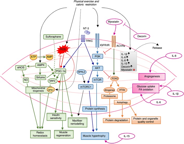
“…organelle disposal pathways [e.g. the insulin-like growth factor 1/protein kinase B (Akt)/mammalian target of rapamycin (mTOR)/forkhead box O (FoxO), and macroautophagy pathways] leading to a failure of protein and organelle quality con-trol 25,26 might contribute to muscle atrophy ( Figure 2).”
Inhibition of JAK-STAT signaling stimulates adult satellite cell function
Price, F. D., von Maltzahn, J., Bentzinger, C. F., Dumont, N. A., Yin, H., Chang, N. C., Wilson, D. H., Frenette, J., & Rudnicki, M. A. (2014) · Nature Medicine · doi:10.1038/nm.3655 · Cited by 433
Fry, C. S., Lee, J. D., Mula, J., Kirby, T. J., Jackson, J. R., Liu, F., Yang, L., Mendias, C. L., Dupont-Versteegden, E. E., McCarthy, J. J., et al. (2015) · Nature Medicine · doi:10.1038/nm.3710 · Cited by 432 · cited 3×
“Yet the studies by Fry et al.(84) and Keefe et al.”
Mitochondria and ageing: Role in heart, skeletal muscle and adipose tissue
Boengler, K., Kosiol, M., Mayr, M., Schulz, R., & Rohrbach, S. (2017) · Journal of Cachexia Sarcopenia and Muscle · doi:10.1002/jcsm.12178 · Cited by 421
Exercise Attenuates the Major Hallmarks of Aging
Garatachea, N., Pareja-Galeano, H., Sanchis-Gomar, F., Santos-Lozano, A., Fiuza-Luces, C., Morán, M., Emanuele, E., Joyner, M. J., & Lucia, A. (2015) · Rejuvenation Research · doi:10.1089/rej.2014.1623 · Cited by 413 · cited 1×
“Thus, physical exercise appears to be the most efficient means to contrast sarcopenia.(65)”
Satellite cells in human skeletal muscle; from birth to old age
Verdijk, L. B., Snijders, T., Drost, M., Delhaas, T., Kadi, F., & van Loon, L. J. C. (2014) · AGE · doi:10.1007/s11357-013-9583-2 · Cited by 399
Cellular dynamics in the muscle satellite cell niche
Bentzinger, C. F., Wang, Y. X., Dumont, N. A., & Rudnicki, M. A. (2013) · The EMBO Reports · doi:10.1038/embor.2013.182 · Cited by 398
Calvo, J. A., Daniels, T. G., Wang, X., Paul, A., Lin, J., Spiegelman, B. M., Stevenson, S. C., & Rangwala, S. M. (2008) · Journal of Applied Physiology · doi:10.1152/japplphysiol.01231.2007 · Cited by 377
Ethical guidelines for publishing in the journal of cachexia, sarcopenia and muscle: update 2017
von Haehling, S., Morley, J. E., Coats, A. J. S., & Anker, S. D. (2017) · Journal of Cachexia Sarcopenia and Muscle · doi:10.1002/jcsm.12261 · Cited by 351 · cited 1×
“guidelines for publishing in the Journal of Cachexia, Sarcopenia and Muscle: update 2017.(152)”
Raponi, É., Agenès, F., Delphin, C., Assard, N., Baudier, J., Legraverend, C., & Deloulme, J. (2006) · Glia · doi:10.1002/glia.20445 · Cited by 347
Lukjanenko, L., Jung, M. J., Hegde, N., Perruisseau-Carrier, C., Migliavacca, E., Rozo, M., Karaz, S., Jacot, G. E., Schmidt, M., Li, L., et al. (2016) · Nature Medicine · doi:10.1038/nm.4126 · Cited by 347
Sandri, M., Barberi, L., Bijlsma, A. Y., Blaauw, B., Dyar, K. A., Milan, G., Mammucari, C., Meskers, C. G. M., Pallafacchina, G., Paoli, A., et al. (2013) · Biogerontology · doi:10.1007/s10522-013-9432-9 · Cited by 340 · cited 1×
“tivation of the ubiquitin ligase atrogin-1 were found to paradoxically cause muscle pathology.(25) Yet a ROS–FoxO cross-talk”
BackgroundMuscle as a paracrine and endocrine organ
Giudice, J., & Taylor, J. M. (2017) · Current Opinion in Pharmacology · doi:10.1016/j.coph.2017.05.005 · Cited by 331 · cited 1×
“contributing to the broad beneficial effects of exercise on cardiovascular, metabolic, and mental health.(62) Among the vari-”
Macrophages: Supportive cells for tissue repair and regeneration
Chazaud, B. (2013) · Immunobiology · doi:10.1016/j.imbio.2013.09.001 · Cited by 326
Nrf2 mediates redox adaptations to exercise
Done, A. J., & Traustadóttir, T. (2016) · Redox Biology · doi:10.1016/j.redox.2016.10.003 · Cited by 308 · cited 1×
“43 ), an event counteracted by a regular exercise that attenuates the age-related changes in Nrf2-mediated pathways and potentially restores the redox homeostasis 28,44 – 46 ( Figure 2).”
Castets, P., Lin, S., Rion, N., Di Fulvio, S., Romanino, K., Guridi, M., Frank, S., Tintignac, L. A., Sinnreich, M., & Rüegg, M. A. (2013) · Cell Metabolism · doi:10.1016/j.cmet.2013.03.015 · Cited by 302
Epidemiology of sarcopenia and insight into possible therapeutic targets
Dennison, E. M., Sayer, A. A., & Cooper, C. C. (2017) · Nature Reviews Rheumatology · doi:10.1038/nrrheum.2017.60 · Cited by 302 · cited 1×
“…muscle strength and agility and increased fatiga-bility inherent to sarcopenic subjects, coupled to a relatively excessive caloric intake, is likely to be one main cause especially if associated with cigarette smoking, alcohol consumption, and low protein and vegetable intake 76 ( Figure 3).”
Yin, H., Pasut, A., Soleimani, V. D., Bentzinger, C. F., Antoun, G., Thorn, S., Seale, P., Fernando, P., van IJcken, W., Grosveld, F., et al. (2013) · Cell Metabolism · doi:10.1016/j.cmet.2013.01.004 · Cited by 293
Baudier, J., Delphin, C., Grünwald, D., Khochbin, S., & Lawrence, J. J. (1992) · Proceedings of the National Academy of Sciences · doi:10.1073/pnas.89.23.11627 · Cited by 285
Lifelong physical exercise delays age-associated skeletal muscle decline
Zampieri, S., Pietrangelo, L., Loefler, S., Fruhmann, H., Vogelauer, M., Burggraf, S., Pond, A., Grim-Stieger, M., Cvecka, J., Sedliak, M., et al. (2015) · The Journals of Gerontology Series A: Biological Sciences and Medical Sciences · doi:10.1093/gerona/glu006 · Cited by 282
Muscle stem cells contribute to myofibres in sedentary adult mice
Keefe, A. C., Lawson, J. A., Flygare, S. D., Fox, Z. D., Colasanto, M. P., Mathew, S. J., Yandell, M., & Kardon, G. (2015) · Nature Communications · doi:10.1038/ncomms8087 · Cited by 279 · cited 3×
“might not concur to sarcopenia while leading to defective regeneration following acute muscle injury and contributing to age-related muscle fibrosis.(84,85) This would imply that pri-”
Stem Cell Review Series: Aging of the skeletal muscle stem cell niche
Gopinath, S. D., & Rando, T. A. (2008) · Aging Cell · doi:10.1111/j.1474-9726.2008.00399.x · Cited by 265 · cited 1×
“of senescent cells being proliferation and differentiation defective.(69,135,136) It is presently unknown whether the elevated levels of S100B found in senescent myoblasts(135) are a consequence or a cause of senescence.”
MyomiR-133 regulates brown fat differentiation through Prdm16
Trajkovski, M., Ahmed, K., Esau, C., & Stoffel, M. (2012) · Nature Cell Biology · doi:10.1038/ncb2612 · Cited by 263 · cited 1×
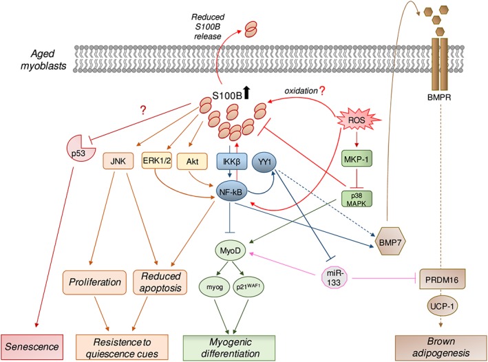
“In turn, S100B stimulates NF-κ B activity with resultant up-regulation of YY1 and YY1-dependent inhibition of miR-133, a promyogenic and anti-adipogenic microRNA, 142 – 144 ultimately leading to transition of myoblasts to brown adipocytes 137 ( Figure 4).”
Mdm4 and Mdm2 cooperate to inhibit p53 activity in proliferating and quiescent cells in vivo
Francoz, S., Froment, P., Bogaerts, S., De Clercq, S., Maetens, M., Doumont, G., Bellefroid, E., & Marine, J.-C. (2006) · Proceedings of the National Academy of Sciences · doi:10.1073/pnas.0508476103 · Cited by 260 · cited 1×
“This potential S100B activity, however, might be mitigated or even abolished by the reported ability of S100B to block the ubiquitin E3 ligases, MDM2 and MDM4,(133) that drive proteasomal degradation of p53.(134) Thus, appropriate levels of S100B in myoblasts appear to be required for myoblasts”
Targeting β1-integrin signaling enhances regeneration in aged and dystrophic muscle in mice
Rozo, M., Li, L., & Fan, C. (2016) · Nature Medicine · doi:10.1038/nm.4116 · Cited by 260
Wang, H., Hertlein, E. K., Bakkar, N., Sun, H., Acharyya, S., Wang, J., Carathers, M., Davuluri, R. V., & D, D. C. G. (2007) · Molecular and Cellular Biology · doi:10.1128/mcb.02020-06 · Cited by 259 · cited 1×
“MyoD and the MyoD-downstream effectors, myogenin and p21, and to up-regulate Yin Yang 1 (YY1)(93) (Figure 4), a transcriptional repressor induced by NF-κB with antimyogenic activity.(119) Interestingly, myoblasts down-regulate”
Mendelson, K. G., Contois, L. W., Tevosian, S. G., Davis, R. J., & Paulson, K. E. (1996) · Proceedings of the National Academy of Sciences · doi:10.1073/pnas.93.23.12908 · Cited by 232 · cited 2×
“phosphatase MKP-1, which inactivates the promyogenic p38 MAPK.(141) Indeed, ROS overproduction in myoblasts causes accumulation of S100B partly owing to MKP-1-dependent inactivation of p38 MAPK and stimulates NF-κB activity, which cause up-regulation of S100B(137) (Figure 4).”
Quantification of GDF11 and Myostatin in Human Aging and Cardiovascular Disease
Schafer, M. J., Atkinson, E. J., Vanderboom, P. M., Kotajarvi, B. R., White, T. A., Moore, M. M., Bruce, C. J., Greason, K. L., Suri, R. M., Khosla, S., et al. (2016) · Cell Metabolism · doi:10.1016/j.cmet.2016.05.023 · Cited by 211 · cited 1×
“Instead, GDF11 is risk factor for age-related frailty and disease in humans.(83)”
Fuggle, N. R., Shaw, S., Dennison, E. M., & Cooper, C. C. (2017) · Best Practice & Research Clinical Rheumatology · doi:10.1016/j.berh.2017.11.007 · Cited by 199
Modulation of reactive oxygen species in skeletal muscle by myostatin is mediated through NF-κB
Sriram, S., Subramanian, S., Sathiakumar, D., Venkatesh, R., Salerno, M. S., McFarlane, C. D., Kambadur, R., & Sharma, M. (2011) · Aging Cell · doi:10.1111/j.1474-9726.2011.00734.x · Cited by 189 · cited 1×
“proteasomal-mediated catabolism of intracellular proteins.(77)”
S100B Protein Regulates Astrocyte Shape and Migration via Interaction with Src Kinase
Brozzi, F., Arcuri, C., Giambanco, I., & Donato, R. (2009) · Journal of Biological Chemistry · doi:10.1074/jbc.m805897200 · Cited by 174 · cited 1×
“120 126 Yet differentiated myoblasts (myocytes) resume expressing S100B under the action of myogenin, 120 and S100B is found expressed in mature myo fi bers as mentioned earlier.”
Proteins of the S100 family regulate the oligomerization of p53 tumor suppressor
Fernández-Fernández, M. R., Veprintsev, D. B., & Fersht, A. R. (2005) · Proceedings of the National Academy of Sciences · doi:10.1073/pnas.0501459102 · Cited by 172
Klotz, L., & Steinbrenner, H. (2017) · Redox Biology · doi:10.1016/j.redox.2017.07.015 · Cited by 165 · cited 1×
“ROS overproduction, owing to altered mitochondrial function and/or defective ROS management, might be one cause of sarcopenia.(41,57,74,75) ROS imbalance might determine the ab-”
Differential response of skeletal muscles to mTORC1 signaling during atrophy and hypertrophy
Bentzinger, C. F., Lin, S., Romanino, K., Castets, P., Guridi, M., Summermatter, S., Handschin, C., Tintignac, L. A., Hall, M. N., & Rüegg, M. A. (2013) · Skeletal Muscle · doi:10.1186/2044-5040-3-6 · Cited by 153
Inhibition of p53 Transcriptional Activity by the S100B Calcium-binding Protein
Lin, J., Blake, M., Tang, C., Zimmer, D. B., Rustandi, R. R., Weber, D. J., & Carrier, F. (2001) · Journal of Biological Chemistry · doi:10.1074/jbc.m104379200 · Cited by 141
Starring or supporting role? Satellite cells and skeletal muscle fiber size regulation
Murach, K. A., Fry, C. S., Kirby, T. J., Jackson, J. R., Lee, J. D., White, S. H., Dupont-Versteegden, E. E., McCarthy, J. J., & Peterson, C. A. (2018) · Physiology · doi:10.1152/physiol.00019.2017 · Cited by 137
Current understanding of sarcopenia: possible candidates modulating muscle mass
Sakuma, K., Aoi, W., & Yamaguchi, A. (2014) · Pflügers Archiv - European Journal of Physiology · doi:10.1007/s00424-014-1527-x · Cited by 133 · cited 1×
“Sarcopenia may be either secondary to chronic inflammatory statuses, diabetes, hormonal alterations, vascular disturbances, renal, respiratory and/or cardiac failure, and immobilization(3) or primary, occurring in otherwise healthy, usually aged persons.(1,2) The pathogenesis of primary”
Stem cells in the hood: the skeletal muscle niche
Pannérec, A., Marazzi, G., & Sassoon, D. A. (2012) · Trends in Molecular Medicine · doi:10.1016/j.molmed.2012.07.004 · Cited by 130 · cited 1×
“In fi ltrating immune cells, among which macrophages play an essential role, 9 – 12 vascular pericytes 13,14 and fi bro/adipogenic precursors (FAPs) 15,16 participate in muscle regeneration ( Figure 1).”
Inhibiting S100B Restores p53 Levels in Primary Malignant Melanoma Cancer Cells
Lin, J., Yang, Q., Yan, Z., Markowitz, J., Wilder, P. T., Carrier, F., & Weber, D. J. (2004) · Journal of Biological Chemistry · doi:10.1074/jbc.m405419200 · Cited by 128
HDAC1 activates FoxO and is both sufficient and required for skeletal muscle atrophy
Beharry, A. W., Sandesara, P. B., Roberts, B. M., Ferreira, L. F., Senf, S. M., & Judge, A. R. (2014) · Journal of Cell Science · doi:10.1242/jcs.136390 · Cited by 128
The many roles of PGC-1α in muscle — recent developments
Chan, M. C., & Arany, Z. P. (2014) · Metabolism · doi:10.1016/j.metabol.2014.01.006 · Cited by 127 · cited 1×
“health and disease regulating endurance, fibre-type switching, and insulin sensitivity.(29) PGC1α is induced by oxida-”
Sorci, G., Riuzzi, F., Arcuri, C., Giambanco, I., & Donato, R. (2004) · Molecular and Cellular Biology · doi:10.1128/mcb.24.11.4880-4894.2004 · Cited by 125
van Dieck, J., Fernández-Fernández, M. R., Veprintsev, D. B., & Fersht, A. R. (2009) · Journal of Biological Chemistry · doi:10.1074/jbc.m901351200 · Cited by 118
Tsoporis, J. N., Izhar, S., Leong-Poi, H., Desjardins, J.-F., Huttunen, H. J., & Parker, T. G. (2010) · Circulation Research · doi:10.1161/circresaha.109.195834 · Cited by 118 · cited 2×
“22 Also, S100B is passively released from injured brain 88,89 and heart 103 and from acutely and chronically injured muscle tissue, 104 behaving like a DAMP.”
The Calcium-binding Protein S100B Down-regulates p53 and Apoptosis in Malignant Melanoma
Lin, J., Yang, Q., Wilder, P. T., Carrier, F., & Weber, D. J. (2010) · Journal of Biological Chemistry · doi:10.1074/jbc.m110.155382 · Cited by 117 · cited 1×
“…to stem cell exhaustion 79 and since S100B inhibits p53 phosphorylation and tetramerization, i.e. its activation, 127 – 130 and reduces p53 levels, 131,132 it is tempting to speculate that high S100B levels might interfere with myo-blast senescence by decreasing p53/p21 CIP1 activity ( Figure 4).”
Cobley, J. N., Sakellariou, G. K., Owens, D. J., Murray, S. W., Waldron, S., Gregson, W. A., Fraser, W. D., Burniston, J. G., Iwanejko, L., McArdle, A., et al. (2014) · Free Radical Biology and Medicine · doi:10.1016/j.freeradbiomed.2014.02.004 · Cited by 108
Saito, T., Ikeda, T., Nakamura, K., Chung, U., & Kawaguchi, H. (2007) · EMBO Reports · doi:10.1038/sj.embor.7400934 · Cited by 105
S100 Calcium Binding Proteins and Ion Channels
Hermann, A., Donato, R., Weiger, T. M., & Chazin, W. (2012) · Frontiers in Pharmacology · doi:10.3389/fphar.2012.00067 · Cited by 104
Lu, L., Zhou, L., Chen, E. Z., Sun, K., Jiang, P., Wang, L., Su, X., Sun, H., & Wang, H. (2012) · PLoS ONE · doi:10.1371/journal.pone.0027596 · Cited by 103
The Danger Signal S100B Integrates Pathogen– and Danger–Sensing Pathways to Restrain Inflammation
Sorci, G., Giovannini, G., Riuzzi, F., Bonifazi, P., Zelante, T., Zagarella, S., Bistoni, F., Donato, R., & Romani, L. R. (2011) · PLoS Pathogens · doi:10.1371/journal.ppat.1001315 · Cited by 100
Activation of mTORC1 in skeletal muscle regulates whole-body metabolism through FGF21
Guridi, M., Tintignac, L. A. J., Lin, S., Kupr, B., Castets, P., & Rüegg, M. A. (2015) · Science Signaling · doi:10.1126/scisignal.aab3715 · Cited by 98
Guan, L., Zhang, L., Gong, Z., Hou, X., Xu, Y., Feng, X., Wang, H., & You, H. (2016) · Hepatology · doi:10.1002/hep.28496 · Cited by 95 · cited 1×
“25 Yet a ROS – FoxO cross-talk occurs in cells whereby ROS and FoxO reciprocally regulate their levels, 40,41 and old sedentary rat muscles show elevated levels of the atrophy genes (atrogenes), atrogin-1 and MuRF-1.”
Zampieri, S., Mammucari, C., Romanello, V., Barberi, L., Pietrangelo, L., Fusella, A., Mosole, S., Gherardi, G., Höfer, C., Löfler, S., et al. (2016) · Physiological Reports · doi:10.14814/phy2.13005 · Cited by 92 · cited 1×
“DAMP, damage-associated molecular pattern; FAPs, fi bro/ adipogenic precursors; IL-4, interleukin-4; SC, satellite cell. muscles 27 likely mediated by preserved expression levels of mitochondrial fusion protein 28 optic atrophy 1 ( Figure 2).”
Riuzzi, F., Sorci, G., Sagheddu, R., & Donato, R. (2012) · Journal of Cell Science · doi:10.1242/jcs.092163 · Cited by 91 · cited 1×
“damage sites, and prolongs the M1 macrophage inflammatory phase.(20) Yet as observed with cultured astrocytes and neurons,(88,89) at high local concentrations, S100B directly causes myoblast apoptosis(116) and RAGE dependently perpet-”
HuR and miR-1192 regulate myogenesis by modulating the translation of HMGB1 mRNA
Dormoy-Raclet, V., Cammas, A., Celona, B., Lian, X. J., van der Giessen, K., Zivojnovic, M., Brunelli, S., Riuzzi, F., Sorci, G., Wilhelm, B. T., et al. (2013) · Nature Communications · doi:10.1038/ncomms3388 · Cited by 89
A Systematic Review of the Biomarker S100B: Implications for Sport-Related Concussion Management
Schulte, S., Podlog, L. W., Hamson-Utley, J. J., Strathmann, F. G., & Strüder, H. K. (2014) · Journal of Athletic Training · doi:10.4085/1062-6050-49.3.33 · Cited by 88 · cited 1×
“marathon, swimming races, and soccer games).(106) These elevated serum S100B levels have been suggested to result from brain concussion known to”
Minetti, G., Feige, J. N., Rosenstiel, A., Bombard, F., Meier, V., Werner, A., Bassilana, F., Sailer, A. W., Kahle, P., Lambert, C., et al. (2011) · Science Signaling · doi:10.1126/scisignal.2002038 · Cited by 87
S100B Expression Modulates Left Ventricular Remodeling After Myocardial Infarction in Mice
Tsoporis, J. N., Marks, A., Haddad, A., Dawood, F., Liu, P. P., & Parker, T. G. (2005) · Circulation · doi:10.1161/01.cir.0000154554.65287.f5 · Cited by 82 · cited 1×
“the peri-infarct heart tissue) and inhibits cardiomyocyte hypertrophic response.(95) Also, bronchial epithe-”
S100B Inhibits Myogenic Differentiation and Myotube Formation in a RAGE-Independent Manner
Sorci, G., Riuzzi, F., Agneletti, A. L., Marchetti, C., & Donato, R. (2003) · Molecular and Cellular Biology · doi:10.1128/mcb.23.14.4870-4881.2003 · Cited by 78 · cited 1×
“ture conditions, S100B inhibits myoblast differentiation and myotube formation and stimulates myoblast proliferation via bFGF/FGFR1 activation.(111,113,114) On the other hand, in”
Arcuri, C., Bianchi, R., Brozzi, F., & Donato, R. (2004) · Journal of Biological Chemistry · doi:10.1074/jbc.m406440200 · Cited by 76
McIlroy, M., McCartan, D. P., Early, S. A., Gaora, P. Ó., Pennington, S. R., Hill, A. D. K., & Young, L. S. (2010) · Cancer Research · doi:10.1158/0008-5472.can-09-3713 · Cited by 73
S100B causes apoptosis in a myoblast cell line in a RAGE‐independent manner
Sorci, G., Riuzzi, F., Agneletti, A. L., Marchetti, C., & Donato, R. (2003) · Journal of Cellular Physiology · doi:10.1002/jcp.10462 · Cited by 71 · cited 2×
“nase (GAPDH) activity by enhanced poly(ADP-ribosyl)ation of GAPDH with no apparent role for RAGE or FGFR.(116) Notably, muscles of S100B KO mice show reduced levels of poly(ADP-ribosyl)ated GAPDH,(118) but there is no information”
BackgroundInfluentialLack of evidence for GDF 11 as a rejuvenator of aged skeletal muscle satellite cells
Hinken, A. C., Powers, J. M., Luo, G., Holt, J. A., Billin, A. N., & Russell, A. J. (2016) · Aging Cell · doi:10.1111/acel.12475 · Cited by 71 · cited 4×
“The initial enthusiasm about the fi nding that growth differentiation factor 11 (GDF11), a myostatin homologue, might support muscle trophism and regeneration and contrast muscle aging 80 was dampened by reports showing that, on the contrary, GDF11 levels increases with age and inhibits muscle regeneration, behaving li…”
InfluentialBricceno, K. V., Sampognaro, P. J., Meerbeke, J. P. V., Sumner, C. J., Fischbeck, K. H., & Burnett, B. G. (2012) · Human Molecular Genetics · doi:10.1093/hmg/dds286 · Cited by 70
Riuzzi, F., Sorci, G., & Donato, R. (2011) · Journal of Cell Science · doi:10.1242/jcs.084491 · Cited by 70 · cited 2×
“factor (bFGF)/FGF receptor 1 (FGFR1) complex depending on its own local concentration, myoblast density, and bFGF availability.(22,104,111,112) In standard (high-density) myoblast cul-”
Oh, S., Komine, S., Warabi, E., Akiyama, K., Ishii, A., Ishige, K., Mizokami, Y., Kuga, K., Horie, M., Miwa, Y., et al. (2017) · Scientific Reports · doi:10.1038/s41598-017-12926-y · Cited by 70 · cited 1×
“and downstream genes and attenuates muscle fatigue via reduction of oxidative stress caused by exhaustive exercise.(47)”
Brzeszczyńska, J., Meyer, A. S., McGregor, R. A., Schilb, A., Degen, S., Tadini, V., Johns, N., LANGEN, R. C. J., Schols, A. M. W. J., Glass, D. J., et al. (2017) · Journal of Cachexia Sarcopenia and Muscle · doi:10.1002/jcsm.12252 · Cited by 69 · cited 2×
“HSPA1A, a stress-induced chaperone acting upon misfolded proteins.(57) These results suggest that reduced cellular de-”
PRDM16: the interconvertible adipo-myocyte switch
Frühbeck, G., Sesma, P., & Burrell, M. A. (2009) · Trends in Cell Biology · doi:10.1016/j.tcb.2009.01.007 · Cited by 67 · cited 1×
“139 Besides inhibiting myoblast differentiation and participating in the process of myoblast aging, elevated S100B levels also promote myoblast – brown adipocyte transition, and myoblasts from sarcopenic subjects show properties of brown (pre)adipocytes.”
Human muscle satellite cells show age-related differential expression of S100B protein and RAGE
Beccafico, S., Riuzzi, F., Puglielli, C., Mancinelli, R., Fulle, S., Sorci, G., & Donato, R. (2010) · AGE · doi:10.1007/s11357-010-9197-x · Cited by 66
Role of reactive oxygen species in the defective regeneration seen in aging muscle
Vasilaki, A., & Jackson, M. J. (2013) · Free Radical Biology and Medicine · doi:10.1016/j.freeradbiomed.2013.07.008 · Cited by 65 · cited 1×
“ROS overproduction, owing to altered mitochondrial function and/or defective ROS management, might be one cause of sarcopenia.(41,57,74,75) ROS imbalance might determine the ab-”
Annexin V, annexin VI, S100A1 and S100B in developing and adult avian skeletal muscles
Arcuri, C., Giambanco, I., Bianchi, R., & Donato, R. (2002) · Neuroscience · doi:10.1016/s0306-4522(01)00330-x · Cited by 64
Serum S100B Protein Is Increased in Fasting Rats
Netto, C. B. O., Conte, S., Leite, M. C., Pires, C., Martins, T. L., Vidal, P. R., Benfato, M. S., Giugliani, R., & Gonçalves, C. (2006) · Archives of Medical Research · doi:10.1016/j.arcmed.2005.11.005 · Cited by 64 · cited 1×
“98 101 Indeed, S100B is secreted constitutively by astrocytes 88 and following stimulation with catecholamines by white adipocytes, 102 and by activated macrophages.”
The Role of Reactive Oxygen Species in Adipogenic Differentiation
de Villiers, D., Potgieter, M., Ambele, M. A., Adam, L. T., Durandt, C., & Pepper, M. S. (2017) · Advances in experimental medicine and biology · doi:10.1007/5584_2017_119 · Cited by 62 · cited 1×
“tioxidants or ROS scavengers suppress ROS effects on adipogenesis.(140) Chronically high ROS levels induce the”
Riuzzi, F., Sorci, G., Beccafico, S., & Donato, R. (2012) · PLoS ONE · doi:10.1371/journal.pone.0028700 · Cited by 60 · cited 2×
“promoting the transition of infiltrating macrophages from M1 (proinflammatory) to M2 (anti-inflammatory and reparative) phenotype.(22,104) Incidentally, promyogenic effects of”
Völkers, M., Rohde, D., Goodman, C. R., & Most, P. (2010) · BioMed Research International · doi:10.1155/2010/178614 · Cited by 58 · cited 3×
“Given the S100B’s localization to the sarcoplasmic reticulum and transverse tubules similar to S100A1,92,93 a member of the S100 family of Ca2+-binding proteins that is 56% identical to S100B,89 one may hypothesize that S100B might regulate ryanodine receptor 1 and/or sarco(endo)plasmic reticulum calcium-ATPase activi…”
BackgroundMorozzi, G., Beccafico, S., Bianchi, R., Riuzzi, F., Bellezza, I., Giambanco, I., Arcuri, C., Minelli, A., & Donato, R. (2017) · Cell Death & Differentiation · doi:10.1038/cdd.2017.132 · Cited by 58
Tubaro, C., Arcuri, C., Giambanco, I., & Donato, R. (2010) · Journal of Cellular Physiology · doi:10.1002/jcp.22035 · Cited by 57
Role of muscle stem cells in sarcopenia
Snijders, T., & Parise, G. (2017) · Current Opinion in Clinical Nutrition & Metabolic Care · doi:10.1097/mco.0000000000000360 · Cited by 56
S100B-RAGE dependent VEGF secretion by cardiac myocytes induces myofibroblast proliferation
Tsoporis, J. N., Izhar, S., Proteau, G. A., Slaughter, G., & Parker, T. G. (2011) · Journal of Molecular and Cellular Cardiology · doi:10.1016/j.yjmcc.2011.08.015 · Cited by 53 · cited 1×
“secretion and VEGF-dependent myofibroblast proliferation potentially contributing to scar formation.(105) In acute muscle injury, a transient release of S100B(104) is required for a rapid”
Dutchak, P. A., Estill-Terpack, S. J., Plec, A. A., Zhao, X., Yang, C., Chen, J., Ko, B., Deberardinis, R. J., Yu, Y., & Tu, B. P. (2018) · Cell Reports · doi:10.1016/j.celrep.2018.04.058 · Cited by 53
Gilquin, B., Cannon, B. R., Hubstenberger, A., Moulouel, B., Falk, E., Merle, N., Assard, N., Kieffer‐Jaquinod, S., Rousseau, D. L., Wilder, P. T., et al. (2010) · Molecular and Cellular Biology · doi:10.1128/mcb.01468-09 · Cited by 52 · cited 1×
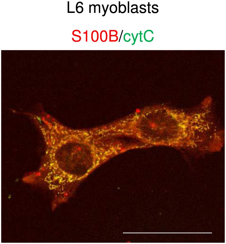
“143 While a physical and functional association of S100B with mitochondria was excluded in a previous study, 151 we found that a fraction of S100B co-localizes with the mitochondrial marker, cytochrome c , in proliferating myoblasts 113 ( Figure 5).”
Is Peripheral Immunity Regulated by Blood-Brain Barrier Permeability Changes?
Bargerstock, E., Puvenna, V., Iffland, P. H., Falcone, T., Hossain, M. S., Vetter, S., Man, S., Dickstein, L. P., Marchi, N., Ghosh, C., et al. (2014) · PLoS ONE · doi:10.1371/journal.pone.0101477 · Cited by 51
Effects of calcium‐binding proteins (S‐100a o , S‐100a, S‐100b) on desmin assembly in vitro
Garbuglia, M., Verzini, M., Giambanco, I., Spreca, A., & Donato, R. (1996) · The FASEB Journal · doi:10.1096/fasebj.10.2.8641565 · Cited by 48 · cited 1×
“found to be associated with Type III intermediate filaments in myoblasts and myotubes in part(91) and to exert an inhibitory effect on desmin assembly in vitro.(94) Functional corre-”
BackgroundLevels of S100B protein drive the reparative process in acute muscle injury and muscular dystrophy
Riuzzi, F., Beccafico, S., Sagheddu, R., Chiappalupi, S., Giambanco, I., Bereshchenko, O., Riccardi, C., Sorci, G., & Donato, R. (2017) · Scientific Reports · doi:10.1038/s41598-017-12880-9 · Cited by 45 · cited 6×
“uates muscle damage by fuelling inflammation as observed during the acute phase of muscular dystrophy.(22) In this latter”
InfluentialExpression of S100B during embryonic development of the mouse cerebellum
Hachem, S., Laurenson, A.-S., Hugnot, J., & Legraverend, C. (2007) · BMC Developmental Biology · doi:10.1186/1471-213x-7-17 · Cited by 44
Zinc Binding to S100B Affords Regulation of Trace Metal Homeostasis and Excitotoxicity in the Brain
Hagmeyer, S., Cristóvão, J. S., Mulvihill, J. J., Boeckers, T. M., Gomes, C. M., & Grabrucker, A. M. (2018) · Frontiers in Molecular Neuroscience · doi:10.3389/fnmol.2017.00456 · Cited by 44 · cited 1×
“98 101 Indeed, S100B is secreted constitutively by astrocytes 88 and following stimulation with catecholamines by white adipocytes, 102 and by activated macrophages.”
Transcription Factor Tfe3 Directly Regulates Pgc‐1alpha in Muscle
Salma, N., Song, J. S., Arany, Z. P., & Fisher, D. E. (2015) · Journal of Cellular Physiology · doi:10.1002/jcp.24978 · Cited by 42 · cited 1×
“…induced by oxidative stress, is a powerful regulator of reactive oxygen species (ROS) removal by increasing the expression of numerous ROS-detoxifying enzymes and factors such as NF-E2-related factor 2 (Nrf2), 30 and is directly regulated by the transcription factor Tfe3 in muscles 31 ( Figure 2).”
Ribeiro, M. B. T., Guzzoni, V., Hord, J. M., Lopes, G. N., Marqueti, R. D. C., de Andrade, R. V., Selistre-de-Araújo, H. S., & Durigan, J. L. Q. (2017) · Scientific Reports · doi:10.1038/s41598-017-09343-6 · Cited by 42
YALVAÇ, M. E., Amornvit, J., Chen, L., Shontz, K. M., Lewis, S., & Sahenk, Z. (2018) · Gene Therapy · doi:10.1038/s41434-018-0009-8 · Cited by 41
Serum S100B level increases after running but not cycling exercise
Stocchero, C. M. A., Oses, J. P., Cunha, G. D. S., Martins, J. B., Brum, L. M. B. D. P., Zimmer, E. R., Souza, D. O., Portela, L. V., & Reischak-Oliveira, Á. (2013) · Applied Physiology Nutrition and Metabolism · doi:10.1139/apnm-2013-0308 · Cited by 38 · cited 1×
“106 108 Extracellular effects of S100B are largely brought about by its interaction with RAGE (receptor for advanced glycation end products, encoded by AGER/ Ager ) 109,110 ; however, at least in the case of muscle injury, S100B can activate either RAGE or the basic fi broblast growth factor…”
S100 proteins interact with the N‐terminal domain of MDM2
van Dieck, J., Lum, J. K., Teufel, D. P., & Fersht, A. R. (2010) · FEBS Letters · doi:10.1016/j.febslet.2010.06.024 · Cited by 38
Kubista, H., Donato, R., & Hermann, A. (1999) · Neuroscience · doi:10.1016/s0306-4522(98)00422-9 · Cited by 35
Muscle PGC-1α modulates satellite cell number and proliferation by remodeling the stem cell niche
Dinulović, I., Furrer, R., Beer, M., Ferry, A., Cardel, B., & Handschin, C. (2016) · Skeletal Muscle · doi:10.1186/s13395-016-0111-9 · Cited by 35 · cited 1×
“tial for mitochondriogenesis promoting oxidative fibre formation at the expense of glycolytic fibre formation,(32) improving exercise performance,(33) increasing muscle mass and strength and resistance to muscle wasting,(34) and augmenting early steps in the activation and proliferation of adult muscle stem cells.(35)…”
Zhang, X., Hamada, J., Nishimoto, A., Takahashi, Y., Murai, T., Tada, M., & Moriuchi, T. (2007) · Journal of Cellular and Molecular Medicine · doi:10.1111/j.1582-4934.2007.00020.x · Cited by 34
Riuzzi, F., Sorci, G., & Donato, R. (2006) · Journal of Cellular Physiology · doi:10.1002/jcp.20580 · Cited by 34 · cited 1×
“ture conditions, S100B inhibits myoblast differentiation and myotube formation and stimulates myoblast proliferation via bFGF/FGFR1 activation.(111,113,114) On the other hand, in”
Age-dependent increase in angiopoietin-like protein 2 accelerates skeletal muscle loss in mice
Zhao, J., Tian, Z., Kadomatsu, T., Xie, P., Miyata, K., Sugizaki, T., Endo, M., Zhu, S., Fan, H., Horiguchi, H., et al. (2017) · Journal of Biological Chemistry · doi:10.1074/jbc.m117.814996 · Cited by 34 · cited 1×
“rophy, accompanied by increased SC activity and inhibition of muscular atrophy, than have wild-type mice.(78)”
Tsuchiya, M., Sekiai, S., Hatakeyama, H., Koide, M., Chaweewannakorn, C., Yaoita, F., Tan–No, K., Sasaki, K., Watanabe, M., Sugawara, S., et al. (2018) · Cell Reports · doi:10.1016/j.celrep.2018.04.067 · Cited by 34 · cited 1×
“performance by priming exercise-dependent glucose transporter 4 translocation.(63) In addition, voluntary exercise was shown to elicit an acute oxidation-induced increase in Ca”
BackgroundTargeting RAGE as a potential therapeutic approach to Duchenne muscular dystrophy
Sagheddu, R., Chiappalupi, S., Salvadori, L., Riuzzi, F., Donato, R., & Sorci, G. (2018) · Human Molecular Genetics · doi:10.1093/hmg/ddy288 · Cited by 34 · cited 1×
“ment of dystrophic muscles with a RAGE blocking antibody results in reduced necrosis and inflammatory infiltrate.(117)”
Replicating myoblasts and fused myotubes express the calcium-regulated proteins S100A1 and S100B
Sorci, G., Bianchi, R., Giambanco, I., Rambotti, M. G., & Donato, R. (1999) · Cell Calcium · doi:10.1054/ceca.1998.0012 · Cited by 33
Tubaro, C., Arcuri, C., Giambanco, I., & Donato, R. (2010) · Biochimica et Biophysica Acta (BBA) - Molecular Cell Research · doi:10.1016/j.bbamcr.2010.11.015 · Cited by 30 · cited 2×
“few hours of culture in differentiation medium is permissive for differentiation as persistence of relatively high S100B levels results in the inhibition of myogenic differentiation.(120)”
Chromatin signaling in muscle stem cells: interpreting the regenerative microenvironment
Brancaccio, A., & Palacios, D. (2015) · Frontiers in Aging Neuroscience · doi:10.3389/fnagi.2015.00036 · Cited by 24
Sorci, G., Riuzzi, F., Arcuri, C., Bianchi, R., Brozzi, F., Tubaro, C., Giambanco, I., & Donato, R. (2010) · Italian Journal of Anatomy and Embryology · doi:10.36253/ijae-1073 · Cited by 21
Riuzzi, F., Sorci, G., Sagheddu, R., Sidoni, A., Alaggio, R., Ninfo, V., & Donato, R. (2014) · Journal of Cell Science · doi:10.1242/jcs.136259 · Cited by 17
Physiological and biochemical characteristics of skeletal muscles in sedentary and active rats
Xu, H., Ren, X., Lamb, G. D., & Murphy, R. M. (2018) · Journal of Muscle Research and Cell Motility · doi:10.1007/s10974-018-9493-0 · Cited by 17 · cited 1×
“sensitivity in Type II fibres, and specific force production was found to be 10–25% lower in muscle fibres of sedentary compared with active rats with accompanying decrease in excitability of skinned fibres.(64) Thus, physical exercise appears to be the most efficient means to contrast sarcopenia.”
Melville, Z., Hernández‐Ochoa, E. O., Pratt, S. J. P., Liu, Y., Pierce, A. D., Wilder, P. T., Adipietro, K. A., Breysse, D. H., Varney, K. M., Schneider, M. F., et al. (2017) · Biochemistry · doi:10.1021/acs.biochem.7b00117 · Cited by 16 · cited 3×
“Given the S100B ’ s localization to the sarcoplasmic reticulum and transverse tubules similar to S100A1, 92,93 a member of the S100 family of Ca 2+ -binding proteins that is 56% identical to S100B, 89 one may hypothesize that S100B might regulate ryanodine receptor 1 and/or sarco(endo)plasmic reticulum calcium-ATPase…”
Deficiency of S100B confers resistance to experimental diabetes in mice
Mohammadzadeh, F., Tsoporis, J. N., Izhar, S., Desjardins, J., & Parker, T. G. (2018) · Experimental Cell Research · doi:10.1016/j.yexcr.2018.02.030 · Cited by 11 · cited 1×
“Further, S100B becomes expressed in activated CD3 T lymphocytes infiltrating pancreatic islets in an animal model of diabetes.(97) Moreover, S100B becomes expressed in breast tumour epithelial cells”
Causes of elevated serum levels of S100B protein in athletes
Donato, R., Riuzzi, F., & Sorci, G. (2012) · European Journal of Applied Physiology · doi:10.1007/s00421-012-2566-7 · Cited by 9
Hosokawa, K., Hamada, Y., Fujiya, A., Murase, M., Maekawa, R., Niwa, Y., Izumoto, T., Seino, Y., Tsunekawa, S., & Arima, H. (2017) · American Journal of Physiology-Endocrinology and Metabolism · doi:10.1152/ajpendo.00328.2016 · Cited by 6 · cited 1×
“not been committed to differentiation resume expressing S100B once transferred back to proliferation medium.(118)”
Background
Cited by
Sorted by citations
Cruz-Jentoft, A. J., & Sayer, A. A. (2019) · The Lancet · doi:10.1016/s0140-6736(19)31138-9 · Cited by 3,905
Sarcopenia: A Contemporary Health Problem among Older Adult Populations
Papadopoulou, S. Κ. (2020) · Nutrients · doi:10.3390/nu12051293 · Cited by 672
Rheumatoid sarcopenia: loss of skeletal muscle strength and mass in rheumatoid arthritis
Bennett, J. L., Pratt, A. G., Dodds, R. M., Sayer, A. A., & Isaacs, J. D. (2023) · Nature Reviews Rheumatology · doi:10.1038/s41584-023-00921-9 · Cited by 174
Myostatin as a Biomarker of Muscle Wasting and other Pathologies-State of the Art and Knowledge Gaps
Bączek, J., Silkiewicz, M., & Wojszel, Z. B. (2020) · Nutrients · doi:10.3390/nu12082401 · Cited by 166
“sarcopenia, the result of chronic inflammatory statuses, diabetes, hormonal alterations, vascular disturbances, renal, respiratory and/or cardiac failure and immobilization [29,30].”
BackgroundInflammaging: The ground for sarcopenia?
Liang, Z., Zhang, T., Liu, H., Li, Z., Peng, L., Wang, C., & Wang, T. (2022) · Experimental Gerontology · doi:10.1016/j.exger.2022.111931 · Cited by 156
Metadata
- Authors
- Riuzzi, F., Sorci, G., Arcuri, C., Giambanco, I., Bellezza, I., Minelli, A., & Donato, R.
- Affiliations
- University of Perugia (IT)
- Corresponding author
- Donato, Rosario
- ORCID
- Venue
- Journal of Cachexia Sarcopenia and Muscle
- Publisher
- Wiley
- Published
- 2018-11-30
- Volume
- 9
- Issue
- 7
- Pages
- 1255-1268
- ISSN
- 2190-5991 (ISSN-L)
- 2190-6009
- PMC
- PMC6351675
- OpenAlex
- W2903461337
- Cited by
- 114
- References
- 152
- Citations by year
- 2026: 7 · 2025: 17 · 2024: 22 · 2023: 16 · 2022: 17 · 2021: 13 · 2020: 16 · 2019: 5 · 2018: 1
- FWCI
- 0.86
- Type
- journal article
- Language
- en
- Open access
- gold
- doi.org
- Licence
- Locations
- Journal of Cachexia Sarcopenia and Muscle (publishedVersion, cc-by-nc-nd)
- Europe PMC (PubMed Central) (publishedVersion, other-oa)
- PubMed Central (publishedVersion, cc-by-nc-nd)
- PubMed (publishedVersion)
- DOAJ (DOAJ: Directory of Open Access Journals) (submittedVersion, cc-by-sa)
- Funders
- Fondazione Cassa di Risparmio di Perugia (2016‐0136.021, 2012.0241.021, 2015.0325.021)
- Associazione Italiana per la Ricerca sul Cancro (17581)
- French Muscular Dystrophy Association (16812, 12992)
- Topics
- Life Sciences › Biochemistry, Genetics and Molecular Biology › Molecular Biology › Muscle Physiology and Disorders
- Health Sciences › Medicine › Physiology (medical) › Nutrition and Health in Aging
- Life Sciences › Biochemistry, Genetics and Molecular Biology › Molecular Biology › S100 Proteins and Annexins
- Keywords
- sarcopenia, S100B, satellite cells, calcium-sensing proteins, reactive oxygen species, aging, muscle stem cells
- MeSH terms
- Humans, Biomarkers, Muscle, Skeletal, Muscle Fibers, Skeletal, Satellite Cells, Skeletal Muscle, Sarcopenia, S100 Calcium Binding Protein beta Subunit
About these checks
Registry, retraction, Cites and statistics results are computed, not judged, with one exception: a printed reference that no index lists is read once by a model before it counts as a citation error. Claims are extracted by a model and kept only when their quote appears verbatim in the text; each cited source is then checked, with a model’s help, for whether it says what the claim relies on. Those verdicts can be wrong — read the claim’s quote and the cited source before relying on one.
Statistics and arithmetic are recomputed, not judged.
Confidence bands are shown only for fields calibrated on our annotated test set. None is calibrated yet, so no band is shown.
What counts as a claim ›How we measure confidence ›How we check this ›
Are you an author, or do you see a problem with this page? Request a review
