Cited by 13Protein Structure and DynamicsMachine Learning in BioinformaticsGeometric and Algebraic Topology

The DOI matches its Crossref record; 3 of the 3 references it cites were found in a registry or an index.

Knot or not? Identifying unknotted proteins in knotted families with sequence‐based Machine Learning model

Open access
Sikora, Maciej, Klimentova, Eva, Uchal, Dawid, Sramkova, Denisa, Perlinska, Agata P., Nguyen, Mai Lan, Korpacz, Marta, Malinowska, Roksana, Nowakowski, Szymon, Rubach, Pawel, Simecek, Petr, Sulkowska, Joanna I.·2024·Protein Science

Published version of a preprint

Abstract

Knotted proteins, although scarce, are crucial structural components of certain protein families, and their roles continue to be a topic of intense research. Capitalizing on the vast collection of protein structure predictions offered by AlphaFold (AF), this study computationally examines the entire UniProt database to create a robust dataset of knotted and unknotted proteins. Utilizing this dataset, we develop a machine learning (ML) model capable of accurately predicting the presence of knots in protein structures solely from their amino acid sequences. We tested the model's capabilities on 100 proteins whose structures had not yet been predicted by AF and found agreement with our local prediction in 92% cases. From the point of view of structural biology, we found that all potentially knotted proteins predicted by AF can be classified only into 17 families. This allows us to discover the presence of unknotted proteins in families with a highly conserved knot. We found only three new protein families: UCH, DUF4253, and DUF2254, that contain both knotted and unknotted proteins, and demonstrate that deletions within the knot core could potentially account for the observed unknotted (trivial) topology. Finally, we have shown that in the majority of knotted families (11 out of 15), the knotted topology is strictly conserved in functional proteins with very low sequence similarity. We have conclusively demonstrated that proteins AF predicts as unknotted are structurally accurate in their unknotted configurations. However, these proteins often represent nonfunctional fragments, lacking significant portions of the knot core (amino acid sequence).

Overview

The claims and statistics of this paper haven’t been checked yet.

Run the check — free, no account needed. It reads the open-access text and checks the sources it cites.

A few free checks a day · papers already verified open instantly · results are public

Integrity signals

Registry

Crossref · 10.1002/pro.4998

Cites

3 of 3 references verified0 citation errors · 0 not checkable

Statistics

Figures

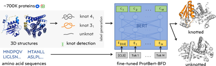
FIGURE 1 Scheme of the machine learning workflow. The entire approach consists of three main steps: 1. Obtaining data from PDB (with support of Knotprot) and AlphaFold (with support of AlphaKnot) predictions for UniProt; 2. Identifying nontrivial topology in the protein backbone using Topoly; 3. Training a machine learning model to predict nontrivial topology from the protein sequence.
FIGURE 1 Scheme of the machine learning workflow. The entire approach consists of three main steps: 1. Obtaining data from PDB (with support of Knotprot) and AlphaFold (with support of AlphaKnot) predictions for UniProt; 2. Identifying nontrivial topology in the protein backbone using Topoly; 3. Training a machine learning model to predict nontrivial topology from the protein sequence.CC BY · PubMed Central
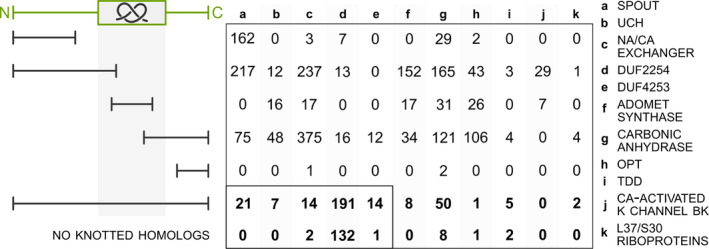
FIGURE 2 Unknotted proteins aligned with their knotted homologs. The schematic on the left represents the sequential alignment between knotted (green) and unknotted (black) proteins. The region with the knot is marked with a gray area. The unknotted proteins that start or end prematurely in comparison to their knotted counterpart are discarded from further analysis (see the section about SPOUT superfamily for details). We focus on cases in which the unknotted protein can be globally aligned with the knotted homolog (even with a deletion in the knot core) or does not have any knotted homologs (the numbers of these proteins are bolded).
FIGURE 2 Unknotted proteins aligned with their knotted homologs. The schematic on the left represents the sequential alignment between knotted (green) and unknotted (black) proteins. The region with the knot is marked with a gray area. The unknotted proteins that start or end prematurely in comparison to their knotted counterpart are discarded from further analysis (see the section about SPOUT superfamily for details). We focus on cases in which the unknotted protein can be globally aligned with the knotted homolog (even with a deletion in the knot core) or does not have any knotted homologs (the numbers of these proteins are bolded).CC BY · PubMed Central
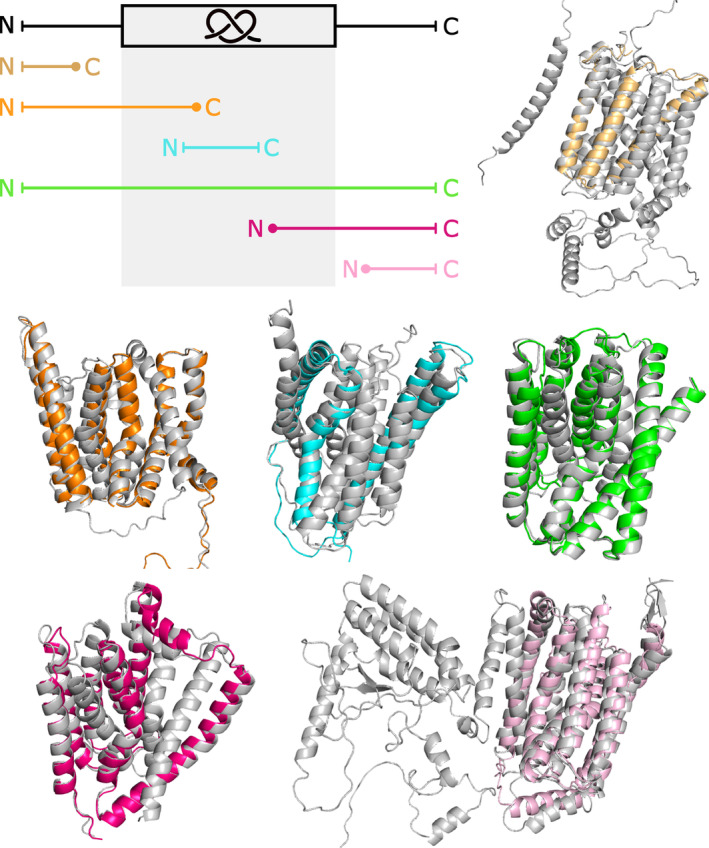
FIGURE 3 Unknotted membrane proteins from Sodium/calcium exchanger family superposed with their knotted homologs. Upper part: unknotted structure (light orange; UniProtKB ID: A0A5K0X7L3) without the entire knot core, unknotted structure (orange; UniProtKB ID: A0A397EWI1) structure lacking the C‐terminal part of the knot core, and unknotted structure (green; UniProtKB ID: A0A562DKV6) with low‐quality part of the knot core. Lower part: unknotted structure (cyan; UniProtKB ID: A0A453N937) without the N‐terminal and C‐terminal part of the knot core, unknotted structure (magenta; UniProtKB ID: A0A378FTV0) without the N‐terminal part of the knot core, and unknotted structure (light pink; UniProtKB ID: K1RUG8) without the entire knot core. All knotted homologs are shown with gray color (UniProtKB IDs: V4U0P6, A0A397A8U0, E6WVS2, A0A453N959, A0A0E1C8E8, and A0A0L8I7H8, respectively).
FIGURE 3 Unknotted membrane proteins from Sodium/calcium exchanger family superposed with their knotted homologs. Upper part: unknotted structure (light orange; UniProtKB ID: A0A5K0X7L3) without the entire knot core, unknotted structure (orange; UniProtKB ID: A0A397EWI1) structure lacking the C‐terminal part of the knot core, and unknotted structure (green; UniProtKB ID: A0A562DKV6) with low‐quality part of the knot core. Lower part: unknotted structure (cyan; UniProtKB ID: A0A453N937) without the N‐terminal and C‐terminal part of the knot core, unknotted structure (magenta; UniProtKB ID: A0A378FTV0) without the N‐terminal part of the knot core, and unknotted structure (light pink; UniProtKB ID: K1RUG8) without the entire knot core. All knotted homologs are shown with gray color (UniProtKB IDs: V4U0P6, A0A397A8U0, E6WVS2, A0A453N959, A0A0E1C8E8, and A0A0L8I7H8, respectively).CC BY · PubMed Central
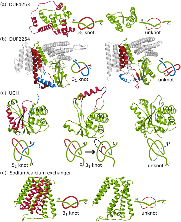
FIGURE 4 Overview of protein families with the potential of having both knotted and unknotted topologies. (a) Family DUF4253. A deletion (in red) aligning to the knot loop in the nontrivial homolog (UniProtKB ID: A0A1F0KRL5) creates an unknotted structure (B0C9T3). (b) DUF2254 family. The C‐terminus (in red) instead of passing through the loop (in blue) as in the knotted structure (A0A848U8E4), turns back creating a slipknot—an unknotted structure (D5C1U6). (c) UCH family. Compared with the standard structure with 52 topology (A0A0E0DGQ1), either we observe a different arrangement of the loop committing one crossing (in red), which simplifies the topology to 31 (A0A0E0DGQ2) or the fragment of N‐terminus is missing, which simplifies the topology to an unknot (A0A178ZI05). (d) Sodium/calcium exchanger transmembrane protein family. In the unknotted protein (A0A2G1VBT6) we observe the deletion of the fragment (in red) corresponding to the loop knot in the nontrivial homolog (A0A2S5ZE01).
FIGURE 4 Overview of protein families with the potential of having both knotted and unknotted topologies. (a) Family DUF4253. A deletion (in red) aligning to the knot loop in the nontrivial homolog (UniProtKB ID: A0A1F0KRL5) creates an unknotted structure (B0C9T3). (b) DUF2254 family. The C‐terminus (in red) instead of passing through the loop (in blue) as in the knotted structure (A0A848U8E4), turns back creating a slipknot—an unknotted structure (D5C1U6). (c) UCH family. Compared with the standard structure with 52 topology (A0A0E0DGQ1), either we observe a different arrangement of the loop committing one crossing (in red), which simplifies the topology to 31 (A0A0E0DGQ2) or the fragment of N‐terminus is missing, which simplifies the topology to an unknot (A0A178ZI05). (d) Sodium/calcium exchanger transmembrane protein family. In the unknotted protein (A0A2G1VBT6) we observe the deletion of the fragment (in red) corresponding to the loop knot in the nontrivial homolog (A0A2S5ZE01).CC BY · PubMed Central
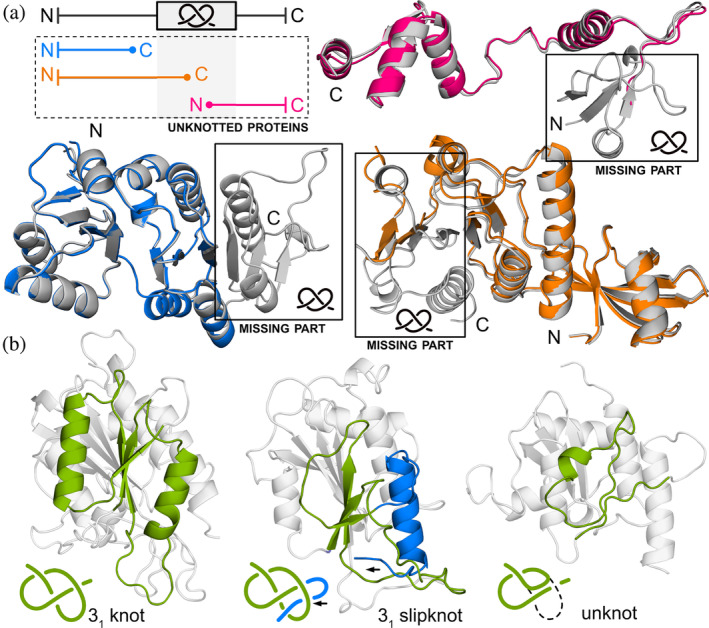
FIGURE 5 Unknotted SPOUT proteins (a) superposed with their knotted homologs. Upper part: unknotted structure (magenta; UniProtKB ID: A0A418H8R5) lacking the N‐terminal part of the knot core. Lower part: unknotted structure (blue; UniProtKB ID: A0A090WL68) without the entire knot core, and unknotted structure (orange; UniProtKB ID: A0A356CS99) without the C‐terminal part of the knot core. All knotted homologs are shown with gray color and their knotted regions are marked (UniProtKB IDs: A0A2X1NEC8, A0A090W861, and A0A3D0W667, respectively). Since all of these unknotted proteins lack at least a portion of the knot core, and the knot core is necessary for the SPOUT protein to function, we presume that these proteins are not truly unknotted. (b) Left: an example of a SPOUT protein with a typical 31 knotted structure (UniProtKB ID: R1DT92). Middle: structure with a 31 slipknot topology (UniProtKB ID: A0A424QRT9). The slipknot is formed by a C‐terminal end (colored blue) threaded back through the knot loop. The structure of the knot (colored green) remains typical for the SPOUT superfamily. Right: unknotted structure due to a deletion in the knot core (UniProtKB ID: R1FAN9). The presented structures are models from the AlphaFold database (4th version).
FIGURE 5 Unknotted SPOUT proteins (a) superposed with their knotted homologs. Upper part: unknotted structure (magenta; UniProtKB ID: A0A418H8R5) lacking the N‐terminal part of the knot core. Lower part: unknotted structure (blue; UniProtKB ID: A0A090WL68) without the entire knot core, and unknotted structure (orange; UniProtKB ID: A0A356CS99) without the C‐terminal part of the knot core. All knotted homologs are shown with gray color and their knotted regions are marked (UniProtKB IDs: A0A2X1NEC8, A0A090W861, and A0A3D0W667, respectively). Since all of these unknotted proteins lack at least a portion of the knot core, and the knot core is necessary for the SPOUT protein to function, we presume that these proteins are not truly unknotted. (b) Left: an example of a SPOUT protein with a typical 31 knotted structure (UniProtKB ID: R1DT92). Middle: structure with a 31 slipknot topology (UniProtKB ID: A0A424QRT9). The slipknot is formed by a C‐terminal end (colored blue) threaded back through the knot loop. The structure of the knot (colored green) remains typical for the SPOUT superfamily. Right: unknotted structure due to a deletion in the knot core (UniProtKB ID: R1FAN9). The presented structures are models from the AlphaFold database (4th version).CC BY · PubMed Central
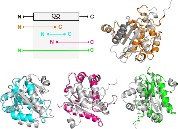
FIGURE 6 Unknotted UCH proteins superposed with their knotted homologs. Upper part: unknotted structure (orange; UniProtKB ID: A0A7S3I8E7) lacking the C‐terminal part of the knot core. Lower part: unknotted structure (cyan; UniProtKB ID: Q06AV7) without the N‐terminal and C‐terminal part of the knot core, unknotted structure (magenta; UniProtKB ID: A0A1A7YJ99) without the N‐terminal part of the knot core, and unknotted structure (green; UniProtKB ID: A0A024WZA) with low‐quality part of the knot core. All knotted homologs are shown with gray color (UniProtKB IDs: A0A078B145, A0A6P6PV05, A0A1A7X9F0, and W7F0M6, respectively).
FIGURE 6 Unknotted UCH proteins superposed with their knotted homologs. Upper part: unknotted structure (orange; UniProtKB ID: A0A7S3I8E7) lacking the C‐terminal part of the knot core. Lower part: unknotted structure (cyan; UniProtKB ID: Q06AV7) without the N‐terminal and C‐terminal part of the knot core, unknotted structure (magenta; UniProtKB ID: A0A1A7YJ99) without the N‐terminal part of the knot core, and unknotted structure (green; UniProtKB ID: A0A024WZA) with low‐quality part of the knot core. All knotted homologs are shown with gray color (UniProtKB IDs: A0A078B145, A0A6P6PV05, A0A1A7X9F0, and W7F0M6, respectively).CC BY · PubMed Central
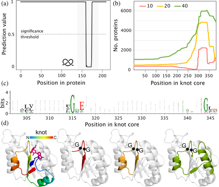
FIGURE 7 Important regions in SPOUT proteins based on machine learning model. (a) An example of how the model's prediction changes with respect to the patch location based on Nep1 protein (patches of length 10; UniProtKB ID: Q57977). Note the one significant drop inside the knot core. (b) Cumulative positions of the patches (with different lengths: 10, 20, and 40 amino acids) based on the knot core alignment. (c) Logo of the C‐terminal part of the knot core showing the conserved glycine motif. (d) Positions of patches in a TrmD structure (based on the N‐terminal domain of AlphaFold model of UniProtKB ID: B6YQ31). Colored regions represent from the left: knotted region (rainbow) and estimated location of the ligand (magenta); 10 (red), 20 (yellow), and 40 (green) amino acid‐long patches, respectively. Black dots show the position of the conserved glycine residues.
FIGURE 7 Important regions in SPOUT proteins based on machine learning model. (a) An example of how the model's prediction changes with respect to the patch location based on Nep1 protein (patches of length 10; UniProtKB ID: Q57977). Note the one significant drop inside the knot core. (b) Cumulative positions of the patches (with different lengths: 10, 20, and 40 amino acids) based on the knot core alignment. (c) Logo of the C‐terminal part of the knot core showing the conserved glycine motif. (d) Positions of patches in a TrmD structure (based on the N‐terminal domain of AlphaFold model of UniProtKB ID: B6YQ31). Colored regions represent from the left: knotted region (rainbow) and estimated location of the ligand (magenta); 10 (red), 20 (yellow), and 40 (green) amino acid‐long patches, respectively. Black dots show the position of the conserved glycine residues.CC BY · PubMed Central

Verification

Claims the paper makes

The claims and statistics of this paper haven’t been checked yet.

Run the check — free, no account needed. It reads the open-access text and checks the sources it cites.

A few free checks a day · papers already verified open instantly · results are public

Self-check

Percentages, sums and reported differences re-computed from the paper’s own numbers.

The self-check needs the paper’s full text.

Citations

Cites

Show all 3 references

Research works printed but not linked: 60

  1. 10.1371/journal.pcbi.1003002 · DOI
  2. 10.1111/j.1742-4658.2009.06990.x · DOI
  3. 10.1038/s41598-019-44928-3 · DOI
  4. 10.1002/pro.4380 · DOI
  5. 10.1038/s41467-017-01691-1 · DOI
  6. 10.1038/nsmb.3282 · DOI
  7. CikI RasamoelinaAD MachM SincakP.Explaining deep neural network using layer‐wise relevance propagation and integrated gradients.2021https://doi.org/10.1109/SAMI50585.2021.9378686 · DOI
  8. 10.1101/2023.03.13.532328 · DOI
  9. 10.1093/nar/gky1140 · DOI
  10. 10.1093/bib/bbaa196 · DOI
  11. 10.1371/journal.pone.0165986 · DOI
  12. 10.1038/s41467-023-42388-y · DOI
  13. 10.1016/j.csbj.2015.08.003 · DOI
  14. 10.1016/j.csbj.2022.07.027 · DOI
  15. 10.1073/pnas.1808312116 · DOI
  16. 10.1021/acs.jpcb.1c00898 · DOI
  17. 10.1090/S0273-0979-1985-15361-3 · DOI
  18. 10.1093/bioinformatics/bts565 · DOI · Paper page
  19. 10.1038/s42003-023-05489-4 · DOI
  20. 10.1016/j.jbc.2023.105553 · DOI
  21. 10.1016/j.sbi.2023.102709 · DOI
  22. 10.1016/j.sbi.2016.10.002 · DOI
  23. 10.1093/bioinformatics/bty334 · DOI
  24. 10.1016/j.jmb.2018.10.012 · DOI
  25. 10.1073/pnas.1007602107 · DOI
  26. 10.1016/j.jmb.2007.07.042 · DOI
  27. 10.1016/j.jmb.2014.09.007 · DOI
  28. 10.1038/nsb0494-213 · DOI
  29. 10.1016/b978-0-12-824406-7.00008-7 · DOI
  30. 10.3389/fmolb.2023.1223830 · DOI
  31. 10.1093/nar/gkac388 · DOI
  32. Paszke A, 2019, Advances in neural information processing systems 32, 8024
  33. 10.1093/nar/gkac993 · DOI
  34. 10.1021/acscatal.0c00059 · DOI
  35. 10.1002/pro.4631 · DOI
  36. 10.1371/journal.pcbi.1007904 · DOI
  37. 10.1371/journal.pcbi.1000864 · DOI
  38. 10.48550/ARXIV.1610.06679 · DOI
  39. 10.1038/268495a0 · DOI
  40. 10.1038/s41598-020-66258-5 · DOI
  41. 10.3390/ijms160818836 · DOI
  42. 10.1101/2023.06.16.545156 · DOI
  43. 10.1007/s00894-022-05094-y · DOI
  44. 10.1371/journal.pcbi.1002504 · DOI
  45. 10.1038/nbt.3988 · DOI · Paper page
  46. 10.1038/s41467-018-04964-5 · DOI · Paper page
  47. 10.1016/j.jbc.2022.102393 · DOI
  48. 10.1073/pnas.0805468105 · DOI
  49. 10.1016/j.sbi.2020.01.007 · DOI
  50. 10.1073/pnas.1205918109 · DOI
  51. 10.1073/pnas.0811147106 · DOI
  52. 10.1021/ja961147m · DOI
  53. 10.1186/1471-2105-8-73 · DOI
  54. 10.1093/nar/gkab1061 · DOI · Paper page
  55. 10.1371/journal.pcbi.0020122 · DOI
  56. 10.1016/j.jmb.2007.02.035 · DOI
  57. 10.1039/D0SC02796K · DOI
  58. 10.1021/jp511029s · DOI
  59. 10.1039/c6mb00214e · DOI
  60. 10.1371/journal.pcbi.1009502 · DOI

Metadata

Authors
Sikora, M., Klimentova, E., Uchal, D., Sramkova, D., Perlinska, A. P., Nguyen, M. L., Korpacz, M., Malinowska, R., Nowakowski, S., Rubach, P., Simecek, P., & Sulkowska, J. I.
Affiliations
  • University of Warsaw (PL)
  • Central European Institute of Technology, Masaryk University Brno Czech Republic
  • Centre of New Technologies, University of Warsaw Warsaw Poland
Corresponding author
Sulkowska, Joanna I.
Venue
Protein Science
Publisher
Wiley
Published
2024-06-18
Volume
33
Issue
7
Pages
e4998-e4998
ISSN
  • 0961-8368 (ISSN-L)
  • 1469-896X
OpenAlex
W4399776685
Cited by
13
References
3
Citations by year
2026: 3 · 2025: 5 · 2024: 5
FWCI
1.77
Type
journal article
Language
en
Open access
Locations
  • Protein Science (publishedVersion, cc-by)
  • bioRxiv (Cold Spring Harbor Laboratory) (acceptedVersion, cc-by-nd)
  • PubMed Central (publishedVersion, cc-by)
  • PubMed (publishedVersion)
  • ReIn UW (University of Warsaw) (submittedVersion, cc-by-nc-nd)
Funders
  • Narodowe Centrum Nauki (UMO‐2018/31/B/NZ1/04016, 2021/43/I/NZ1/03341)
  • Grantová Agentura České Republiky (23‐04260L)
Topics
  • Life Sciences › Biochemistry, Genetics and Molecular Biology › Molecular Biology › Protein Structure and Dynamics
  • Life Sciences › Biochemistry, Genetics and Molecular Biology › Molecular Biology › Machine Learning in Bioinformatics
  • Physical Sciences › Mathematics › Geometry and Topology › Geometric and Algebraic Topology
Keywords
knotted proteins, sequence-based prediction, protein topology, AlphaFold, UniProt
MeSH terms
Machine Learning, Amino Acid Sequence, Models, Molecular, Protein Conformation, Proteins, Databases, Protein

About these checks

Registry, retraction, Cites and statistics results are computed, not judged, with one exception: a printed reference that no index lists is read once by a model before it counts as a citation error. Claims are extracted by a model and kept only when their quote appears verbatim in the text; each cited source is then checked, with a model’s help, for whether it says what the claim relies on. Those verdicts can be wrong — read the claim’s quote and the cited source before relying on one.

Statistics and arithmetic are recomputed, not judged.

Confidence bands are shown only for fields calibrated on our annotated test set. None is calibrated yet, so no band is shown.

What counts as a claim ›How we measure confidence ›How we check this ›

Are you an author, or do you see a problem with this page? Request a review

How to cite

Cite CoChat for the checks on this page, and the paper for its own findings.

This page
CoChat. (2026). Citation check: Knot or not? Identifying unknotted proteins in knotted families with sequence‐based Machine Learning model. https://app.cochat.ai/papers/10.1002/pro.4998
The paper
Sikora, M., Klimentova, E., Uchal, D., Sramkova, D., Perlinska, A. P., Nguyen, M. L., Korpacz, M., Malinowska, R., Nowakowski, S., Rubach, P., Simecek, P., & Sulkowska, J. I. (2024). Knot or not? Identifying unknotted proteins in knotted families with sequence‐based Machine Learning model. Protein Science. https://doi.org/10.1002/pro.4998